

 PDF
PDF ePub
ePub Citation
 Print
Print


INTRODUCTION
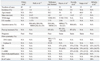
Glycogen rich clear cell carcinoma (GRCC) of the breast is known as a rare subtype of invasive ductal carcinoma, and involves a poor prognosis. It was first described by Hull, et al.1 in 1981. Since then, fewer than 150 cases have been reported. Many researchers have attempted to characterize GRCC, according to, electron microscope (EM),1,2 flow cytometry,3 or clinical data,4-6 the results of which have been summarized in Table 1.
Hull, et al.1 confirmed intracytoplasmic glycogen of GRCC by EM and stated that the H&E and EM features of GRCC were very similar to the morphology of 13-week old fetal developing mammary gland. In diagnosing GRCC, tumor cells that contain abundant fine glycogen granules in their clear cytoplasm, are stained by periodic acid stain (PAS), but not stained by diastase-periodic acid stain (d-PAS). The incidence of GRCC is estimated to be 1 to 3% of breast carcinoma. Opinions differ with among researchers concerning the prognosis of GRCC, ranging from favorable to unfavorable.2-6 Immunohistochemical (IHC) staining for estrogen receptor (ER) is usually positive. However, IHC for progesterone receptor (PR) and c-erbB2 has been shown to vary.5-8 Other than hormone receptors, no organized study concerning the immunophenotype of GRCC has been published. Here, we present three cases of GRCC and their immunohistochemical profiles.
MATERIALS AND METHODS
Clinical data
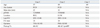
Table 2 summarizes the clinical data. There were only three cases found during the last 10 years. One case was lost to follow up after diagnosis by biopsy. The ages at diagnosis were 49 to 69 years old and follow up data were 14 months and 18 months. They were currently disease-free states. They were treated with total and partial mastectomy followed by chemotherapy and radiation therapy.
Immunohistochemical (IHC) staining
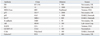
The antibodies used to conduct this study are listed in Table 3. Paraffin blocks were sectioned to a thickness of 4 um and tissues were attached on silane coated slides. The slides were baked at 60℃ for 60 minutes and washed in xylene for deparaffinization. For hydration of sections, the slides were bathed in gradient alcohol and distilled water. The antigen retrieval step was followed by boiling slides in 10 mM citrate buffer (pH6.0) for 10 minutes. Afterwards, the slides were immersed in 3% H2O2 for 15 minutes to block intrinsic hydro-peroxidase activity. The slides were incubated with primary antibodies for 2 hours at room-temperature, followed by incubation with secondary antibody (Enzyme-conjugated polymer, EnVision Detection kit, Denmark) for 15 minutes at room-temperature. A NovaRED substrate kit (VECTOR Laboratory, Burlingame, CA, USA) was applied to develop color following the manufacture's protocol. Harris hematoxylin was used for counter staining.
Interpretation of immunohistochemical staining
IHC slides were evaluated by light microscopy. The scoring intensity of IHC staining was based on a three-grade system; 1 positive (weak), 2 positive (intermediate) and 3 positive (strong). The grade intensity of positive stainig was assessed as follows; nuclear positivity for ER, PR, androgen receptor (AR), p53, p63, and ki-67; cytoplasmic positivity for gross cystic disease fluid protein 15 (GCDFP-15); nuclear and cytoplasmic positivity for c-kit and galectin3; and cytoplasmic membrane positivity for c-erbB2, e-cadherin, and epidermal growth factor receptor (EGFR). Additionally, a proportional index was evaluated to describe the percentage of positivity for a few of the antibodies, including hormone receptors (ER, PR, and AR) and p53.
RESULTS
Three patients were diagnosed with invasive glycogen rich clear cell carcinoma from 2000 to 2011 at our institution. On gross examination, GRCCs appeared as well-circumscribed, irregular and solid masses. No difference was found in comparison to conventional invasive ductal carcinoma. Microscopically, GRCCs consisted of solid nests of clear cells with fine meshwork vasculatures. The individual cancer cells showed clear cytoplasm with fine granules and relatively low nuclear and cytoplasmic ratio. Nuclei were somewhat wrinkled. Nuclear membranes were irregularly thickened and their nucleoli were prominent. All of them contained PAS positive and d-PAS labile granules in the cytoplasm of more than 90% of tumor cells (Fig. 1). Mucin was not detected upon alcian blue and mucicarmin staining. Tests for GCDFP-15 were negative. The tumors were, therefore, considered to involve no apocrine differentiation.
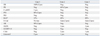
The three cases revealed three different immuno-phenotypes (Table 4). Case 1 showed positivity for ER and negativity for PR. Case 2 was positive for PR, but negative for ER. Case 3 presented with triple negative invasive carcinoma. All three cases were negative for c-erbB2. Androgen receptor was positive in the ER positive case 1. EGFR, E-cadherin, and CD117 (c-kit) showed positivity in two cases (cases 2 and 3). Among these three cases, p53-positive case exhibited a low proliferative index (ki-67: 15%), while p53-negative cases showed high proliferative index (ki-67: 50-60%).
DISCUSSION
GRCC is a very rare entity that exhibits distinctive clear cells with intracytoplasmic glycogen. However, no trials have been conducted to reveal the distinct characteristics of GRCC, including clinical behavior, biologic features, immunophenotypes, and genetics.2-6,8
Because of the rarity, we accumulated only three cases of GRCC out of more than 2000 cases of carcinoma over the last 10 years. Twelve different antibodies were evaluated in the three cases, the immunophenotypes of which prove not to be uniform. Based on immuno-scoring, we were able to characterize the expression patterns of targeted proteins into three groups. The first group of proteins shared the same immuno-scoring and were related to cell surface molecules, including EGFR, c-kit, and E-cadherin. The second group was related to hormone receptors, including ER and AR. The third group was characterized by p53 related proliferation.
EGFR and c-kit are both cell surface receptors important to the PI3K through AKT and RAS to MAPK signal transduction pathways, and have been shown to be involved in cell survival and proliferation,9,10 Double expression of EGFR and c-kit in breast carcinoma is known as a predictive marker of poor prognoses.11 Among our cases, ki-67, marker of cell proliferation, was also synchronously up-regulated along with up-regulation of EGFR and c-kit. Taken together, in predicting poor prognoses, expression of both EGFR and c-kit sould be associated with high mitotic index. In our GRCC cases, case 1 was both EGFR and c-kit negative with a low mitotic index, while the other two (cases 2 and 3) were positive for both with a high mitotic index. The expression pattern of EGFR and c-kit was concordant with E-cadherin and discordant with ki-67. Most studies on E-cadherin have focused on lobular carcinoma.12 The role of E-cadherin in ductal carcinoma is still unknown. The function of E-cadherin is considered related with cell adhesion molecules; thus, loss of expression would be associated with a high incidence of invasion and metastasis. A few researchers stated that up-regulation of E-cadherin would be associated with a better prognosis in invasive ductal carcinoma.13 However, the mechanism is unclear.
P53 is a well known nuclear protein that participates in cell proliferation. In benign conditions, p53 is not detectable by IHC staining. Cancer progression demonstrates accumulations of non-functional and non-degraded p53 protein in nuclei, resulting in immuno-positivity of p53 protein. Mutated p53 protein cannot suppress genetic errors leading to cancer development. Nevertheless, p53 over-expression has been considered a poor prognostic marker and accompanied by a high mitotic index in invasive ductal carcinoma.14 Our three cases demonstrate just the opposite. Case 1 showed strong positivity for p53 with low mitotic index, while cases 2 and 3 showed negativity for p53 with high mitotic index. These findings support the possibility of an association of cell proliferation with the signal transduction of surface molecules (EGFR, c-kit and E-cadherin), not with mutation of p53 protein, in GRCC.
Case 3 presented as a triple negative carcinoma and also expressed p63 protein. Case 3 was a basal-like breast cancer and was predicted to involve a poor prognosis. Increased proliferative activity as determined by ki-67 (60%) and over-expression of EGFR supported the possibility of a poor prognosis. The diagnosis of this case was confirmed only 3 months prior to the preparation of this manuscript; therefore, follow up data are currently not available. Based on IHC results, this case will require continued attention for future treatment. Presently, researchers agree that if GRCC is comparable to the conventional ductal carcinoma of the same TNM stage, the prognosis of the two types of carcinomas would be the same.7
As only three cases were available for study, the drawing of any conclusions would be limited. However, these cases did reveal different IHC characteristics for GRCC. We believe that the cases presented herein substantiate a need to re-examine the general features of GRCC. While the immunophenotypes of GRCC were not uniform, they appeared to be very similar to conventional ductal carcinoma.

 XML Download