

 PDF
PDF ePub
ePub Citation
 Print
Print


INTRODUCTION
Neuromuscular disease (NMD) is characterized by progressive weakening of skeletal, respiratory, and or bulbar-innervated muscles.1 NMD patients have decrements in vital capacity (VC), lung and chest wall compliance, and coughing capacity. These patients cannot fully expand their chests, which leads to stiffening of the joints and tissues of the rib cage.1,2 This in turn results in a reduction in chest wall compliance.2 Reduction in lung and chest wall compliance may also be related to alterations in the elastic properties of lung tissues by chronically limited range of activity.3 Normal breathing consists of varying tidal volumes with intermittent deep breaths or sighs.1,4 Even in people with normal lungs, periodic hyperinflation is required to prevent closure of lung units.1 For NMD patients, who cannot expand the lungs fully due to respiratory muscle weakness, periodic deep insufflation by air-stacking is essential.1
Air stacking involves the use of a manual resuscitator or a volume-cycled ventilator to deliver volumes of air that are consecutively held by glottic closure until no more air can be retained.5 The maximum lung volume that can be held by air stacking is the maximum insufflation capacity (MIC).6 In 2000, Kang and Bach1 demonstrated that the MIC could significantly exceed VC for patients with NMD. However, patients presenting weak or no glottic closure due to bulbar muscle dysfunction or indwelling tracheostomy cannot air stack. Lung insufflation can only be given by bypassing glottis dysfunction in these patients, and the maximum passive lung insufflation volume achieved in this manner is defined as the lung insufflation capacity (LIC).7
In this study, we designed a device that provides external control of an artificial glottic opening and closure for patients with bulbar innervated muscle weakness or who have indwelling tracheostomy tubes. We attempted to investigate the effectiveness of air stacking by replacing dysfunctional glottises with the newly designed device.
MATERIALS AND METHODS
Patients
The study consisted of 37 subjects with bulbar innervated muscle weakness or indwelling tracheostomy tubes. Bulbar-innervated muscle weakness was defined by clinically apparent dysarthria and/or dysphagia and impaired glottic function. Exclusion criteria were: inability to cooperate due to severe cognitive impairment; medical instability; and severe overriding lung and airway disease.
Methods
Artificial external glottic device (AEGD)
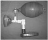
An AEGD has a connection and a control part (Fig. 1). The connection part is a T-shaped plastic adaptor. The control part is designed to artificially modulate glottic opening and closure.
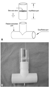
The connection part
As shown in Fig. 2, the connection part consists of a patient connection port, the insufflation port, and the exhalation port. The patient connection port is connected directly to the patient's airway through a tracheostomy tube or an oronasal mask (Fig. 2A). A manual resuscitator delivers air via the insufflation port, which is located in the middle of the connection part and at a right angle. It has a one-way valve (Fig. 2A). The exsufflation port is connected to the control part.
The control part
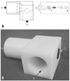
The control part of the AEGD artificially modulates glottic opening and closure by pushing a button. The tip of the button protrudes from the outer wall of the control part. Pushing the button permits the air to flow from the patient to the spirometer connected to the control part. When the button is pressed, the concave portion of the bar is exposed to the connecting tube, and the patient can exhale. A spring is inserted in the connecting axis of the bar, and this axis is designed to pass through the center of the hole in a disc (Fig. 3). When the button is not pressed, airflow from the connecting tube is completely interrupted by the bar, which is fixed due to the recoil of the spring and support action of the disc hole. Therefore, patients are permitted to exhale after passive lung insufflations by pressing the AEGD button to open.
Clinical evaluation
This study was approved by the institutional review board, and informed consent was obtained from all of the study candidates. All lung volumes were measured by Micro Spirometer (Micro Medical Ltd., Rochester, Kent, UK) in a sitting position, and recorded as the greatest observed value in at least three attempts.
Outcome measurements were as follows:
1) For the measurement of VC, all subjects were asked to take a deep breath, and then exhale the maximally held volume of air into the spirometer.
2) MIC was then measured as described.1 The maximum value that was observed in at least three attempts was recorded.
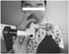
3) LIC with AEGD (LICA) was measured by using the same manual resuscitator with MIC after connecting an AEGD to the measurement apparatus (Fig. 4). The largest value in three or more attempts was recorded as the LICA.
Data analysis
A paired t-test was used to determine the statistical significance between VC and LICA, and MIC and LICA. The Kruskal-Wallis method was used to determine the statistical significance among VC, MIC, and LICA. Data was analyzed using SPSS 12.0b, and a p-value <0.05 was considered statistically significant.
RESULTS
Thirty-seven patients (24 males, 13 females; mean age, 48; range, 20-76 years) met the criteria, and were included in this study. Mean±standard deviation values of all the patients totaled were 794±391 mL for VC, 1,084±260 mL for MIC, and 1,668±493 mL for LICA measurements.
When candidates were divided by diagnosis (Table 1), only 6 from the ALS group and 1 from the cervical spinal cord injury group could somewhat air stack. The remaining 30 patients failed to air stack due to glottic dysfunction, thus MICs were checked as zero. AEGD either maximized the ability to air stack or enabled passive lung insufflation even for those who initially failed. Consequently, LICA significantly exceeded the MIC for all of the candidates (p<0.05).
In accordance with underlying pulmonary function, 37 patients were subdivided into groups 1, 2 or 3 (Table 2). Group 1 included three patients who presented severe inspiratory and expiratory muscle weakness with tracheostomies. Since their respiratory muscles were too weak to even take a deep breath in and out, VCs and MICs were essentially 0 mL. Group 2 included 27 patients whose MICs were initially 0 mL. After applying the AEGD, LICAs were successfully measured in both groups. The other 7 patients who had the minimal residual glottic function to hold and stack a little volume of air were named as Group 3. Although MIC-VC difference was greater than 0 mL, when using AEGD, the LICA was greater than MIC or VC in this group (p<0.05).
DISCUSSION
Just as limb range of motion helps avert limb contractures, assisted lung insufflations help to maintain compliance for patients with diminished VCs.1 However, NMDs with glottic dysfunction and/or tracheostomized patients can not benefit from air stacking, because they cannot prevent leakage of inhaled air from the lungs.8,9 Besides the inability to maintain pulmonary compliance, these patients also present difficulty with coughing due to respiratory muscle weakness and/or glottic dysfunction. Thus effective cough mechanisms can be hindered, which in turn results in the failure of eliminating airway secretions.10,11
Bach had already defined a simple technique to provide deep lung insufflations, and called the deep passive lung insufflations attained this way as LIC.7 He used a manual resuscitator connected to a spirometer, and simply occluded the expiratory port of the spirometer with fingers while providing an additional volume of air with the manual resuscitator bag. Through this method, he was not only able to give deep lung insufflations, but also quantified the volumes.
In this study, on the other hand, we developed a clean, mechanical device to substitute for dysfunctional glottises. Despite the methodological difference, our handheld device is conceptually identical to Bach's simple method in providing deep lung insufflations and quantifying them. Although Bach's method is convenient in that does not require any device, AEGD is a clean technique that might provide more precise control and measurement of the glottic function by preventing possible air leaks. In addition, quantification of coughing ability was also possible with AEGD by changing the measurement apparatus to a peak flow meter. Recruited candidates are another distinguishing factor. Although the previous study7 presented a larger number of patients, the majority of the patients had relatively intact bulbar muscle function whose ability to air stack was spared. However, in this study, air stacking was essentially impossible in more than two thirds of the patients. About 70% of the candidates had tracheostomy tubes, and the remaining patients were bulbar-dominant ALS who showed severe glottic dysfunction.
Limitations of this study include the fact that in the process of connecting the AEGD with measuring instruments, the value could be underestimated, because the exhaled air passes the connection system at a right-angled. Consequently, the fully assembled apparatus for measurement had a relatively long pathway for expiratory flow. Speed of the flow can also be a problem. Thus, measured MICA values may actually be greater than those reported in this study.
In conclusion, NMD patients need to practice regular deep lung insufflations. The artificial external glottic device enables passive lung insufflations even for patients with severe bulbar-innervated muscle impairment or tracheostomy tubes, whose ability to air stack is poor.


 XML Download