

 PDF
PDF ePub
ePub Citation
 Print
Print


INTRODUCTION
Human leukocyte antigen G (HLA-G) is a non-classical HLA class I molecule expressed by extravillous trophoblast cells at the feto-maternal interface during pregnancy.1 Messenger RNA of HLA-G can be alternatively spliced into at least seven transcripts, which encode four membrane bound isoforms (G1, G2, G3, and G4) and three soluble isoforms (G5, G6, and G7).2 The proteolytic cleavage of surface isoform HLA-G1 generates the soluble HLA-G1 form (sHLA-G1). Both full-length membrane-bound HLA-G1 and two soluble forms (sHLAG1 and HLA-G5) have been shown to exert various immunosuppressive effects such as inhibiting the cytotoxic activity of CD8 positive T lymphocytes and natural killer (NK) cells, inducing the apoptosis of NK and activating cytotoxic T cells.1 Several lines of investigation indicate that the HLA-G molecule is involved in maternal acceptance of the semi-allogenic fetus during pregnancy and in the development of tolerance.2
Although sHLA-G is primarily expressed in the invading trophoblast cells, it can be detected in plasma or serum not only from pregnant women, but also from non-pregnant women. A low serum level of sHLA-G1 in the preovulatory period and during the first nine gestational weeks has been suggested as a risk factor for early abortion after in vitro fertilization (IVF).3 Complications during pregnancy, such as severe pre-eclampsia, spontaneous abortion, intrauterine growth retardation, and premature birth, are associated with a low or undetectable level of sHLA-G1 in the maternal blood circulation.4
Accumulating evidence supports the fact that preimplantation human embryos can secrete sHLA-G into culture supernatants and the levels may be predictive of their ability to implant.2,5 Soluble HLA-G is also found in follicular fluid (FF) where its presence has been linked to the presence in corresponding embryo culture supernatants.6 An immunocytochemical study revealed that granulosa cells are responsible for production of sHLA-G.6,7 Soluble HLA-G molecules are detected also in supernatants of cumulus-oocyte complex (COC) that generates mature oocytes, but not in supernatants of COCs that correspond to immature oocytes.8
Because FF represents an essential and specific microenvironment for the regulation of ovarian function, a relationship has been proposed between specific FF components and IVF outcome. However, the role of sHLA-G within FF remains still unknown. In the present study, we investigated the direct relationship between FF levels of sHLA-G and fertilizability of the corresponding oocyte as well as the morphological quality of corresponding embryo.
MATERIALS AND METHODS
Our study involved 63 infertile women participating in IVF procedure between March 2007 and March 2010. The cycle was included when at least one mature oocyte was obtained and the corresponding FF from a single follicle was available. The averaged age was 34.4±3.3 years (range 27-40). Fifty-one women were nulliparous including 33 who were nulligravid. Thirty women had no history of previous IVF treatment. The indications of IVF were identified as tubal (n=25), unexplained (n=20), male (n=7), diminished ovarian reserve (n=5), polycystic ovary syndrome (n=3), endometriosis (n=2), and uterine factor (n=1). The study was approved by the Institutional Review Board of Seoul National University Hospital to use stored FF samples.
Patients were stimulated with recombinant FSH (Gonal-F; Merck-Serono, Geneva, Switzerland). The pituitary was suppressed by the gonadotropin-releasing hormone (GnRH) agonist long protocol (n=5) or GnRH antagonist protocol (n=58). For the GnRH agonist long protocol, daily injections of 0.1 mg triptorelin acetate (Decapeptyl; Ferring, Saint-Prex, Switzerland) were started in the mid-luteal phase of the previous cycle. After confirmation of pituitary down-regulation, gonadotropin injections were initiated on cycle day 2 or 3. The initial gonadotropin dose was fixed for the first 4 or 5 days, followed by adjustment on the basis of individual follicular growth and E2 levels until triggering. For the GnRH antagonist protocol, gonadotropin was started on cycle day 2 or 3. When the leading follicle reached a diameter of 14 mm, cetrorelix 0.25 mg (Cetrotide; Merck-Serono) was added daily until triggering.
Follicular growth was monitored by transvaginal ultrasound and serum E2 levels, which were started on day 4 or 5 of the stimulation and repeated as appropriate. When at least three follicles reached 17 mm or two follicles reached 18 mm in diameter, exogenous hCG (Ovidrel, Merck-Serono) was administered and oocytes were retrieved transvaginally 36 hrs later. At the time of oocyte retrieval, the single dominant follicle (>17 mm in diameter) was punctured. If one intact mature oocyte was present and the FF was not contaminated by visible blood, the FF was isolated. To prevent contamination with other follicles, FF was collected from only one follicle for each ovary, and different needle was used for each side of the ovary. All FF samples were centrifuged immediately at 1,300 g for 10 min and the supernatants were stored at -80℃ until the assay.
IVF and subsequent culture of embryo were performed as previously reported.9 In each study subject, ejaculated sperms were used for fertilization. The mean of the total motile sperm count was 53.0×10⁶. Intracytoplasmic sperm injection (ICSI) was used in nine cycles. Normal fertilization was defined as those showing two distinct pronuclei. Non-fertilized oocyte or those with three pronuclei was considered as failed fertilization. Subsequently, individual embryo quality was evaluated at day 3 and graded by morphological criteria;
Grade A: symmetrical blastomeres and no fragmentation
Grade B: slightly uneven blastomeres and <20% fragmentation
Grade C: uneven blastomeres and >20% fragmentation
Grade D: uneven blastomeres and >50% fragmentation
Good-quality embryos were defined as those with morphologic grade A or B. The embryo score was also calculated by multiplication of morphological grade of the embryo (Grade A=4, Grade B=3, Grade C=2, Grade D=1) by the number of blastomeres.
All well-developed embryos were transferred to each patient's uterus at day 3. The luteal phase was supported with daily intramuscular progesterone initially for 14 days starting on the day of oocyte retrieval and continued for another 6-8 weeks in cases where a pregnancy was achieved. A clinical pregnancy was defined by the presence of an intrauterine gestational sac with pulsating fetal heart beat 3-4 weeks after oocyte retrieval.
The levels of soluble HLA-G in FF were estimated by enzyme-linked immunosorbent assay (ELISA) using commercial sandwich ELISA kit (RD194070100R, BioVender, Czech Republic). The kit with monoclonal antibody could measure shed HLA-G1 and HLA-G5 in variable specimens. According to procedure described in manufacturer's instruction, the samples were incubated with microplate wells pre-coated with monoclonal anti-sHLA-G antibody. After 60 min of incubation and washing, monoclonal anti-human beta2-microglobulin antibody labeled with horseradish peroxidase was added to the wells and incubated for 60 min with captured sHLA-G. A calibration curve was constructed by plotting absorbance values against concentrations of calibrators, and concentrations of each sample were determined using the calibration curve. The FF was not diluted, and single measurement was performed in each sample. The limit of detection was 3 U/mL, and the intra- and inter-assay coefficients of variation were 5.2% and 5.5%, respectively.
Statistical analysis was performed using MedCalc 4.15 (MedCalc Software, Mariakerke, Belgium). Since the sub-group data did not show a normal distribution, the Wilcoxon test was used to compare the medians. Data are presented as the median (25th percentile, 75th percentile). The Chi-square test was used to compare proportions. The Spearman correlation test was used to assess degree of association between different parameters. Sensitivity and specificity were calculated, and the receiver operating characteristic (ROC) curve was plotted by changing the criteria of sensitivity and specificity. A p-value of <0.05 (two-tailed) was considered statistically significant.
RESULTS
The overall fertilization rate was 84.1% (53/63). Using the ICSI procedure, seven of nine oocytes were normally fertilized (77.8%), and the rate did not differ when compared with conventional IVF (85.2%, p=0.946). Soluble HLA-G was not detected in 15 FF samples. In the group with positive FF sHLA-G (n=48), the median concentration was 37.2 U/mL (6.5, 135.5), and the fertilization rate was 85.4%. This rate was not statistically different from the negative FF sHLA-G group (80.0%, p=0.927).
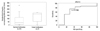
When fertilization occurred, the median concentration of positive FF sHLA-G was 35.5 U/mL (6.1, 118.2). In contrast, the median concentration was 173.3 U/mL (55.1, 187.8) when fertilization did not occur and the difference was not statistically significant (p=0.140). However, by using ROC curve analysis, high levels of sHLA-G (>117.758 U/mL) could predict the failure of fertilization with a statistical significance
(AUC 0.676, 95% CI 0.525-0.804, sensitivity 71.4%, specificity 75.6%, positive likelihood ratio (LR) 2.93, negative LR 0.38) (Fig. 1).
Among those with both a positive FF sHLA-G and fertilization (n=41), the sHLA-G level could not predict the formation of a good quality embryo (AUC 0.512, 95% CI 0.351-0.671, sensitivity 93.3%, specificity 27.3%, positive predictive value 77.8%, negative predictive value 60.0%). When the corresponding embryos had grade A or B, the median concentration of positive FF sHLA-G was 32.6 U/mL. The level did not show a statistical difference when compared with grade C or D embryos (38.4 U/mL, p=0.906). In addition, the concentration of FF sHLA-G did not correlate with the quality score of its matched embryo (r=-0.147, p=0.361).
DISCUSSION
Our results suggest that the concentration of sHLA-G in the FF of a dominant follicle can serve as a predictor of normal fertilization, but not of embryo quality. Contrary to our expectations, a low level of sHLA-G in FF was closely related to higher fertilizability of the corresponding oocyte. When FF sHLA-G was 117.758 U/mL or lower, the fertilization rate was 93.9%; in contrast, the fertilization rate was 66.7% among subjects above the cut-off value (p=0.042). However, FF sHLA-G appears not to be the sole factor influencing the fertilization process because 80.0% of non-detectable cases also resulted in normal fertilization.
The role of sHLA-G within FF remains largely unknown. Although immunocytochemical study indicates that granulosa cells can produce sHLA-G molecules, FF sHLA-G could possibly originate from peripheral circulation.6,8 Since sHLA-G has various immunosuppressive effects, a relatively high concentration of FF sHLA-G might lead to a strong immunosuppressive environment, resulting in repression of pro-inflammatory cytokines within FF. Indeed, it has been reported that higher FF IL-1beta levels are associated with normal fertilization.10 Taken together, the close relation between high FF sHLA-G and low fertilization potential of the corresponding oocyte could be linked with low IL-1beta production within FF, which could prevent cytoplasmic maturation and normal fertilization. However, further investigation could elucidate whether immuno-modulating substances can directly affect oocyte quality.
We could detect FF sHLA-G in 76.2% of the study subjects. The positivity appeared to vary depending on the use of different assay kits. Rizzo, et al.6 reported a 38% detection rate, while other researcher, using the same antibody, observed a positivity of 47%.7 However, none of them reported the relation with oocyte fertilization potential and embryo quality.
The assessment of oocyte quality is rapidly becoming one of the major objectives of embryologists in human IVF.11 The potential use of non-invasive markers for selecting the best quality embryos in IVF is of immense importance, especially in countries like the UK where there is an increasing move towards single embryo transfer to reduce multiple pregnancy rates. Currently, oocyte morphology is the most popular method for selecting competent oocytes.
In the present study, we could not assess the direct relation between oocyte morphology and FF concentration of sHLA-G, because only good quality oocytes were studied. In conclusion, our findings indicate that FF sHLA-G testing could be used as an additional tool to assist oocyte selection. Further studies are required to fully understand the nature of sHLA-G expression.
 XML Download