

 PDF
PDF ePub
ePub Citation
 Print
Print


INTRODUCTION
A single coronary artery originating from the left or right coronary ostium occurs infrequently. Clinical outcomes for these anomalies are unfavorable. Congenital coronary artery anomalies are often detected incidentally by coronary angiography or autopsy findings. Unfortunately, these anomalies are not detectable on an electrocardiogram, chest X-ray, or thoracic echocardiography.1
The classification of coronary anomalies has been developed in accordance with the origin, course, and termination of the anomalous vessel. Certain anomalies are related to syncope, angina pectoris, exertional dyspnea, heart disease, and sudden death.2 Patients who have coronary anomalies could also have other cardiac malformations, however, may not be aware of this risk or the implications of those potential malformations.
This report describes a young woman who unexpectedly died shortly after the onset of symptoms and was identified postmortem as an undiagnosed congenital coronary anomaly.
CASE REPORT
According to the police report, a 19-year-old woman who majored in Security Services in college died while attending a picnic party. She had a lunch and climbed a mountain for almost two hours. She then rested for an hour. After resting, she suddenly lost consciousness while too competitively playing the stretch-the-legs game as a team member, Resuscitation was performed by her colleagues. She was then immediately taken to a hospital where she failed to recover and died. Her family reported that she had previously been in good health. However, her colleagues stated that she had one recent episode of exertional dyspnea while rafting and was absent from school for three days. Unfortunately, we have no any her medical records for further evaluation or treatment of this symptom.
Autopsy findings
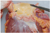
She was approximately 160 cm tall and weighed 66 kg. Upon external examination, there were defibrillator related bruises on the chest and two needle punctures in the cubital fossa. Internally, the lungs weighed 904 g and 923 g, respectively. There were petechial subpleural hemorrhages on the interlobular surfaces. The heart showed slightly hypertrophic (weighed 349 g). There were petechial hemorrhages on the posterior interventricular groove. The right and left coronary arteries simultaneously opened to a single bifurcated ostium on the left sinus of Valsalva (Fig. 1). The left coronary artery was dominant. The left coronary artery and subsequent branches followed a normal route and distribution. However, the right coronary artery arose from the left sinus of Valsalva and proceeded between the ascending aorta and the pulmonary artery (Fig. 2). Both coronary arteries had neither arteriosclerotic change nor thromboemboli in the lumen, and acute myocardial lesions like infarction or myocarditis were not observed. Histologically, every organ showed congested, although there were neither pathological changes nor congenital abnormalities that could possibly be the cause of her death. In the wall of the coronary arteries, no pathological lesions such as atherosclerosis, angiitis or fibromuscular dysplasia were observed. There was no evidence of any specific disease or injury. No toxic agents or alcohol components were detected in the blood or gastric contents.
DISCUSSION
Coronary artery anomalies are rare and detected incidentally in less than 0.3% of autopsies. The incidence of these anomalies is estimated to range from 0.2% to 1.2%.3,4 Coronary artery anomalies that entail a risk of sudden death cause non-specific symptoms and are rarely detected by angiography. Types of coronary artery anomalies include the following: a coronary artery originating from the wrong aortic sinus, the emission of a single coronary artery from the aortic sinus, the left coronary artery originating from the pulmonary trunk, a high take-off coronary ostium from the aortic wall, and a stenosis of the coronary ostium. Most coronary anomalies are usually of little or no clinical significance. However, several types can result in sudden death.
Among single coronary artery anomalies, a left coronary artery originating from the right sinus of Valsalva has more frequently been reported. However, the present case had an even rarer variant; a right coronary artery originating from the left sinus of Valsalva. A coronary artery which originates from the wrong aortic sinus may ultimately result in abnormal coronary circulation. Since the heart as well as the cardiac conduction system in the present case entirely depended on the single coronary artery for oxygenated blood supply, this can cause sudden cardiac death, which is often the first manifestation of the disorder. An anomalous coronary ostium originating from the wrong aortic sinus has a slit-like shape, which differs from a normal round coronary ostium. Because the contralateral coronary artery returns to its normal position, the aorto-coronary junction may form an acute angle, which can result in a slit-like ostium and a flap-like closure of the orifice. This occurrence can lead to bad flow in the beginning of the coronary stream.5 An anomalous coronary artery that arises from the contralateral sinus may course between the aorta and the pulmonary trunk, anterior to the pulmonary trunk and posterior to the aorta. The risk is due to coronary artery squeezing by virtue of the increased cardiac output during diastolic expansion of the vessels.6 Active exercise may increase the preexisting acute angulations of the coronary artery anomaly.
In the present case, the deceased woman had the right coronary artery originating from the left sinus of Valsalva. The artery coursed between the pulmonary trunk and the aorta to the right of the atrioventricular groove. The cause of the woman's death seems to be due to a relatively marked decrease of oxygenated blood through this abnormal coronary circulation, since it is highly likely that her unusual, abrupt physical and psychological excitation changed the blood flow pattern. Such an unbalanced blood circulation probably caused the dysfunction of the cardiac conduction system with the blood supply coming only from single coronary artery ostium, and this eventually led her to sudden cardiac arrest.
The purpose of this report is to recommend the improvement of diagnosis and therapeutic management of coronary artery anomalies in young patients and to highlight their medicolegal importance in cases of sudden death.

 XML Download