

 PDF
PDF ePub
ePub Citation
 Print
Print


INTRODUCTION
The causative factors of secondary coxarthrosis include the sequelae of pediatric hip joint lesions such as the developmental dysplastic hip, Legg-Calve-Perthes (LCP) disease and a slipped capital femoral epiphysis, and also include inflammatory arthritis of the hip, infection, trauma and osteonecrosis of the femoral head.1 Hip joint diseases that develop in children lead to apoplasia. The surgery that is done for cases with mild deformation is virtually no different from the typical surgery that is done for standard primary total hip replacement. At the other extreme, the problems presented by severe forms of acetabular hypoplasia, femoral underdevelopment and gross anatomic anomalies, including increased femoral anteversion, a narrow femoral canal, leg length discrepancy and high riding great trochanter, are very challenging when performing total hip replacement surgery using a standard femoral stem. Various methods have been attempted to solve these problems,2-4 and one of them is the use of a proximal modular femoral stem.
We analyzed the clinical and radiological outcomes of total hip arthroplasty using the S-ROM proximal modular femoral stem (DePuy Johnson and Johnson Co., Warsaw, IN, USA).
MATERIALS AND METHODS
Materials
Among all patients who underwent total hip arthroplasty using the S-ROM stem at our medical institution between January 2001 and March 2004, 42 patients (45 hips) were enrolled in the current study. For these patients, the mean follow-up period was 80 months (range: 60 to 96 months). The mean age was 48.5 years (range: 21 to 65 years). There were 28 women and 14 men. The inclusion criteria for this study were cases with a dysplastic hip or deformed femur due to previous hip disease. Hips with normal proximal femoral geometry were excluded. The preoperative diagnoses were classified based on the preoperative radiological findings of the hip joint and the patient histories, which included 26 cases of developmental dysplastic hip, 13 cases of the sequelae of LCP disease, two cases of slipped capital femoral epiphysis, three cases of sequelae of pyogenic arthritis and one case of congenital coxa vara. In accordance with Crowe's classification5 system based on the severity of femoral subluxation, there were 13 cases of type 1, 16 cases of type 2, 10 cases of type 3, and 6 cases of type 4.
The surgery was performed through a posterolateral approach. Femoral shortening osteotomy was concomitantly performed in five cases. The insertion of the acetabular cup in the proper anatomical location was the primary goal of the surgery. Yet in two cases, the acetabular cup was inserted more superiorly. The fixation of the acetabular cup was performed compressively by under-reaming it to 1 to 2 mm smaller than its actual size. Variable sizes of acetabular cup were used, and the mean value was 50.2 mm (range: 42 to 58 mm). The femoral stems used herein include 20 cases with a 13 mm stem, 12 cases with a 15 mm stem, ten cases with an 11 mm stem and three cases with a 9 mm stem. The articular surface used herein was metal-metal articulation in 22 cases and ceramic-ceramic articulation in 23 cases. A 28 mm head was used in all cases.
Clinical assessment
Clinical assessment was performed based on the preoperative and postoperative Harris hip scores.6 Scores of >90 points were assessed as 'Excellent'; scores of 80-90 points were assessed as 'Good'; scores of 70-80 points were assessed as 'Fair'; and scores of <70 points were assessed as 'Poor'. The presence of thigh pain was confirmed. This clinical study was approved by the Institutional Review Board of Inha University Hospital.
Radiological assessment
For radiological assessment, radiography was taken with a certain method and attempts were made to minimize error. Changes in radiological findings around the acetabular cup and femoral stem were monitored on radiography that was performed immediately after surgery, six months post-operation, and thereafter on a yearly basis. The osteolysis seen around the acetabular cup was analyzed based on three areas that were classified by DeLee and Charnley.7 Vertical migration of the acetabular cup was defined as the measurement of the vertical distance between the inferior border and that of the ipsilateral tear drop. The horizontal migration was defined as the horizontal distance between the center of the lateral border and Kohler's line. The cases in which the acetabular cup had migrated by more than a 5-degree angle or more than 2 mm were assessed as unstable fixation.
The initial degree of fixation of the femoral stem was determined by measuring the ratio between the femoral prosthesis and the intramedullary canal in the upper part of the lesser trochanter and the distal part of the implant on the anterior and posterior view, which were taken postoperatively in accordance with the classification system recommended by Engh and Bobyn.8 Based on these criteria, the cases in which the degree of canal filling exceeded 80% in the bone marrow cavity in the proximal area and those in which the contact between the cortical bone of the femur and the implant was achieved within a 1 mm distance were determined to be 'Fair'. The final degree of femoral fixation was determined based on the methods of Engh, et al.9 To do this, the stabilization scores were measured. This led to the assessment of stability based on such parameters as stable bone ingrowth, stable fibrous ingrowth and unstable fixation. The locations of osteolysis and radiolucent lines were analyzed in accordance with the classification system proposed by Gruen.10
RESULTS
Clinical assessment
The preoperative Harris hip score was 52.2 points on average. At the final follow-up, the mean Harris hip score was 85.5 points. Of all the cases, 21 cases were assessed as 'Excellent', 20 cases were 'Good', 4 cases were 'Fair' and none of the cases were 'Poor'. Overall, clinical findings of more than 'Good' were presented at the final follow-up for 41 cases (91.9%). Three cases of thigh pain were presented, and two cases of these had a radiolucent line around the femoral stem. In all three cases, the thigh pain disappeared after two years post-operation and the radiolucent lines around the stem did not progress.
Radiological assessment
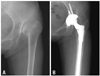
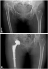
In two cases, there were progressive radiolucent lines and loosening around the acetabular cup. Of these, one case underwent monitoring of the clinical course and the other case was diagnosed as the loosening of an infected cup. The latter case was treated with revision surgery. Excluding these two cases, the remaining 43 cases showed stable fixation. There were no findings that were suggestive of osteolysis of the acetabular cup. In all cases, the femoral stem showed stable fixation (Figs. 1 and 2). The initial degree of the femoral fixation was also measured in accordance with the classification system proposed by Engh and Bobyn.8 This showed that 41 cases had canal filling of >90% and four cases had canal filling ranging from 80% to 90%. These findings indicate that all cases had satisfactory canal filling. The stability of the femoral stem was assessed based on the classification system proposed by Engh, et al.9 This showed that 37 cases had bony stability and eight cases had fibrous stability. There were no cases of instability. Fibrous fixation was defined in the cases in which the reactive radiolucent line was observed in Gruen zones II and VI until postoperative year 2. Thereafter, the radiolucent line did not progress further, but rather was stabilized. In ten cases, a reactive radiolucent line was observed in Zone I around the femoral stem. Yet it was assumed that these lines were local reactive lines that did not extend to the areas below the sleeve. Upon final follow-up, radiolucency of the calca femorale was observed in 16 cases (35.5%). Progressive bone loss of the proximal femur due to stress shielding was not shown.
In accordance with the classification system proposed by Brooker, et al.,11 Types I and II heteotopic ossification were identified in four cases. However, their clinical outcomes were all determined to be 'Excellent'. A hard bearing surface was used in all cases. Measuring the wear of the articular surface at the final follow-up radiography was impossible. There were no problems found associated with the articular surface.
Complications
The postoperative complications included two cases of hip dislocation. No further recurrence was noted following manual reduction. Partial palsy of the sciatic nerve was observed in one case, but this recovered to normal level at ten months post-operation. Intraoperatively, periprosthetic fracture on the proximal femur occurred in one case. Stable fixation was obtained by cerclage wiring. Infected cup loosening developed in one case at three years post-operation. This case was treated with revision surgery. At three year post-operation, this case showed a good clinical outcome with no recurrent episodes of infection.
DISCUSSION
The terminal point of the natural course of hip dysplasia is secondary degenerative arthritis. Acetabular dysplasia exerts an excessive amount of stress to the articular cartilage at the weight-bearing portion. Patients who complain of pain and functional disturbance and who are refractory to conservative treatment are indicated for total hip arthroplasty.
Arthroplasty becomes problematic in cases in which growth failure of the hip is concurrently present, due to developmental dysplasia of the hip joint or pediatric hip diseases, because of the deformities of the anatomical structures around the hip joint, including limb shortening, deformed acetabulum, deformed femur, and muscle atrophy around the hip joint. In particular, because femoral deformity shortens the femoral neck, the greater trochanter is located superiorly to the femoral head and the anteverion angle is increased. The narrow canal of the bone marrow cavity in the femur poses difficulty when selecting appropriate joint prostheses.2,3,5,12,13
A proximal modular femoral stem (S-ROM stem) is composed mainly of three parts: the stem, sleeve and head. Each stem and sleeve are available to select the optimal type and size. It is known that 10,398 combinations of the type and size are available on a theoretical basis.14,15 Due to these advantages, the S-ROM modular stem is highly advantageous for total hip arthroplasty in patients with arthritis and who concurrently have anatomical deformities. Particularly in cases with developmental dysplasia of the hip joint in which the anterior and posterior diameters on the left and right side of the proximal femur are small, the degree of curvature is low and the bone marrow cavity is narrow, the S-ROM stem can provide the maximum correction force because it forms maximum filling to the deformed proximal femur at the initial phase. The S-ROM modular stem is also advantageous in that it can be easily used for correcting the femoral anteversion angle and leg length discrepancy and for offset correction during the surgery.
It has been reported that the clinical outcomes of total hip arthroplasty using the S-ROM modular stem showed no difference from or that they were more excellent than the conventional type of total hip arthroplasty.16,17 Cameron16 reported that the clinical outcomes were more than 'Good' in 97.9% of the total patients. Park, et al.17,18 also reported that the clinical outcomes were 92.25 points at 2-year follow-up and 95.90 points at 5-year follow-up. Also in our series, the Harris hip score improved from 52.5 points preoperative to 85.5 points postoperative. At the final follow-up, the clinical outcomes were more than 'Good' in 41 cases (91.9%). On the other hand, thigh pain following cementless total hip arthroplasty has been reported to have a relatively lower incidence. Presumably, this might be true because flexibility of the femoral bone marrow cavity could be generated because of the decrease in comparison with the cylindrical stem with the same diameter due to the presence of a slot with a tubular shape in the distal area.19 In the current study, thigh pain developed in three cases. Of these, two cases had a radiolucent line detected around the femoral stem. In all three cases, the thigh pain disappeared two years post-operation.
The S-ROM modular stem was designed to generate maximal contact between the stem and the endosteum in the metaphysis and the diaphysis. In the proximal part, canal filling could be maximized with the use of a sleeve. In the distal part, the S-ROM modular stem has a flute-like shape and therefore tolerates torsional force. For inserting the femoral stem into the bone marrow cavity, the contact surface between the stem and the proximal femur can be maximized and the stability can also be maximized. Thus, the anterior length of the stem can be stabilized.
Also in the current study, the femoral stem showed stable fixation in all cases. According to Cameron,20 the distal flute-like shape of the S-ROM modular stem can provide the fixation effect in cases in which the degree of curvature of the proximal femur was excessive or when shortening osteotomy was performed. In the current study, shortening osteotomy of the femur was performed in five cases. This was effective in securing the initial fixation force in the distal femur. Accordingly, early rehabilitation was possible for these cases.
With regards to the change in the proximal femur due to stress shielding following a cementless total hip arthroplasty, Rosenthall, et al.21 compared the bone loss using a Dexa scan. According to these authors, the bone loss on the proximal side was significantly decreased by using an S-ROM as opposed to an anatomic medullary locking stem (DePuy Orthopaedics, Warsaw, IN, USA). Particularly, regarding bone loss in the cortex on the distal area of the lesser trochanter and the medial side of the femoral head, the S-ROM showed a lesser extent of bone loss than that of the AML. In the current study, a decreased shadow of the calcar femorale was observed in 16 cases (35.5%) at the final follow-up. However, no progressive bone loss of the proximal femur due to stress shielding was observed in our series.
The possibility of developing osteolysis due to dissociated metal particles is one of the concerns when using a modular stem. Tanzer, et al.22 reported that osteolysis occurred in 42% of the patients who underwent total hip arthroplasty using the S-ROM modular stem. Cook, et al.23 reported that great care must be taken when using a modular stem because metal particles are formed and can cause osteolysis, leading to loosening of the prosthesis and pain, in cases in which fatigue loading was exerted on a repeated basis. Other studies24,25 have also reported that the metal particles produced by the movement of the modular stems could lead to third body wear of the bearing surface. Krygier, et al.26 reported that the modular connecting area was stable despite the dissociation of metal particles. These authors also noted that it remains unclear as to whether metal particles produce third-body erosion or if this is due to an immunologic reaction. Chandler, et al.27 reported that there was no erosion between the sleeve and stem during revision surgery using S-ROM. According to a review of the literature, Spitzer15 also reported that there were no areas showing erosion in the connecting site of the modular stems. The stability of the S-ROM has been demonstrated in prior reports. Although our current study conducted mid to long-term follow-up observation, all the stems were stable and no osteolysis of the distal femur was observed. We believe these results are related to the design of this prosthesis. The design of this stem offers maximal proximal and distal filling with modularity and rotational stability. Use of the S-ROM stem is recommended for performing arthroplasty in patients with coxarthrosis and a dysplastic hip.
 XML Download