

 PDF
PDF ePub
ePub Citation
 Print
Print


INTRODUCTION
Extracts from various allergenic sources have been used for the diagnosis and allergen-specific immunotherapy of type I hypersensitivity since 1911.1,2 However, the quality of commercial allergen extract has been repeatedly reported to be variable.3-6
Biological standardization, which mainly determines overall IgE-binding potency, is generally carried out to assess the quality of allergen extracts. The Center for Biologics Evaluation and Research (CBER) in the US Food and Drug Administration (FDA) maintains 19 US reference standards for potency; six Hymenoptera venoms, two house dust mites, two cat dander extracts, one short ragweed pollen, and eight grass pollens.7 More specifically, grasses and mite allergen extracts are standardized on the basis of overall allergens with pooled serum by ELISA. Allergen potency of cat and ragweed allergen extracts is evaluated by determination of specific allergen with a sheep antibody by immunodiffusion. Allergenicity of Hymenoptera venoms are mainly assessed by total protein content and enzymatic activities. All manufacturers in the US are required to demonstrate batch-to-batch consistency and compliance with the standard. However, in-house reference (IHR) preparations from the companies are utilized for standardization in European countries.8 Therefore, each manufacturer uses a different unit for labelling the allergenic activity, making it difficult to interchange products between different companies.
SELECTION OF ALLERGEN SOURCE MATERIALS
Selection of raw materials is the first step for the preparation of an allergen extract. The quality of an allergen extract directly reflects the identity and purity of the allergenic source materials. Samples should be collected by qualified personnel. The content of all relevant allergens should be verified by appropriate methods. It may be necessary to culture or cultivate plants and animals of the allergen source in order to control the possible contamination of microbes, which can produce natural adjuvant-like susbstances in the allergen extract. Interestingly, it has been reported that allergen extracts prepared even from the same species of house dust mite, Dermatophagoides pteronyssinus, obtained from manufacturers in geographically distinct regions such as Sweden, Chile, and Australia, were found to exhibit remarkably different IgE reactivity.9 In contrast, the populations of mite-allergic patients in different geographic regions also showed different IgE reactivity to purified natural and recombinant allergens.10
The extraction procedure should reflect the physiological conditions of the airway. Generally, raw source materials are freeze-dried, and proteins are extracted using buffers resembling the physiological conditions of human airway. Extracted proteins are dialyzed to eliminate chemicals and hapten molecules. Effort should be made to suppress proteolytic degradation and contamination and the growth of microorganisms.
ALLERGEN UNIT
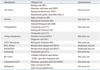
Commonly used allergen units are summarized in Table 1. The Noon unit and the protein nitrogen unit (PNU) were widely used for allergen products. A Noon unit denotes the quantity of water-soluble protein extractable from 1 µg of pollen.1 An allergen extract prepared from one gram of pollen in 10 mL of extraction buffer contains 100,000 Noon units per mL. Pollen derived from different sources is assumed to have the same allergenic activity. The weight per volume system is still widely used. PNU was introduced because most allergens were found to be proteins. PNU corresponds to 0.01 µg phosphotungstic acid-precipitable nitrogen, which is approximately 0.06 µg of protein.11 Recently, histamine equivalent in prick testing (HEP), biologic unit (BU), and bioequivalent allergen unit (BAU) have come to be widely used on standardized products. Ten HEP is equivalent to the allergen concentration, which elicits the same wheal size on a skin prick test as 10 mg/mL of histamine dihydrochloride.12 BU represents 1/1,000 of HEP since 10,000 BU/mL is equivalent to 10 HEP. However, the detailed method determining the unit differs between manufacturers even though most of the companies utilize the skin prick test.4 BAU is based on intradermal skin tests.13 The 3-fold dilution (0.05 mL) is calculated to induce a sum of erythema of 50 mm (D50). The allergen extract with a D50 of 14th dilution is arbitrarily assigned 100,000 BAU/mL [BAU/mL=100,000×3(D(D50-14)]. However, these approaches can't be applied to the chemically modified allergens (allergoids) or hypoallergenic allergens (which are currently not commercially available).14
For in vitro standardization, competitive IgE-binding inhibition test (e.g., ELISA or RAST) has proven to be reliable and is most commonly applied for measurement of total allergenic activity.15 The relative potency may be calculated by comparing activity with the potency of reference allergen extracts. Various biochemical and immunological methods such as SDS-PAGE, immunoelectrophoresis, circular dichroism spectrum analysis, immunoblotting, mass-spectrometric analysis, dot blot analysis, and proteomic approaches, etc. could be performed to check and control the consistent composition and activity of allergen product. However, IgE-reactivity does not reflect ability to cause allergic symptoms. The basophil activation test utilizes the same mechanism as a skin test and is expected to more closely reflect the biological activity of allergens, although it has not been widely utilized until now.16,17
ALLERGEN STANDARD
Production of allergen standard is essential for the standardization of allergen extracts. In fact, international reference standards were produced under the auspices of WHO according to the guidelines established by the Allergen Standardization Subcommittee under the International Union of Immuunological Societies (IUIS) for short ragweed (Ambroisia artemisiifolia), timothy grass (Phleum pratense), birch (Betula verrucosa) pollen, house dust mite (Dermatophagoides pteronyssinus), and dog (Canis familiaris) between 1984 and 1989. These allergen extracts are obtainable from the National Institute of Biological Science and Control (NIBSC), London. However, these standard materials have not been widely utilized for the standardization of commercial products because they were determined based on IgE potency tests, which use various serum pools and cannot distinguish the ratios of major allergens that are not identical between companies.
MAJOR ALLERGEN CONTENT
An alternative approach is necessary to overcome these problems caused by using allergen extracts containing a mixture of antigens and toxic substances such as endotoxin, enterotoxin, and/or proteases, which may play a role as an allergic adjuvant. The use of combinations of optimized concentrations of purified allergens, recombinant or native proteins, seems to be advantageous.18,19 Measurement of the concentration of individual major allergens is reported to correlate with the biological potency and IgE reactivity of allergen extracts.20,21 Furthermore, clinical trials using cocktails of recombinant allergens have demonstrated clinical efficacy, supporting the importance of major allergen content for immunotherapy.22,23 It also facilitates direct comparison between allergen products.24
The CREATE project (Development of Certified Reference Materials for Allergenic Products and Validation of Methods for their Quantification) was initiated by the European Commission from nine European Union member states in order to evaluate the potential of purified natural or recombinant allergens as certified reference materials, and to validate available two-site ELISAs for the measurement of major allergens.25 So far, two reference materials (rBet v 1 and rPhl p 5a) and relevant ELISA assays are now being validated for future establishment as international references under the guidance of the European Directorate for the Quality of Medicine (EDQM) after extensive biological and biochemical investigation. Currently, these two reference materials (rBet v 1 and rPhl p 5a) are under further validation by BSP090 project. The CREATE project leads us to conclude that standardization with major allergens is possible, even though recombinant allergens and monoclonal antibody-based immunoassays are not always ideal. Still, standardization based on major allergen content seems to be reasonable for determining the optimal dose of allergen vaccine, inducing a clinically relevant effect in the majority of subjects without unacceptable side effects. It is generally thought that 5-20 µg of major allergen (50-250 µg/yr) for house dust mite, cat, ragweed, and Hymenoptera venom are optimal for subcutaneous allergen specific immunotherapy. Much higher concentrations are needed for sublingual immunotherapy.
SEQUENCE POLYMORPHISMS OF ALLERGENS
A mixture of similar molecules with some variation in the amino acid sequences, isoallergens or variants often constitutes allergenic proteins.26 This phenomenon, often called polymorphism, may be caused by the presence of several gene alleles or families. Sequence diversity of major allergens is one of the important factors affecting complete allergen characterization and standardization.21,27 RNA editing as well as multiple genes were found to produce sequence variations, which may influence its allergenicity in case of cockroach allergen, Bla g 4.28 Importantly, mite group 2 allergens and birch pollen allergen Bet v 1 showed sequence diversity, which could influence IgE-binding reactivity, T cell response, and cytokine production from various immune cells.29-32 Extraction procedure and storage can also influence allergen contents. Different isoforms could be included in different batches of allergen extract. IgE or IgG recognition could be affected even by a single amino acid substitution on the surface of the allergens.33
Sequence polymorphism also influences reactivity to monoclonal antibodies. Commonly used immunoassay kits to quantify major allergens in Korea could underestimate the quantity of dominant isoforms in Korea.34 In case of Der f 2, amino acid sequence variations were described from Korean isolates of house dust mite.35 Differences in the capture antibody (monoclonal or polyclonal), storage buffer, or microplates showed up to 2-fold differences in two-site ELISA. Furthermore, distinct combinations of materials or incubations from different steps also could produce up to 5-fold differences in Can f 1 and egg white analyses.36 Epidemiological studies using immunoassay kits are also believed to underestimate the allergen content in dust samples. It is necessary to investigate the sequence polymorphism of major allergens that are thought to play important roles in the sensitization of Korean allergic patients. Studies of the sequence polymorphism and identification of major isoforms in each country/geographical region are thought to be necessary for more accurate determination of allergen content from a given environment or allergen extract by using immunoassay kits.37
NATURAL ADJUVANT IN ALLERGENS EXTRACT
Some component of allergen extract could activate innate and/or adaptive immunity. In particular, house dust mites have been known to contain many substances that could induce innate immunity, subsequently affecting the development of allergic responses.38 Some proteins in allergen extract, whether they are allergens themselves or not, can activate the various human cell and induce inflammatory responses.39
Some common contaminants from the indoor environment can play a role in the development of allergic disorders through their adjuvant-like activity. Several lines of evidence have been described on the relationship between pathogen-associated molecular patterns (PAMPs).40 Endotoxin has been implicated as an important modulator of allergic responses.41,42 Natural adjuvant derived from various microbes may affect allergy diagnosis and immunotherapy as well as pathogenesis of allergic disorders. Interestingly, the major house dust mite allergen Der p 2, homologous to MD-2, was shown to reconstitute lipopolysaccharide-induced TLR4 signalling in MD-2-deficient mice.43
Endotoxin, one of the well-described cell wall components of gram-negative bacteria, is known to induce inflammatory responses in human cells, subsequently affecting allergic diseases.44 Several lines of evidence have shown that sequence variation of TLR-2 and -4 genes are associated with the development of allergic disorders.45 In a normal indoor environment, 300-18,000 ng/g dust of endotoxin was detected.44 This concentration implies that a significant amount of endotoxin is breathed in and can have influence on immune reactions. Exposure to the high level of endotoxin is thought to protect against the allergy. However, low level of endotoxin is believed to worsen allergic symptoms.
β-glucan, a component of fungal cell walls, is another common component of the indoor environment. It is also known to modulate allergic responses, influencing both the Th1 and Th2 responses.46,47 Notably, 0.4-41.8 ng/mL of β-glucan was detected in standardized allergen extracts.48
Various enzyme activities have been detected from the allergen extracts and house dust.49,50 Mite group 1, 3, 6, and 9 allergens are cysteine protease, trypsin-like serine protease, chymotrypsin-like serine protease, and collagenolytic serine protease, respectively.51 Proteins with cysteine or serine protease activity are well known to induce Th2 immune response. Protease allergens can cleave CD23 (low affinity IgE receptor) of activated B cells52 and CD25 (IL-2 receptor) of T cells.53 Tight junctions in the lung epithelium could be degraded and cause various cells to release inflammatory cytokines.54,55 Degraded epithelium is vulnerable even to bystander allergens.56,57 Protease allergens also can cleave several component of the complement system, generating active anaphylatoxins, C3a, C4a, and C5a.58-60 Furthermore, protease-activated receptor 2 is activated and induces proinflammatory responses.61,62 Human basophils are known to be directly activated by cysteine protease (Der p 1 and Der f 1) and to release cytokines, which can induce allergic inflammation.63 Some serine protease inhibitors and defects of the protease gene are known to be associated with allergic conditions.54 Interestingly, cystatin A, a cysteine protease inhibitor in secreted sweat, has been shown to eliminate the activity of mite group 1 allergens.64 It is also conceivable that some proteases originating from symbionts or parasites of allergen sources (house dust mite or cockroach) can influence allergic inflammation.65-68 In fact, various microorganisms, which may secrete or excrete proteases, have been isolated from the cockroach hindgut.69
Flagellin, a principal constituent of bacterial flagellum, is one element that may elicit an inflammatory response.70,71 However, much remains to be investigated.
Chitin, a major component of the exoskeletons of arthropods, fungal walls and the microfilarial sheath of parasites, is the second most abundant polysaccharide in nature. Inhalation or infection of chitin-containing pathogen could activate chitinases and chitinase-like protein in humans and play a role in the pathogenesis of Th2 inflammation.72,73 Interestingly, mite allergen Der f 15 and 18 show significant homology to chitinases. It would be interesting to explore the role of chitinases in the pathogenesis of allergic diseases and to examine the chitin content and chitinase activity in allergen extracts.
Superantigen, endotoxin B from Staphylococcus in skin, is also known to be associated with the development of atopic dermatitis by activating T cells.74
DNA fragments derived from microorganisms may have various effects on the pathogenesis of allergic disorders. Herpes simplex is known to induce production of TNF-α by activation of TLR-9.75 DNA fragments may contain the CpG motif, which could be protective against allergy.76 Bacterial DNA are detectable even from house dust mite cultures for allergen extract preparation,77 indicating that DNA fragments are included in the extracts.
It is noteworthy that many adjuvant-like activities are derived from a microbial component. It is known that commensals in intestine and skin can influence allergic disorders.78 Recently, it has been reported that there are a large number of microbiota that can affect allergic diseases.79 Viral infection can also exacerbate the symptoms via TLR-3, -7, -8, -9 activation.80 Concomitant infectious diseases and hygiene are important not only for the development of allergic disorders and exacerbation of the symptoms, but also for diagnosis and allergy immunotherapy. Currently, it is thought to be almost impossible to remove all those elements that may play adjuvant-like roles. Therefore, it should be emphasized that every effort must be made to minimize possible contamination and suppress microbial growth. It is desirable to check the activity and content of substances that may play adjuvant-like roles.
ALLERGEN STANDARDIZATION IN KOREA
The allergen extracts produced at the Research Center for the Standardization of Allergic Diseases, Department of Internal Medicine and Institute of Allergy, Yonsei University College of Medicine, Seoul, Korea, exhibit good allergen potency, in terms of in vitro (ELISA inhibition) and in vivo (intradermal skin test) tests, when compared with commercially available extracts, even though immunosassays detected low levels of major allergens. Comparisons were performed between endotoxin levels of house dust mite extracts both commercially available and produced in our laboratory. The endotoxin level was determined to be comparable with commercial products even without the addition of phenol (unpublished data).
It is urgently necessary to develop standard references and immnoassays for the detection of major allergens, such as Japanese hop (Humulus japonicus) (Fig. 1A),81 spider mites (Tetranychus urticae and Panonychus citri),82 non-biting midge (Chironomus spp) (Fig. 1B),83 Pharaoh ant (Monomorium pharaonis),84 and Asian needle ant (Pachydonyla chinensis),85 which are frequent causes of allergic illness in Korea.
CONCLUDING REMARKS
The recent research trend for allergen standardization has changed from IgE potency test to characterization of major allergens and assays for the quantification of major allergen concentrations. Still, measurement of biological potency of extracts remains the basis of allergen standardization. Further study is required to characterize the major allergens that are thought to be important in Korea. Studies on the correlation between major allergen concentrations and the potency of allergen extracts will facilitate the development of advanced allergen-specific immunotherapeutics.

 XML Download