

 PDF
PDF ePub
ePub Citation
 Print
Print


INTRODUCTION
A lipogranuloma is an inflammatory reactive process associated with exogenous or endogenous lipids involving the dermis and subcutaneous layer.1 A lipogranuloma has been described as an iatrogenically induced chronic inflammatory change, and has frequently been reported in the penis and scrotum of young adult males.1 A lipogranuloma of the breast is a rare condition. This soft tissue reaction has been reported to develop after injection of oily substances or after release of a silicone gel from a breast implant.2
Osseous metaplasia is a finding of the extraskeletal bone in soft tissue and may resemble a neoplasm in a clinical and imaging appearance.3-5 Osseous metaplasia can also develop from several conditions such as trauma, post-traumatic hematoma and soft-tissue tumors, especially slow growing types and tumors treated with radiation.3
We report a case of a lipogranuloma with osseous metapalsia after post-traumatic treatment with traditional oriental medicine, called "Bu-Hwang."
CASE REPORT
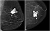
A 66-year-old woman visited our hospital with a painless lump in the left breast. The patient had a history of trauma in the left breast three years earlier. The patient had fallen and damaged her left breast. At that time, the patient had received an oriental medicine, traditional alternative treatment, called "Bu-Hwang" for the bruise and swelling in the left breast. After several months, the patient felt a painless lump in the left breast, and thereafter, the breast lump gradually increased in size for the next three years. The patient visited our hospital for evaluation of a palpable lump in the breast. At that time, a breast surgeon noticed no abnormalities on the chest wall, except for the breast lesion. Routine mammography was performed and it showed the presence of a relatively well-demarcated bizarre shaped lesion with brighter density as compared to calcification in the upper medial portion of the left breast. The maximum diameter of the lesion, as depicted on mammography, was approximately 3.5 cm (Fig. 1). The mass was excised at a breast surgery clinic at the request of the patient for cosmetic reasons and discomfort. Unfortunately, the mass was excised without an ultrasound examination. The mass was surgically excised and a histopathological examination showed a metaplastic focus of bone formation, multiple stromal fat vacuoles, scattered lymphocytes and foreign body type multinucleated giant cells forming a granuloma (Fig. 2). The pathologist confirmed that there were osteocytes in the newly-formed bony trabeculae (Fig. 3), and also verified epithelioid cells within the granulomatous changes, which support the diagnosis of a lipogranuloma with osseous metaplasia rather than fat necrosis. The pathologist also found that breast gland tissue was continuous with the lesion, suggesting that this lesion arose in the breast tissue. These findings were consistent with a lipogranuloma with osseous metaplasia in the breast.
DISCUSSION
A lipogranuloma of the female breast is a relatively rare condition. Lipogranulomas are nonallergic foreign body reactions typically found in areas where lipid material has been injected either traumatically or for a specific purpose.2 Several types of lipogranulomas have been reported to develop after injection of lipid substances for cosmetic purposes and after trauma or specific therapy.1,2,6,7 In the breast, this type of soft tissue reaction has been reported. Lipogranulomas can develop after release of a silicone gel from breast implants and after phenothiazine therapy.2,8
Our patient had undergone oriental medicine alternative therapy called "Bu-Hwang" to reduce pain and bruising. "Bu-Hwang" is a traditional oriental alternative medicine therapy and is performed by puncturing the skin and depleting the blood by giving strong negative pressure using a Bu-Hwang cup. According to the tradition, it is considered to facilitate gas exchange for achievement of blood purification. We think that this procedure can draw parenchymal soft tissue or fat tissue into the subcutaneous level. A lipogranuloma is known as iatrogenically caused inflammatory change that occurs mostly in the penis and scrotum of a young adult male.1 Our patient denied having a history of exogenous foreign body injection or topical use of drugs. In this case, we suspect that the lipogranuloma originated from endogenous fat degeneration by the strong negative pressure that pulled the parenchymal fat into the subcutaneous layer. It is known that trauma is related directly to or by an allergic mechanism for the development of a lipogranuloma.6 It seems that "Bu-Hwang" can occasionally be a type of traumatic process.
In our case, the final pathological diagnosis was related to osseous metaplasia. The terms "osseous metaplasia" or "heterotopic bone formation" are used to express bone formation in abnormal locations.4 Osseous metaplasia may develop after trauma, or with a post-traumatic hematoma and soft-tissue tumors, especially slow growing tumors treated with radiation.3 Several cases of osseous metaplasia have been reported that were related to conditions, including tubular adenoma of the colon, diffuse large B-cell lymphoma and amyloid-producing dyscrasia.5,9 Few studies have reported benign breast lesions that were seen as osseous metaplasia.10 It is generally agreed that bone in breast tissue occurs in a variety of mammary lesions and may have more than one mechanism of formation.3
Mammography of nonneoplastic osseous metaplasia of the breast has been reported only once, depicting extensive focal ossification of both breasts in association with chronic nonspecific mastitis.10 Imaging findings of osseous metaplasia for a neoplasm depend on the nature of the underlying neoplasm. In the case of a fibroadenoma or cystsarcoma phylloides, mammography and sonography demonstrate a well-defined, oval lesion with coarse calcification.3 Our case also showed well-defined dense ossification, however, the bizarre shape in this case might have been produced by artificial external pressure due to the "Bu-Hwang" procedure. It is difficult to determine if osseous metaplasia developed from a tumorous condition such as a lipogranuloma or originated from a traumatic process. The suggested mechanism for the origin of osseous metaplasia remains obscure. In this case, post-traumatic fat degeneration after "Bu-Hwang" oriental medicine alternative therapy caused the formation of the lipogranuloma and osseous metaplasia. To the our knowledge, this is the first report of a lipogranuloma with osseous metaplasia on the breast.


 XML Download