

 PDF
PDF ePub
ePub Citation
 Print
Print


INTRODUCTION
Laparoscopic renal surgeries are increasingly being used for treatment of both benign and malignant conditions, because laparoscopic surgery has several advantages over open surgery.1-3 However, bleeding, hernias, internal organ damage, preand undesirable cosmetic effects such as scars are complications that can occur at the working ports in the conventional laparoscopic approaches.
To overcome these limitations of conventional laparoscopy, laparoendoscopic single-site surgery (LESS) for minimally invasive urology has been the focus of numerous investigations, and consequently, the use of LESS in urology as an alternative to conventional laparoscopy has increased substantially over the past two years with the refinement of laparoscopic instruments.4-8
Although a variety of LESS procedures have been described in the literature, we have primarily performed LESS in cases requiring extirpative procedures, considering that a minilaparotomy or auxiliary incision of the abdominal wall is required to remove the specimen in conventional laparoscopic surgery. To minimize the incision length, we performed a modified umbilical incision as described by Casciola, et al.9 and used home-made transumbilical ports, because commercial multilumen ports typically used in LESS approaches are not available in Korea.7,10-12
Thus, this study was designed to verify the clinical utility of LESS nephrectomy using a modified umbilical incision and a home-made transumbilical port in initial consecutive cases requiring extirpative surgery based on a single surgeon experience.
MATERIALS AND METHODS
Patient selection
Between March 2009 and October 2009, initial consecutive 18 patients who underwent various LESS nephrectomies for treatment of benign and malignant conditions, performed by a single laparoscopic surgeon (Ham WS), were enrolled in this study. Our institution's institutional review board approved this study. Preoperatively, all patients consented to LESS.
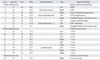
For simple nephrectomy cases, a diuretic renogram confirmed < 15% function in the kidney of interest with a normal contralateral kidney. Radical nephrectomy was performed for all cases with a clinically-confirmed T2 or lower tumor stage without evidence of lymph node enlargement or renal vein involvement for which nephron-sparing surgery by either an open or laparoscopic approach was not possible. We performed a nephroureterectomy for one patient with an enhancing ureteral mass. If a patient's overall condition was suitable for laparoscopy, we performed LESS, irrespective of the patient's body mass index or previous operation history. The characteristics of patients included in this study are summarized in Table 1.
Operation technique
Modified umbilical incision
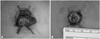
Under general anesthesia, the patient was positioned in a modified lateral decubitus position. A modified umbilical incision was made three-quarters of the way along the circumference of the umbilicus along the umbilical ring.9 A suitable fasciotomy beneath the umbilical skin incision was performed for intact specimen extraction (Fig. 1). For cases with a large kidney and tumor seen during the preoperative imaging study, the skin incision length was extended to 4.0 cm in the cranio-caudal direction before port placement.
Home-made transumbilical port placement
An Alexis wound retractor® (Applied Medical, Rancho Santa Margarita, CA, USA) was placed in position through the incision with the bottom ring (green) inside the abdomen after the umbilical skin incision was carried into the peritoneum. We used an extra-small Alexis wound retractor® for pediatric patients, and a small Alexis wound retractor® for adult patients. A size 7 sterile surgical glove was snapped onto the external ring (white), and the three fingers of the glove were secured to the ends of the trocars (three 5-mm trocars for pediatric patients and three 12-mm trocars for adult patients) with silk thread and a rubber band.13-15 After insufflation of the peritoneum with CO2 to 12 mm Hg, a 30° 5-mm or 10-mm rigid laparoscope was inserted through the 5-mm or 12-mm trocars. We used a flexible fiber light cable 8062™ with a 90° angled instrument attachment (Richard Wolf, Knittlingen, Germany) to reduce the interactions between the laparoscope and instruments.
Laparoendoscopic single-site nephrectomy procedure
The standard laparoscopic transperitoneal nephrectomy technique was performed with the combined use of conventional and articulating laparoscopic instruments (Autonomy LaparoAngle™, Cambridge Endo, Framington, MA, USA). To minimize the external crowding and clashing of instruments, we also used standard and bariatric length conventional laparoscopic instruments. For adult patients, we used conventional laparoscopic instruments for most dissection and retraction maneuvers, and articulating instruments were used selectively, whereas, articulating instruments were used primarily throughout the procedures for pediatric patients.
After colon mobilization, the ureter was identified and traced to the renal hilum. Renal pedicle control was performed using vascular EndoGIA staplers (Ethicon, Cincinnati, OH, USA) or Hem-o-Lok™ (Weck Closure Systems, Research Triangle Park, NC, USA) clips with additional metal clips. The upper pole and retroperitoneal attachments were then divided with a Harmonic Scalpel™ (Ethicon, Cincinnati, OH, USA) and stapler or metal clips. We did not perform additional laparoscopic port placement for liver retraction, even for right-sided cases. Hemostasis was confirmed, and the kidney was extracted intact in a laparoscopic retrieval bag through the incision. For pediatric patients, the small renal pedicles supplying the dysplastic kidney were clipped using the metal clips, and the specimen was removed intact through the home-made port without the laparoscopic retrieval bag. For cases with a large kidney and tumor, the initial skin incision (4.0 cm) was extended in the cranio-caudal direction until the specimen in the laparoscopic retrieval bag could be extracted intact without severe resistance.
For nephroureterectomy, the ureter was dissected to the level of the bladder after the nephrectomy was completed. The bladder cuff was dissected circumferentially around the ureteric orifice, and we then performed laparoscopic stapling of the distal ureter and bladder cuff using a pure extravesical approach.16
Postoperative management and outcomes
Postoperatively, all adult patients received continuous intravenous patient-controlled analgesia (IV-PCA) as well as IV tramadol as needed. Thirty minutes before the end of the operation, the IV-PCA was connected to the patients. The IV-PCA regimen comprised 1,000 µg of fentanyl in normal saline 100 mL (10 µg/mL) and tramadol (50 mg/A) was administered in a bolus if pain was 5 or more on the visual analog scale (VAS). Although the IV-PCA was maintained maximally for 48 hr, it was discontinued earlier if the patient desired or the patient had a VAS pain score of ≤ 2. Three pediatric patients received only oral ibuprofen syrup according to their age. All patients were discharged at their own discretion after consideration of their general condition. The demographics, operation time, estimated blood loss, perioperative complications, duration of hospital stay, incision length, and final pathological results of all patients were recorded.
RESULTS
LESS nephrectomy was performed in nine right-sided and nine left-sided cases. All procedures were successfully completed using only two instrument ports throughout the procedure, even for the right-sided cases, without conversion to conventional laparoscopy or an open approach. Two intraoperative injuries (diaphragm injury and renal vein injury) occurred, but were controlled immediately by either primary closure or clipping without specific problems.
Simple nephrectomy for a nonfunctional kidney was performed in eight cases; the final pathology in all these cases was consistent with chronic pyelonephritis induced by a ureteropelvic junction obstruction or a stone (dysplastic kidney in three pediatric patients) without any evidence of malignancy. Nine patients had radical nephrectomy for enhancing renal masses, and one patient had a nephroureterectomy for a ureteral mass. Except for one case with a mixed epithelial and stromal tumor, final pathological analysis revealed that eight cases were renal cell carcinomas (six clear cell, one papillary, and one chromophobe) that could be broken down into pathological stages as follows: T1a (n = 2), T1b (n = 3), T3a (n = 2), and T3b (n = 1). One patient with a ureteral mass was diagnosed with ureteral transitional cell carcinoma, high grade, Ta.
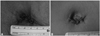
Most specimens could be extracted intact through a modified umbilical incision of minimal length: 2.5 cm for adult patients and 1.5 cm for pediatric patients (Fig. 2). For four cases with a large kidney (longest kidney length: range, 14.0-18.0 cm) and tumor (size range, 4.0-7.0 cm), the specimens could be extracted intact using an extended skin incision of 5.0-6.0 cm length (Table 2).
From postoperative day 1, the patients reported tolerable levels of pain (median VAS pain score: 2, range 1-4). Only five patients (33.3%) required additional IV tramadol on the day of the operation and 12 patients (80%) discontinued IV-PCA before 48 hr. Median duration of hospital stay was 5 days (range 2-8).
DISCUSSION
Although the transition from multiple port access surgery to LESS as a step toward scar-free surgery may represent a compoparadigm shift in reconstructive and extirpative surgery,11 LESS procedures have been performed only in a very limited number of high-volume centers worldwide,6-8,17,18 and the advantages of LESS over conventional laparoscopic surgery have yet to be proven.10,19 Two major factors limiting the generalization of LESS in medical centers without prior experience in LESS may be technical challenges12 and higher costs because of the requirement for more sophisticated instruments.11
However, as mentioned earlier, if surgery could be conducted and completed through a single, small incision while being true to oncological principles in cases requiring extirpative procedures, the patient would undoubtedly benefit. Therefore, we believe that LESS is a very attractive surgical technique, despite the limitations discussed above.11,12 Furthermore, the benefits of LESS could be maximized in cases requiring extirpative procedures.
Therefore, we performed LESS nephrectomies for various cases in which an intact specimen was required. We used a transumbilical approach, which provides a familiar anatomical view of the kidney and potentially affords maximal cosmetic benefits.20 Furthermore, to minimize the incision length, we adopted a modified umbilical incision.9 In our study, most specimens could be extracted intact through a minimal umbilical incision extended three-quarters of the way along the circumference of the umbilicus of minimal length. For cases with a large kidney and tumor, the initial skin incision was extended to 4.0 cm in a cranio-caudal direction before port placement, thereby allowing us to proceed with the operation without severe instrument crowding, even though we used conventional laparoscopic instruments for most dissection and retraction maneuvers.
Because commercial multilumen ports typically used in LESS approaches are not available in Korea,4,7,11,12,18 we have used home-made transumbilical ports.13-15 The basic structure of our home-made port is very similar to that of R-Port™ (Advanced Surgical Concepts, Wicklow, Ireland) in that it consists of two parts: the retractor component and the valve component. Therefore, our home-made port can be used also in performing single-port transvesical simple prostatectomy, and digital assistance for adenoma enucleation can be done by simply removing the valve component, without having to remove the port itself.21-23 Moreover, we were able to freely adjust the incision length using various sizes of Alexis wound retractors® at great savings, although additional time was required to make a valve component. Several merits of home-made port was well summarized by Han, et al.15 however, it is also true that durability of our port can be limitation during some lengthy procedures, compared to those of commercial multilumen ports. Therefore, it would be best to use a proper multilumen port according to the preference of surgeon and the situation of each hospitals.
From the aspects of surgical techniques, it would be an inevitable fact that LESS is more technically challenging than conventional laparoscopy. Especially in our study, we used only two instrument trocars, even for right-sided cases, because we felt that applying an additional port on the abdominal wall would undermine the concept underlying LESS, and the use of three instrument trocars through our small single incision would cause severe extracorporeal instruments crowding. Liver retraction was not problematic. We intentionally dissected toward the upper pole,after the control of renal pedicles, and were able to finish the dissection in the area below the liver without severe difficulties.
Current LESS techniques are in their infancy, and advances in instruments and surgical techniques are required for innovation. Nevertheless, based on our experience, LESS is feasible with minimal incision and safe procedure even for medical centers without prior LESS experience and specialized instruments, however, the ultimate question is whether the use of LESS translates to patient benefits: a prospective multi-institutional clinical trial is essential to confirm whether incremental benefits do indeed exist.12
In conclusion, LESS nephrectomy using a modified umbilical incision and a home-made port is feasible with both minimal incision and cost-effective, especially for medical centers without prior LESS experience and specialized instruments. Our technique may be more useful for extirpative procedures in which a specimen needs to be removed intact, because the incision length can be freely adjusted; however, prospective studies are required to confirm our findings.

 XML Download