Journal List > Yonsei Med J > v.52(2) > 1030771

Joung, Chen, and Lin: The Role of the Calcium and the Voltage Clocks in Sinoatrial Node Dysfunction

Abstract

Recent evidence indicates that the voltage clock (cyclic activation and deactivation of membrane ion channels) and Ca2+ clocks (rhythmic spontaneous sarcoplasmic reticulum Ca2+ release) jointly regulate sinoatrial node (SAN) automaticity. However, the relative importance of the voltage clock and Ca2+ clock for pacemaking was not revealed in sick sinus syndrome. Previously, we mapped the intracellular calcium (Cai) and membrane potentials of the normal intact SAN simultaneously using optical mapping in Langendorff-perfused canine right atrium. We demonstrated that the sinus rate increased and the leading pacemaker shifted to the superior SAN with robust late diastolic Cai elevation (LDCAE) during β-adrenergic stimulation. We also showed that the LDCAE was caused by spontaneous diastolic sarcoplasmic reticulum (SR) Ca2+ release and was closely related to heart rate changes. In contrast, in pacing induced canine atrial fibrillation and SAN dysfunction models, Ca2+ clock of SAN was unresponsiveness to β-adrenergic stimulation and caffeine. Ryanodine receptor 2 (RyR2) in SAN was down-regulated. Using the prolonged low dose isoproterenol together with funny current block, we produced a tachybradycardia model. In this model, chronically elevated sympathetic tone results in abnormal pacemaking hierarchy in the right atrium, including suppression of the superior SAN and enhanced pacemaking from ectopic sites. Finally, if the LDCAE was too small to trigger an action potential, then it induced only delayed afterdepolarization (DAD)-like diastolic depolarization (DD). The failure of DAD-like DD to consistently trigger a sinus beat is a novel mechanism of atrial arrhythmogenesis. We conclude that dysfunction of both the Ca2+ clock and the voltage clock are important in sick sinus syndrome.

INTRODUCTION

The sinoatrial node (SAN) automaticity is responsible for initiating the heart rhythm. SAN function is therefore essential for normal cardiac physiology. Although it has been shown more than 40 years ago that spontaneous diastolic depolarization of SAN cells periodically initiates action potentials to set the rhythm of the heart, the mechanism of heart rhythm generation is still unclear. Sick sinus syndrome is an abnormality involving the generation of the action potential by the SAN and is characterized by an atrial rate inappropriate for physiological requirements. The sick sinus syndrome occurs in 1 of every 600 cardiac patients older than 65 years and accounts for approximately half of implantations of pacemakers in the United States.1 A better understanding of the mechanisms of SAN automaticity and sick sinus syndrome is therefore clinically important.

Voltage and calcium clocks in the SAN automaticity

The mechanism of spontaneous diastolic depolarization has traditionally been attributed to a "voltage clock" mechanism, mediated by voltage-sensitive membrane currents, such as the hyperpolarization-activated pacemaker current (If) regulated by cyclic adenosine monophosphate (cAMP).2,3 The funny channel becomes activated at the end of the action potential (at voltages from - 40/- 50 mV to - 100/- 110 mV), which corresponds to the range where diastolic depolarization occurs. It then depolarizes the membrane to a level where L-type Ca2+ channel open to initiate the action potential.4,5 If is a mixed Na+-K+ inward current activated by hyperpolarization and modulated by the autonomic nervous system. Because the membrane ionic channels open and close according to the membrane potential, this process is referred to as membrane voltage clock. The major role of If has been reinforced by the fact that mutations in the If channel are associated with reduced baseline heart rate,6 and drugs which blocks If (such as ivabradine) do the same.7 However, while point mutation of hyperpolarization-activated cyclic nucleotide-gated channel 4 (HCN4) is associated with baseline sinus bradycardia, the maximum heart rate achieved during exercise is normal.8 The latter finding implies that If is not the only mechanism of SAN automaticity, especially during sympathetic activation.
Recently, the spontaneous sarcoplasmic reticulum (SR) Ca2+ release was suggested as an additional mechanism of sinus rhythm generation also known as the Ca2+ clock. The involvement of intracellular calcium (Cai) cycling in heart rhythm generation was first suggested from the observation that application of ryanodine slowed subsidiary pacemakers in cat right atrium.9 Subsequent work showed the involvement of the electrogenic Na-Ca exchange current (INCX) in pacemaker activity and also raised the possibility that increased Ca2+ release followed by activation of INCX could play a role in the positive chronotropic effect of β-adrenergic stimulation in latent pacemaker cells.10-12 Lakatta, et al. suggested that the spontaneous rhythmic SR Ca2+ release in SAN cells, manifested as Ca2+ sparks, work as the "Ca2+ clock". The elevated Cai causes diastolic depolarization via INCX activation, which coordinately regulates sinus rate along with the voltage clock.13-15
The idea of Ca2+ clock suggests that the mechanism of automaticity is the same as that of delayed afterdepolarization (DAD), which occurs when there is SR Ca2+ overload. This suggests that the SAN must exist normally in a state of calcium overload. Vinogradova, et al.15 also demonstrated that a high basal level of protein kinase A (PKA) activity in the SAN might contribute to a state of Ca2+ overload. The disease associated with Ca2+ clock malfunction was also reported in patients with a genomic deletion of ryanodine receptor 2 (RyR2) exon-3. Patients with that mutation develop catecholaminergic polymorphic ventricular tachycardia, along with SAN and atrioventricular node dysfunction, atrial fibrillation and atrial standstill.16 It is possible that Ca2+ clock malfunction contribute to the bradycardia and atrial arrhythmias in these patients.

Pacemaker hierarchy and the importance of Ca2+ clock in intact SAN

The cardiac automaticity at the organ level is a very complex phenomenon and, beside cellular mechanisms, integrative factors are involved in cardiac pacemaking. The intact SAN is a heterogeneous structure that includes multiple different cell types interacting with each other.17-19 The relative importance of the voltage and Ca2+ clocks for pacemaking in different regions of the SAN, and in response to neurohumeral stimuli such as β-agonists, may be different. Indeed, activation maps in intact canine right atrium (RA) showed that SAN impulse origin is multicentric,20 and sympathetic stimulation predictably results in a cranial (superior) shift of the pacemaking site in human and dogs.21,22 Based on evidence from isolated SAN myocytes, late diastolic Cai elevation (LDCAE) relative to the action potential upstroke is a key signature of pacemaking by the Ca2+ clock.10-15 It is possible that the same phenomenon could provide insights into the relative importance of the Ca2+ and voltage clock mechanisms in pacemaking in intact SAN tissue. We therefore designed a study to determine the role of Ca2+ and voltages clocks on the heart rhythm generation and on the mechanisms of pacemaker hierarchy in the intact SAN.23

Heterogeneous Cai dynamics in intact SAN

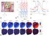
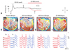
At baseline, the spontaneous diastolic SR Ca2+ release, which is manifested by the LDCAE, was observed in only a small percentage of the preparations. However, LDCAE occurred in all preparations during isoproterenol infusion, associated with a superior shift of the leading pacemaker site, coincident with the appearance of robust LDCAEs (Fig. 1) in this region. Most importantly, the site of maximum LDCAE slope always co-localized with the leading pacemaking site, suggesting a shift in which the voltage clock now lagged the Ca2+ clock (Fig. 2). This observation indicates a strong association between LDCAE and pacemaking during β-adrenergic stimulation, and provides new insights into pacemaker hierarchy in the canine RA.20-22
The Cai Dynamics of SAN were characterized not only by the earliest onset of LDCAE but also by the fastest Cai reuptake as compared with other RA sites. The baseline 90% Cai relaxation time was shorter at superior SAN than at other RA sites. This resulted in the formation of the Cai sinkhole, which was facilitated by a rapid decline (short relaxation time) of the Cai fluorescence at the superior SAN during isoproterenol infusion and suggests that Cai reuptake by SR is the fastest in the superior SAN (Fig. 1D). The key protein regulator of SR Ca2+ uptake is phospholamban, which inhibits SERCA2a in its dephosphorylated state. There was a significantly lower SERCA2a/phospholamban ratio at SAN sites than at RA sites, suggesting more phospholamban molecules are available to regulate SERCA2a molecules in SAN than in RA. Isoproterenol infusion phosphorylates phospholamban and relieves phospholamban inhibition of SERCA2a, which may account for more robust Ca2+ uptake in SAN than in RA during isoproterenol infusion.23

Mechanisms of diastolic depolarization of SAN

The LDCAE was closely related with the SR Ca2+ release. Caffeine sensitizes the ryanodine receptor 2 to activation, resulting in increased SR Ca2+ release.24 The superior shift of LDCAE and the pacemaking site was also consistently observed with caffeine infusion. The ryanodine, which block ryanodine receptors, caused a dose-dependent suppression of sinus node activity, and impaired isoproterenol-induced LDCAE. The combination of ryanodine and thapsigargin also suppressed the sinus node activity, and impaired isoproterenol-induced LDCAE. In contrast, the If blocker, ZD 7288 (3 µmol/L) did not prevent LDCAE in the superior SAN.
Multiple time- and voltage-dependent ionic currents have been identified in cardiac pacemaker cells which contribute to diastolic depolarization, including ICa-L, ICa-T, IST and various types of delayed rectifier K currents.25 Many of these membrane currents are known to respond to β-adrenergic stimulation. Some of these currents, such as ICa-L, also promote LDCAE and the acceleration of sinus rate by the Ca2+ clock as well as the voltage clock. In intact SAN, both SR inhibitors and If blockade slowed sinus rate under basal conditions, as well as blunted the isoproterenol-induced increase in sinus rate. Therefore, the interdependence and synergy between the two clocks are evident.

Impaired Ca2+ clock after β-adrenergic stimulation in AF dogs

Atrial fibrillation (AF)-induced remodeling of ionic currents has been well documented in the atrium.26,27 The typical electrophysiological remodeling in AF includes action potential duration (APD) shortening, the downregulation of L-type Ca2+ channel (ICa-L) caused by atrial cardiomyocyte Ca2+ loading,28,29 downregulation of Ito30 and upregulation of IKACh and IK1.26,27 The ionic current remodeling could reduce the slope of phase 0, hyperpolarize the Vm and reduce the heart rate. However, the mechanism of tachycardia induced-SAN dysfunction is unclear. Yeh, et al.31 recently reported that If downregulation may contribute to the association between SAN dysfunction and supraventricular tachyarrhythmias. However, normal functioning SAN depends not only on membrane ionic currents but also on the rhythmic Ca2+ releases from the SR.10-15
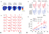
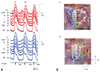
In a recent study, we found that the SAN dysfunction in AF is associated with Ca2+ clock malfunction, characterized by unresponsiveness to isoproterenol and caffeine, as well as downregulation of RyR2 in SAN. Fig. 3A shows a typical isoproterenol response of RAs from normal dogs. Isoproterenol infusion increased heart rate to and shifted the leading pacemaker site to superior SAN with a robust LDCAE [arrows in Cai tracing of Fig. 3A(b)]. This finding was consistently observed in all normal RAs during isoproterenol infusion. However, LDCAE increase in superior SAN was completely absent in AF dogs [Fig. 3B(b)]. Also, the heart rate was increased by the acceleration of the ectopic focus from inferior RA [Fig. 3B(a)]. The Cai tracing showed no LDCAE anywhere in the mapped region with isoproterenol dose ranging from 0.01 to 10 µmol/L.32,33

Sympathetic stimulation and tachybradycardia syndrome

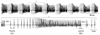
It is known that heart failure is frequently associated with SAN remodeling, resulting in decreased SAN reserve.34 We performed nerve recording in a canine model of pacing-induced heart failure and found intermittent tachybradycardia episodes.35 Interestingly, the prolonged (>3 s) sinus pauses were triggered not by vagal activation but by short bursts of sympathetic activity. Typically, a burst of sympathetic activity is associated with tachycardia. When there is sympathetic withdrawal, the tachycardia terminates, followed by prolonged pauses during which no activation was observed. The molecular mechanism of this association, however, remains unclear.
In a recent study, we developed various model of sick sinus syndrome with pharmacological manipulation of Ca2+ and membrane ion clock. Combined malfunction of both membrane and Ca2+ clocks underlie the mechanisms of long sinus pauses. Prolonged (- 1 hour) isoproterenol infusion simulates the persistently elevated sympathetic tone, which is typical in patients with heart failure but can also occur in normal individuals. The persistently elevated sympathetic tone by itself does not induce tachybradycardia. However, If blockade in the presence of prolonged sympathetic stimulation could produce tachybradycardia (Fig. 4). This finding highlights the importance of If current in maintaining normal SAN function in conditions with persistently increased sympathetic tone, such as heart failure. Similar to the experimental condition, important hallmarks of heart failure include both chronically elevated sympathetic tone35 and a concomitant reduction of If.36 The chronically increased sympathetic tone is known to have profound effects on the cardiac contractile function and arrhythmogenesis.37 However, the effects of chronic prolonged catecholamine stimulation on SAN remains poorly understood.
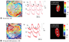
Our study was the first to map Ca2+ clock function in the SAN during prolonged isoproterenol infusion. LDCAE slope and 90% Cai relaxation time reached peak at 5 ± 2 min after isoproterenol infusion, and decreased after prolonged infusion. This finding occurred with the shift of the leading pacemaker site from superior to inferior SAN (Fig. 5). SNRT and cSNRT were increased after ZD 7288 infusion in the presence of prolonged isoproterenol infusion. These findings provide new insights into the mechanism of SAN dysfunction commonly found in patients with heart failure.38
Our results from an in vitro tachybradycardia model indicate that chronically elevated sympathetic tone results in abnormal pacemaking hierarchy in the RA, including suppression of the superior SAN and enhanced pacemaking from ectopic sites.39

Subthreshold DAD and a new mechanism of atrial arrhythmia

Automaticity and triggered activity are thought to be two distinct mechanisms for the initiation of heart beats. Automaticity occurs spontaneously and can be a source of both normal and abnormal heart beats, while triggered activity is pacing-induced and is almost always pathological. A mechanism of triggered activity is spontaneous (non-voltage gated) sarcoplasmic reticulum (SR) Ca release, which causes Na-Ca exchanger current (INCX) activation and membrane depolarization, resulting in delayed afterdepolarization (DAD).40 When DAD reaches threshold, it initiates triggered activity and arrhythmia (reverse excitation-contraction coupling).41,42 Recent studies, however, showed that rhythmic spontaneous Ca release ("Ca clock")43-45 may work together with hyperpolarization-activated membrane currents ("membrane clock") to generate normal sinus rhythm, a prototypical example of normal automaticity. These findings suggest that SAN activity may share mechanisms that underlie both automaticity and triggered activity, i.e., INCX activation.11,12,14,40,46-49 Consistent with this hypothesis, Bogdanov, et al.49 showed that in single isolated SAN cells, spontaneous SR Ca release in conditions of impaired INCX may result in membrane potential (Vm) oscillation without leading to regenerative action potential. However, whether or not DADs can occur in the intact SAN remains unknown.
We demonstrated that in intact RA preparation, failure of subthreshold DAD to reach threshold allowed latent pacemakers elsewhere to activate the atrium, resulting in atrial arrhythmia (Fig. 6). In these arrhythmic episodes, a beat that closed the longer PP interval, rather than a premature beat, was from an ectopic focus. This phenomenon was also compatible with the concept of parasystole in which the SAN was a source of normal rhythm while the ectopic pacemaker was the parasystolic focus. When the SAN failed to generated a rhythm to inhibit (or pre-excite) the parasystolic focus, the latter was able to exit and capture the entire RA. Shinohara, et al.50 recently used the same intact RA preparation to study the mechanisms of pacemaking of the ectopic pacemakers. They found that while spontaneous SR Ca release underlies isoproterenol-induced increase of superior SAN activity, the atrial ectopic pacemaker is less dependent on the Ca clock and more dependent on the membrane clock for its automaticity. These ectopic pacemakers outside the SAN therefore can effectively serve as backup pacemakers when SAN fails. The co-existence of two pacemaking sites resulted in atrial arrhythmias observed in the present study.

CONCLUSIONS

The voltage and Ca2+ clocks jointly regulate SAN automaticity. In various models of sick sinus syndrome, the dysfunction of both clocks was consistently observed. Our results from normal and pathological SANs strongly support the notion that the Ca2+ clock and the voltage clocks work synergistically to generate SAN automaticity.

Figures and Tables

Fig. 1
Activation pattern of SAN and surrounding RA during isoproterenol infusion of 0.3 µmol/L. (A) Isochronal map of Vm. The number on the each isochronal line indicates time (ms). White shaded area is the SAN. (B) The Vm (blue) and Cai (red) recordings from the superior (a), middle (b), inferior (c) SANs and RA (d) presented in A. (C) Magnified view of Cai and Vm tracings of superior SAN. Note the robust LDCAE (solid arrow) before phase 0 of action potential (0 ms), which in turn was much earlier than onset of p wave on ECG. (D) The Vm and Cai ratio maps at times from - 60 ms before to 180 ms after phase 0 action potential of C. The LDCAE (broken arrows in frame - 40 and - 20 ms) was followed by the Cai sinkhole during early diastole (solid arrow in frame 180 ms). This figure was reproduced with permission from Joung, et al.23 SAN, sinoatrial node; RA, right atrium; LDCAE, late diastolic Cai elevation.
ymj-52-211-g001
Fig. 2
Co-localization of LDCAE and the leading pacemaker site. (A) Upward shift of the leading pacemaker site with LDCAE during isoproterenol infusion. (a) Cai ratio maps of SAN at each sinus rate. (b) Corresponding Cai tracings from superior (1, 2), middle (3, 4) and inferior (5, 6) SAN. At 95 bpm, the sites 4 and 5 had most prominent LDCAEs (asterisks). As sinus rate gradually increased, the sites of Cai elevation progressively moved upward. At the maximum sinus rate of 173 bpm, the site 2 had the most apparent LDCAE. (B) Differential responses of different SAN sites to isoproterenol. (a) The Cai and Vm tracings from inferior, middle, and superior SAN sites at different sinus rates. (b) The LDCAE and DD slopes of superior SAN at different sinus rates. This figure was reproduced with permission from Joung, et al.23 SAN, sinoatrial node; LDCAE, late diastolic Cai elevation; DD, diastolic depolarization.
ymj-52-211-g002
Fig. 3
Complete absence of LDCAE in AF dogs during isoproterenol infusion. (A) Isoproterenol response of normal dogs. In normal dogs, isoproterenol increased the heart rate and shifted the leading pacemaker site to the superior SAN with robust LDCAE (arrows). (B) Isoproterenol response of AF dogs. The LDCAE was complete absent in AF dogs during isoproterenol infusion. (a) RA Vm isochronal map. (b) Cai tracings from superior (S), mid (M), and inferior (I) SANs. (c) SAN LDCAE isochronal map. The unit of numbers on RA Vm isochronal map is ms. The earliest activation of RA was considered as 0 ms. This figure was reproduced with permission from Joung, et al.32,33 SAN, sinoatrial node; AF, atrial fibrillation; RA, right atrium; LDCAE, late diastolic Cai elevation.
ymj-52-211-g003
Fig. 4
Tachybradycardia produced by prolonged isoproterenol and ZD 7288 infusion. The figure shows pseudo ECG from canine isolated RA. Upper panel, eight episodes of tachybradycardia. Lower panel, expanded view of a section marked by asterisk (*) showing 4.0 s and 3.5 s pauses. This figure was reproduced with permission from Joung, et al.39 RA, right atrium.
ymj-52-211-g004
Fig. 5
The effect of prolonged low dose isoproterenol infusion (n = 8). (A) Heart rate trends after prolonged (> 1 hr) isoproterenol infusion of 0.01 µmol/L. Heart rate was increased maximum at 5 min, and decreased after 10 min. ZD 7288 infusion of 3 µmol/L decrease the heart rate again almost to the baseline level. B through E show Vm isochronal map of SAN and surrounding RA (upper panels), and Cai (red) and Vm (blue) tracings from superior (a and b), middle (c and d) and lower (e and f) SAN. (B) Baseline. Note the diastolic depolarization (asterisks) in SAN. C, Isoproterenol infusion for 5 min. Note the LDCAE (asterisks) from superior SAN. (D) Isoproterenol infusion for 1 hr. Note the disappearance of LDCAE with shifting of the leading pacemaker site to inferior SAN (site e). (E) ZD 7288 infusion for 30 min. The pacemaker site shifted to the more inferior SAN (site f). The unit of numbers on Vm isochronal map is ms. This figure was reproduced with permission from Joung, et al.39 SAN, sinoatrial node; LDCAE, late diastolic Cai elevation.
ymj-52-211-g005
Fig. 6
The intermittent pattern of subthreshold DADs. These tracings were obtained during isoproterenol infusion (0.03 µmol/L) in the first RA preparation. (A) Optical signals of Cai (red) and Vm (blue) from superior (a and b), middle (c and d), and inferior (e and f) SAN, and RA (g). There were 3 consecutive activations in this figure. Among them, the first 1) and third beats show LDCAE (arrows) followed by the initiation of sinus beats from the same sites. In contrast, the second beat 2) showed both LDCAE on Cai tracings and subthreshold DADs on Vm tracings (asterisks). The downslope of the subthreshold DADs were observed because they failed to trigger an action potential. (B) Vm isochronal maps of the first (1) and second (2) beats. The white shaded area is the SAN. The first beat 1) was from SAN. Because subsequent LDCAE in the SAN (asterisks in Panel A) failed to trigger a sinus beat, an ectopic pacemaker was able to take over and activate the mapped region 2). This figure was reproduced with permission from Joung, et al.51 RAA, right atrial appendage; SVC, superior vena cava; A, anterior; P, posterior; S, superior; I, inferior, DAD, delayed afterdepolarization; SAN, sinoatrial node; RA, right atrium; LDCAE, late diastolic Cai elevation.
ymj-52-211-g006

ACKNOWLEDGEMENTS

This work was supported by NIH Grants P01 HL78931, R01 HL78932, 71140, faculty research grants of Yonsei University College of Medicine (6-2009-0176, 6-2010-0059, 7-2009-0583, 7-2010-0676), Basic Science Research Program through the National Research Foundation of Korea funded by the Ministry of education, science and technology (2010-0021993), the Korean Healthcare Technology Research and Develop grant number A085136 (BJ), a Medtronic-Zipes Endowment (PSC) and by an AHA Established Investigator Award (SFL).

Notes

The authors have no financial conflicts of interest.

References

1. Adán V, Crown LA. Diagnosis and treatment of sick sinus syndrome. Am Fam Physician. 2003. 67:1725–1732.
2. Brown HF, Difrancesco D, Noble SJ. How does adrenaline accelerate the heart? Nature. 1979. 280:235–236.
crossref
3. Baruscotti M, Bucchi A, DiFrancesco D. Physiology and pharmacology of the cardiac pacemaker ("funny") current. Pharmacol Ther. 2005. 107:59–79.
crossref
4. DiFrancesco D. Pacemaker mechanisms in cardiac tissue. Annu Rev Physiol. 1993. 55:455–472.
crossref
5. DiFrancesco D. The pacemaker current (I(f)) plays an important role in regulating Sa node pacemaker activity. Cardiovasc Res. 1995. 30:307–308.
crossref
6. Milanesi R, Baruscotti M, Gnecchi-Ruscone T, DiFrancesco D. Familial sinus bradycardia associated with a mutation in the cardiac pacemaker channel. N Engl J Med. 2006. 354:151–157.
crossref
7. Bucchi A, Barbuti A, Baruscotti M, DiFrancesco D. Heart rate reduction via selective 'funny' channel blockers. Curr Opin Pharmacol. 2007. 7:208–213.
crossref
8. Nof E, Luria D, Brass D, Marek D, Lahat H, Reznik-Wolf H, et al. Point mutation in the HCN4 cardiac ion channel pore affecting synthesis, trafficking, and functional expression is associated with familial asymptomatic sinus bradycardia. Circulation. 2007. 116:463–470.
crossref
9. Rubenstein DS, Lipsius SL. Mechanisms of automaticity in subsidiary pacemakers from cat right atrium. Circ Res. 1989. 64:648–657.
crossref
10. Li J, Qu J, Nathan RD. Ionic basis of ryanodine's negative chronotropic effect on pacemaker cells isolated from the sinoatrial node. Am J Physiol. 1997. 273:H2481–H2489.
11. Ju YK, Allen DG. Intracellular calcium and Na+-Ca2+ exchange current in isolated toad pacemaker cells. J Physiol. 1998. 508:153–166.
crossref
12. Hüser J, Blatter LA, Lipsius SL. Intracellular Ca2+ release contributes to automaticity in cat atrial pacemaker cells. J Physiol. 2000. 524(Pt 2):415–422.
crossref
13. Vinogradova TM, Bogdanov KY, Lakatta EG. Novel perspectives on the beating rate of the heart. Circ Res. 2002. 91:e3.
crossref
14. Maltsev VA, Vinogradova TM, Lakatta EG. The emergence of a general theory of the initiation and strength of the heartbeat. J Pharmacol Sci. 2006. 100:338–369.
crossref
15. Vinogradova TM, Lyashkov AE, Zhu W, Ruknudin AM, Sirenko S, Yang D, et al. High basal protein kinase A-dependent phosphorylation drives rhythmic internal Ca2+ store oscillations and spontaneous beating of cardiac pacemaker cells. Circ Res. 2006. 98:505–514.
crossref
16. Bhuiyan ZA, van den Berg MP, van Tintelen JP, Bink-Boelkens MT, Wiesfeld AC, Alders M, et al. Expanding spectrum of human TYR2-related disease: new electrocardiographic, structural, and genetic features. Circulation. 2007. 116:1569–1576.
17. Verheijck EE, van Kempen MJ, Veereschild M, Lurvink J, Jongsma HJ, Bouman LN. Electrophysiological features of the mouse sinoatrial node in relation to connexin distribution. Cardiovasc Res. 2001. 52:40–50.
crossref
18. Lancaster MK, Jones SA, Harrison SM, Boyett MR. Intracellular Ca2+ and pacemaking within the rabbit sinoatrial node: Heterogeneity of role and control. J Physiol. 2004. 556:481–494.
crossref
19. Tellez JO, Dobrzynski H, Greener ID, Graham GM, Laing E, Honjo H, et al. Differential expression of ion channel transcripts in atrial muscle and sinoatrial node in rabbit. Circ Res. 2006. 99:1384–1393.
crossref
20. Boineau JP, Miller CB, Schuessler RB, Roeske WR, Autry LJ, Wylds AC, et al. Activation sequence and potential distribution maps demonstrating multicentric atrial impulse origin in dogs. Circ Res. 1984. 54:332–347.
crossref
21. Boineau JP, Schuessler RB, Mooney CR, Wylds AC, Miller CB, Hudson RD, et al. Multicentric origin of the atrial depolarization wave: the pacemaker complex. Relation to dynamics of atrial conduction, P-wave changes and heart rate control. Circulation. 1978. 58:1036–1048.
crossref
22. Schuessler RB, Boineau JP, Wylds AC, Hill DA, Miller CB, Roeske WR. Effect of canine cardiac nerves on heart rate, rhythm, and pacemaker location. Am J Physiol. 1986. 250:H630–H644.
crossref
23. Joung B, Tang L, Maruyama M, Han S, Chen Z, Stucky M, et al. Intracellular calcium dynamics and acceleration of sinus rhythm by beta-adrenergic stimulation. Circulation. 2009. 119:788–796.
crossref
24. Vinogradova TM, Bogdanov KY, Lakatta EG. beta-Adrenergic stimulation modulates ryanodine recepr Ca(2+) release during diastolic depolarization to accelerate pacemaker activity in rabbit sinoatrial nodal cells. Circ Res. 2002. 90:73–79.
crossref
25. Dobrzynski H, Boyett MR, Anderson RH. New insights into pacemaker activity: promoting understanding of sick sinus syndrome. Circulation. 2007. 115:1921–1932.
26. Verkerk AO, Wilders R, Coronel R, Ravesloot JH, Verheijck EE. Ionic remodeling of sinoatrial node cells by heart failure. Circulation. 2003. 108:760–766.
crossref
27. Nattel S, Maguy A, Le Bouter S, Yeh YH. Arrhythmogenic ion-channel remodeling in the heart: heart failure, myocardial infarction, and atrial fibrillation. Physiol Rev. 2007. 87:425–456.
crossref
28. Van Wagoner DR, Pond AL, Lamorgese M, Rossie SS, McCarthy PM, Nerbonne JM. Atrial L-type Ca2+ currents and human atrial fibrillation. Circ Res. 1999. 85:428–436.
29. Workman AJ, Kane KA, Rankin AC. The contribution of ionic currents to changes in refractoriness of human atrial myocytes associated with chronic atrial fibrillation. Cardiovasc Res. 2001. 52:226–235.
crossref
30. Van Wagoner DR, Pond AL, McCarthy PM, Trimmer JS, Nerbonne JM. Outward K+ current densities and Kv1.5 expression are reduced in chronic human atrial fibrillation. Circ Res. 1997. 80:772–781.
crossref
31. Yeh YH, Burstein B, Qi XY, Sakabe M, Chartier D, Comtois P, et al. Funny current downregulation and sinus node dysfunction associated with atrial tachyarrhythmia: a molecular basis for tachycardia-bradycardia syndrome. Circulation. 2009. 119:1576–1585.
crossref
32. Efimov IR, Fedorov VV, Joung B, Lin SF. Mapping cardiac pacemaker circuits: methodological puzzles of the sinoatrial node optical mapping. Circ Res. 2010. 106:255–271.
33. Joung B, Lin SF, Chen Z, Antoun PS, Maruyama M, Han S, et al. Mechanisms of sinoatrial node dysfunction in a canine model of pacing-induced atrial fibrillation. Heart Rhythm. 2010. 7:88–95.
crossref
34. Sanders P, Kistler PM, Morton JB, Spence SJ, Kalman JM. Remodeling of sinus node function in patients with congestive heart failure: reduction in sinus node reserve. Circulation. 2004. 110:897–903.
crossref
35. Ogawa M, Zhou S, Tan AY, Song J, Gholmieh G, Fishbein MC, et al. Left stellate ganglion and vagal nerve activity and cardiac arrhythmias in ambulatory dogs with pacing-induced congestive heart failure. J Am Coll Cardiol. 2007. 50:335–343.
crossref
36. Zicha S, Fernández-Velasco M, Lonardo G, L'Heureux N, Nattel S. Sinus node dysfunction and hyperpolarization-activated (HCN) channel subunit remodeling in a canine heart failure model. Cardiovasc Res. 2005. 66:472–481.
crossref
37. Rubart M, Zipes DP. Mechanisms of sudden cardiac death. J Clin Invest. 2005. 115:2305–2315.
crossref
38. Sanders P, Berenfeld O, Hocini M, Jaïs P, Vaidyanathan R, Hsu LF, et al. Spectral analysis identifies sites of high-frequency activity maintaining atrial fibrillation in humans. Circulation. 2005. 112:789–797.
crossref
39. Joung B, Shinohara T, Zhang H, Kim D, Choi EK, On YK, et al. Tachybradycardia in the isolated canine right atrium induced by chronic sympathetic stimulation and pacemaker current inhibition. Am J Physiol Heart Circ Physiol. 2010. 299:H634–H642.
crossref
40. Bers DM. Calcium cycling and signaling in cardiac myocytes. Annu Rev Physiol. 2008. 70:23–49.
crossref
41. ter Keurs HE, Zhang YM, Miura M. Damage-induced arrhythmias: reversal of excitation-contraction coupling. Cardiovasc Res. 1998. 40:444–455.
crossref
42. Boyden PA, ter Keurs HE. Reverse excitation-contraction coupling: Ca2+ ions as initiators of arrhythmias. J Cardiovasc Electrophysiol. 2001. 12:382–385.
crossref
43. Lakatta EG, DiFrancesco D. What keeps us ticking: a funny current, a calcium clock, or both? J Mol Cell Cardiol. 2009. 47:157–170.
crossref
44. Maltsev VA, Lakatta EG. Synergism of coupled subsarcolemmal Ca2+ clocks and sarcolemmal voltage clocks confers robust and flexible pacemaker function in a novel pacemaker cell model. Am J Physiol Heart Circ Physiol. 2009. 296:H594–H615.
45. Lakatta EG, Maltsev VA, Vinogradova TM. A coupled SYSTEM of intracellular Ca2+ clocks and surface membrane voltage clocks controls the timekeeping mechanism of the heart's pacemaker. Circ Res. 2010. 106:659–673.
crossref
46. Zhou Z, Lipsius SL. Na(+)-Ca2+ exchange current in latent pacemaker cells isolated from cat right atrium. J Physiol. 1993. 466:263–285.
47. Hata T, Noda T, Nishimura M, Watanabe Y. The role of Ca2+ release from sarcoplasmic reticulum in the regulation of sinoatrial node automaticity. Heart Vessels. 1996. 11:234–241.
crossref
48. Rigg L, Terrar DA. Possible role of calcium release from the sarcoplasmic reticulum in pacemaking in guinea-pig sino-atrial node. Exp Physiol. 1996. 81:877–880.
crossref
49. Bogdanov KY, Vinogradova TM, Lakatta EG. Sinoatrial nodal cell ryanodine receptor and Na(+)-Ca(2+) exchanger: molecular partners in pacemaker regulation. Circ Res. 2001. 88:1254–1258.
50. Shinohara T, Joung B, Kim D, Maruyama M, Luk HN, Chen PS, et al. Induction of atrial ectopic beats with calcium release inhibition: Local hierarchy of automaticity in the right atrium. Heart Rhythm. 2010. 7:110–116.
crossref
51. Joung B, Zhang H, Shinohara T, Maruyama M, Han S, Kim D, et al. Delayed Afterdepolarization in intact Canine Sinoatrial Node as a Novel Mechanism for Atrial Arrhythmia. J Cardiovasc Electrophysiol. 2010. (In press).
crossref
TOOLS
Similar articles