

 PDF
PDF ePub
ePub Citation
 Print
Print


INTRODUCTION
The third variable (V3) region of human immunodeficiency virus type 1 (HIV-1) gp120 has been termed the principal neutralizing determinant of the virus1 and is involved in HIV-1 viral entry.2,3 For efficient entry into target cells, HIV-1 requires specific chemokine receptors (CCR5, CXCR4) as well as the primary receptor, CD4.4,5 The specific amino acids located in the V3 loop of HIV-1 gp120 play an important role in determining cellular tropism and viral preference for the specific chemokine co-receptor for viral entry.6 HIV-1 strains that enter T cell lines prefer CXCR4 as a co-receptor, resulting in the syncytium-inducing phenotype.7 CCR5 and, less commonly, CCR3 or CCR2b, are preferred by HIV-1 strains that enter macrophages, resulting in the macrophage-tropic phenotype.8 However, V3-specific antibody is rarely detected in the serum of patients with AIDS for V3 itself is not highly immunogenic.9 Therefore, the V3 loop can be a useful target for vaccine development against HIV-1. Several approaches have been attempted to intensify the immunogenicity of V3, such as substitution of amino acids,10,11 fusion with HBV surface Ag12 or toxoid,13 and construction of multimers.14
Bacille Calmette-Guérin (BCG) is a live, attenuated bovine tubercle bacillus widely used to immunize against tuberculosis. BCG has many advantages as a vaccine vehicle, such as low toxicity, adjuvant potential, low cost, and long-lasting immune-inducing capacity. Since Stover, et al.15,16 developed a shuttle vector (pMV261) that could replicate in BCG and Escherichia coli, many researchers have constructed recombinant BCG vaccines expressing foreign antigens with sufficient immunogenicity to induce humoral and cellular immunity that could protect the host from viral, bacterial, or parasitic infections.17,18 Whereas, current development of HIV-1 vaccine is strongly recommended to induce humoral and cellular immunity against HIV-1 after vaccination.19-21 In this aspect, recombinant BCG can be a fascinating vaccine vector for the development of HIV-1 vaccine.19-21
In the present study, recombinant BCG (rBCG-mV3) was constructed which expressed the trimeric HIV-1 V3 loop (mV3), and the immunogenicity of rBCG-mV3 has been determined in experimental animals.
MATERIALS AND METHODS
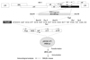
All of the experiments were performed based on the process shown in Fig. 1.
Plasmid construction and expression
V3 was amplified by polymerase chain reaction (PCR) from cDNA of HXB2 with PCR primers covering the V3 region. Sense 5' ACC CGG GAT CCA TGT ACA AGA CCC AAC AAC-3' (BamHI), antisense 5' CTA CA CAA TTC AAG GTT ACA ATG TGC TT-3' (EcoRI), sense 5' CTA CA GAA TTC ATT AAT TGT ACA AGA CC-3' (EcoRI), antisense 5' CAA GT CTG CAG AAT GTT ACA ATG TGC TT-3' (PstI), sense 5' CAA GT CTGCAG ATT AAT TGT ACA AGA CC-3' (PstI) and antisense 5' GCA TTA AGC TTA AAT GTT ACA ATG TGC TTG TC-3' (HindIII) primers were designed from the sequence of HXB2, as reported previously.22 PCR-amplified cDNA were eluted from agarose gel and digested with BamHI, EcoRI, PstI, and HindIII. The PCR fragments of the 135bp of V3 DNA was ligated, and reamplified with the BamHI sense & HindIII antisense primer set. The 405bp of V3 trimer (mV3) fragments were ligated into pRSET B (Invitrogen, Carlsbad, CA, USA) and pMV261, and henceforth referred to as pRSET-mV3 and pMV-mV3, respectively. Recombinant pRSET-mV3 plasmid was introduced into an E. coli strain containing an episome of T7 RNA polymerase, BL21 (DE3). The pRSET-mV3/BL21 (DE3) clone was cultured in modified M9 broth (1% Bacto-tryptone, 0.4% glucose, 0.5% NaCl, 1 mM MgSO4, 0.3% KH2PO4, 0.6% NaH2PO4, and 0.1 mM NH4Cl). Recombinant mV3 synthesis was induced by isopropyl-beta-D-thiogalactopyranoside (IPTG) at a final concentration of 1 mM. pMV-mV3 recombinant plasmid was transformed into the Mycobacterium bovis BCG 1173-P2 Pasteur strain, as described previously.17 Cells were plated on Middlebrook 7H10 agar (Difco Laboratory, Detroit, MI, USA) containing albumin, dextrose, catalase (ADC) enrichment, and kanamycin (25 µg/mL). Kanamycin-resistant colonies were sub-cultured in Middlebrook 7H10 liquid medium containing ADC enrichment and kanamycin for 1 week. mV3 expression was induced by treating the rBCG-mV3 positive clones at 45℃ for 2 hours, as described previously.17 To extract plasmid DNA from the positive BCG clones, the cell pellets were resuspended and incubated with 400 µL of glucose-Tris-EDTA/20 mg lysozyme at 37℃ for 2-3 hours. Cells were disrupted by adding 300 µL of lysis buffer (2.5% SDS and 0.3% NaOH), and 250 µL of Sol III [3M potassium acetate (pH 5.2)] to the reaction mixture. Supernatant was precipitated by adding the same volume of isopropanol.
Anti-mV3 antiserum preparation
The pRSET-mV3-transformed E. coli BL21 (DE3) culture was harvested, resuspended in lysis buffer [50 mM Tris-Cl (pH 8.0), 1 mM EDTA and 100 mM NaCl], and then lysed by an ultrasonic dismembrator. The lysate was dissolved in buffer A [6 M guanidine HCl, 0.1 M Na-phosphate and 0.01 M Tris-Cl (pH 8.0)], centrifuged, and the supernatant was applied to the 50% slurry of a Ni+-NTA agarose column (Quiagen, Chatsworth, CA, USA) at a flow rate of 10-15 mL/hour. The column was sequentially washed with 10 column-volumes of buffer A, 5 column-volumes of buffer B [8 M urea, 0.1 M Na-phosphate, and 0.01 M Tris-Cl (pH 8.0)], and 3 column-volumes of buffer C [buffer B (pH 6.8)]. Purified proteins were eluted with buffer D [buffer B (pH 4.5), supplemented with 200 mM EDTA], dialyzed against phosphate buffered saline (PBS), and then re-dissolved in 0.2 M sodium bicarbonate buffer containing 0.02% SDS (pH 7.4) overnight. Small amounts of the eluted samples were tested for purity on 12% SDS-PAGE. Ten 6-week-old female BALB/c mice were used to raise antiserum. Recombinant mV3 protein was mixed with the same volume of Freund complete adjuvant by sonication. A protein-adjuvant emulsion containing 25 µg of immunogen was injected subcutaneously into each mouse. The first immunization was followed by two booster injections every 2 weeks with 20 µg of the protein mixed with incomplete adjuvant. The final immunization was given 2 weeks after the second booster by injecting 10 µg of recombinant protein via the tail vein. Blood was collected from the ophthalmic vein of immunized mice, from which antiserum was prepared and used for Western blot analysis.
Analysis of mV3 expression in rBCG
As described previously,17 recombinant BCG-mV3 transformants were cultured in Middliebrook 7H9 broth media containing 25 µg/mL of kanamycin. When the cells were grown to 1×10⁶ cells/mL, the culture was heat-induced, and then harvested at the requested time points. The cells were washed in PBS plus 0.05% Tween 80 and resuspended in 1/20-volume of radioimmunoprecipitation assay buffer. Culture lysates were analyzed by Western blot hybridization with anti-mV3-antiserum prepared from the previous step and goat-antimouse IgG (Sigma Chemical Co., St. Louis, MO, USA).
Immunization of BALB/c mice with rBCG-mV3
BALB/c female mice, 5-6 weeks old, were inoculated intraperitoneally with heat-induced rBCG-mV3 and control BCG at a concentration of 1×10⁷ cells/mouse.
Genetic stability of the rBCG-mV3 in vivo
Immunized BALB/c mice were killed 4, 8, and 16 weeks after inoculation. Spleens were homogenized in PBS with 0.05% Tween 80 (pH 7.5) and were then cultured on the Middlebrook 7H11 agar plate supplemented with oleic acid, albumin, dextrose, catalase (OADC) enrichment (Difco Laboratory), and 25 µg/mL of kanamycin. Kanamycin-resistant colonies were subcultured in Middlebrook 7H9 liquid medium containing ADC enrichment and kanamycin. Lysates from these cultures were tested for mV3 expression by Western blot, and plasmid DNA extracted from the recovered BCG clone was tested for its integrity by PCR with mV3-covering PCR primer set.
Antibody titration and IgG isotyping
Blood was obtained from the ophthalmic vein from BALB/c mice every other week beginning 5-6 weeks after immunization, and antiserum was assessed by ELISA to measure mV3-specific antibodies, as described previously.17 EIA plates were coated with 0.5 µg/mL of the purified recombinant mV3 overnight. After the plates were washed 3 times with 0.2% Tween 20 in PBS (PBS-T) and blocked with 2% blotto/0.5% bovine serum albumin (BSA), 100 µL of the serum samples (1 µL of serum in 100 µL binding buffer) were added to each well and incubated at 37℃ for 1 hour. Plates were washed 3 times with PBS-T buffer, and incubated with alkaline phosphatase-conjugated goat antimouse IgG secondary antibody (Sigma Chemical Co.). P-nitrophenyl phosphate solution was added to each well, and then read by an ELISA reader (Molecular Devices Inc., St. Louis, MO, USA) at 405 nm. Analysis of mV3-specific antibodies was performed with a mouse Ig isotyping kit (Pharmingen, San Diego, CA, USA).
DTH response in immunized guinea pigs
As described previously,17 guinea pigs were administered 1×10⁸ cells of live rBCG-mV3 and control BCG intraperitoneally. Four weeks later, the mice were boosted with the same amount of cells via the same route. Two weeks after boosting, the animals were skin tested by intradermal injection of purified recombinant mV3 protein and purified protein derivative of tuberculin (PPD). Delayed type hypersensitivity (DTH) responses were evaluated by measuring erythema 48 hours after skin injection.
T cell proliferation assay
Spleens were removed from mice 10 weeks and 5 months after the primary injection. The splenocytes of each immunization group (n = 4) were pooled, and 2×10⁵ cells/well in RPMI-1640 containing 2% mouse serum were cultured in 96-well plates in the presence or absence of antigen for 120 hours, labeled with [3H]-thymidine (1 mCi/well) for 8 hours prior to harvesting, and then counted on a scintillation counter.
RESULTS
Recombinant mV3 protein and polyclonal anti-mV3 antibody
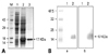
Recombinant mV3 proteins were purified from pRSET B-mV3/E. coli BL21 (DE3) using a Ni+-NTA resin column (Fig. 2A). Polyclonal antibody specific to mV3 protein was obtained from BALB/c mice immunized with the recombinant protein (Fig. 2B). In order to determine whether or not the mV3 protein shared similar properties with the other V3 proteins, recombinant protein was assessed by Western blot analysis with anti-gp120 polyclonal antibodies. The mV3 protein was detected by mouse anti-V3 and rabbit anti-gp120 polyclonal antibodies (Fig. 2B). These results suggest that V3-specific antibodies induced by mV3-immunization are likely to interact with wild type V3 motif.
Genetic stability of rBCG-mV3 in vitro and in vivo
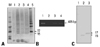
pMV-mV3 recombinant plasmid was transformed into BCG by electroporation, as described in the Materials and Methods. The rBCG-mV3 stably expressed mV3 over 6 weeks when examined every 2 weeks (Fig. 3A). BALB/c mice were immunized with rBCG-mV3. The rBCG-mV3 isolated from the mice 8 weeks after immunization still harbored the inserted mV3 (Fig. 3B) and expressed mV3 protein (Fig. 3C). These data indicate that the rBCG-mV3 is genetically stable, not only in vitro, but also in vivo.
Recombinant BCG-mV3 was efficient in inducing V3-specific humoral immunity which persisted for at least 14 weeks
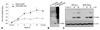
After a single heat shock, the rBCG-mV3 was injected into BALB/c mice. Six weeks later, V3-specific antibody was assessed in the antiserum collected from the immunized mice every 2 weeks. As shown in Fig. 4A, mV3-specific polyclonal antibodies were induced and maintained for at least 14 weeks. The amount of V3-specific polyclonal antibodies rapidly increased in the immunized mice, reached a maximum at week 10, and then lasted for over 4 weeks (Fig. 4A). The specific antibodies completely disappeared when examined 10 months after primary inoculation (data not shown). The specificity of polyclonal antibodies to mV3 was further evidenced by Western blot analysis with purified mV3 and control BCG lysates (Fig. 4B). However, HIV-1 replication was not efficiently blocked by the antiserum (Fig. 4C).
Recombinant BCG-mV3 elicited mV3-specific DTH responses in guinea pigs
In general, recombinant BCG is well-known to induce strong DTH responses against BCG and introduced foreign antigens in immunized animals.23,24 The mV3-specific DTH was assessed in the guinea pigs immunized with rBCG-mV3. Two weeks after the second inoculation, animals were tested for the reciprocal DTH response to mV3 protein and PPD. The DTH responses to mV3 in the immunized animals were positive 6 weeks after primary inoculation (Table 1). Recombinant BCG-mV3 induced relatively weak DTH response against mV3 protein compared with that against PPD in immunized guinea pigs, which was attributable to the amount of mV3 weakly expressed in rBCG-mV3.
Cellular immune responses in mice immunized with rBCG-mV3
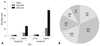
Sometimes there is a discrepancy between the DTH response and T cell proliferation. The T cell responses in immunized mice were examined. The splenocytes of mice immunized with rBCG-mV3 or control BCG were isolated 10 weeks and 5 months after immunization, and subjected to a T cell proliferation assay. The stimulation index (SI) was determined, as described in the Materials and Methods. The SI in response to mV3 in mice immunized with rBCG-mV3 was about three times as strong as that in mice immunized with control BCG (BCG-PMV), and the SI to mV3 assessed 5 months after immunization was markedly enhanced compared with that assessed 10 weeks after immunization (Fig. 5A). These results imply that rBCG-mV3 is able to produce and maintain the memory T cells against mV3 in immunized animals during induction of cellular immunity against BCG.
mV3-specific IgG2a was prevalent in anti-sera
It is well established that IgG1 and IgG3 represent the Th2 response,25,26 and IgG2a results from the Th1 response.26 Based on IgG isotyping analysis, V3-specific IgG2a was prevalent in the sera of mice immunized with recombinant BCG-mV3, whereas IgG1 and IgG3 isotypes were relatively lower (Fig. 5B). This means that rBCG-mV3 was very effective in inducing Th1-oriented cell-mediated immunity in immunized mice.
DISCUSSION
A rBCG-mV3, expressing the trimer of HIV-1 V3, was constructed by introducing pMV-mV3 plasmid into BCG as a vaccine vector. Based on the information that Gly-Pro-Gly-Arg-Ala-Phe residues (GxGRxF) are highly conserved in HIV-1 variants27,28 and play a crucial role in the interaction with chemokine receptor,29 these sequences have been included into the V3-trimer.
Not only humoral immunity but also Th1-oriented cellular immunity are essential for protection against HIV-1 infection.30 BCG is well-known for its safety and efficacy in the induction of strong cell-mediated immunity.31 However, even a BCG as a live attenuated vaccine caused tuberculosis-like disease in simian immunodeficiency virus (SIV)-infected monkeys is probably due to a loss of T-cell immunity.32 Therefore, it is advisable to develop a recombinant BCG as a preventative vaccine rather than therapeutic vaccine against HIV-1.33
In the present study, we have examined the humoral and Th1-oriented cell-mediated immunities in mice and guinea pigs immunized with recombinant BCG expressing multiple HIV-1 V3 domains. The mV3 protein expressed in BCG was detected by Western blot analysis with mouse anti-mV3 serum and anti-gp120 polyclonal antibodies (Fig. 2B). Even though V3 has high sequence variations among HIV-1 variants, it still has some conserved region which can be targeted for vaccine development.34
The mV3 proteins expressed in E. coli were easily detected by polyclonal antibody against V3 protein, but hardly detected when expressed in rBCG-mV3 due to the limited amount of expression (Fig. 3). Nonetheless, the immune response to mV3 was not as weak as shown in mice immunized the rBCG-mV3 (Figs. 4 and 5). The rBCG-mV3 injected into mice survived over 8 weeks and produced the mV3 protein for the same period in vitro and in vivo (Fig. 3). Thus, a single immunization of rBCG-mV3 would be enough to induce specific immunity without an additional boosting injection. mV3-specific antibodies lasted longer than 14 weeks after immunization (Fig. 4).
The mV3-containing the conserved sequence (GxGRxF-GPGRAF) was expected to produce a neutralizing antibody against various HIV-1 subtypes. rBCG-mV3 induced substantial amounts of anti-mV3 antibody in the serum of immunized mice. Unfortunately, the V3-specific antiserum, obtained from mice immunized with rBCG-mV3, was not able to inhibit the HIV-1 replication in vitro, indicating that the antiserum does not contain any neutralizing antibody. This was due to the lack of appropriate V3-conformational epitope after expression in BCG. However, rBCG-mV3 was very effective in inducing V3-specific antibodies which might be involved in opsonization of the virus in vivo, followed by facilitating virus clearance in vaccinated animals. rBCG-mV3 also induced a strong humoral immunity against control BCG itself (Fig. 4B). However, we are unable to rule out the possibility that the construction of recombinant BCG to express HIV-1 V3 multimer might cause some changes of the characteristics of original BCG.
Cell-mediated immunity plays a key role in the inhibition of AIDS progression.19,20,34,35 In the present study, antigen-specific DTH response in guinea pigs (Table 1) and high titer of V3-specific IgG2a in recombinant BCG-immunized mice (Fig 5B) suggest that recombinant BCG also induced strong cell-mediated immunity and immune memory against the introduced foreign gene. While it is well-known that BALB/c mice have a bias tendency towards Th2 immunity, recombinant BCG is such a good Th1 immune stimulator that it pushes for Th1 development even in BALB/c mice, as shown previously in IL-12-mediated Th1 development in BALB/c mice.36 However, to make it clearer, DTH response in immunized BALB/c mice remains to be traced by measuring footpad swelling, as reported previously.37
In summary, recombinant BCG expressing mV3 efficiently induced humoral and cellular immunities against the V3 domain. Particularly noteworthy is that rBCG-mV3 induced long-lasting mV3-specific cellular immunity in immunized animals.
 XML Download