

 PDF
PDF ePub
ePub Citation
 Print
Print


INTRODUCTION
Drug eluting stent (DES) has successfully been introduced to interventional cardiology in order to prevent in-stent restenosis. Nevertheless, concerns about the safety of DES still exist. Especially, late stent thrombosis (LST) is an unsolved problem in the DES era, which was rarely seen with bare metal stents (BMS).1 The risk of LST is associated with patient and lesion characteristics, non-compliance of anti-platelet drugs and incomplete stent apposition, number and the length of stents, coronary dissection, etc.2,3 Incomplete neointimal coverage of stent struts is the most important predictor of LST.4,5 We report a case of LST despite complete neointimal coverage on well-opposed stent struts and continued dual-antiplatelet therapy more than 1 year.
CASE REPORT
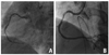
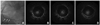
A 57-year-old male with a history of hypertension, diabetes mellitus, normal renal function and a 60 pack years smoking received percutaneous coronary intervention at the right coronary artery with the impression of stable angina pectoris in September 2007 (Fig. 1). Two sirolimus-eluting stents (SES) [3.0×28 mm, 2.75×28 mm (Cypher, Cordis Corp., Johnson & Johnson Co., Warren, New Jersey, USA)] were implanted at the distal part of the right coronary artery. Transthoracic echocardiography demonstrated normal left ventricular systolic function (ejection fraction=56%) with regional wall motion abnormality at the inferior wall. One-year follow-up angiogram showed that 2 stents were all patent. Intravascular ultrasound (IVUS) and optical coherence tomography (OCT) revealed complete neointimal coverage of stent struts (Fig. 2).
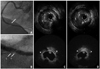
Until recently, he had good compliance with dual antiplatelet agents and other medications, and did not suffer from chest pain. However, the result of treadmill test was positive. Therefore, he underwent another follow-up angiogram to evaluate silent ischemia in January 2010. The follow-up angiogram revealed 2 patent stents implanted in the right coronary artery. However, abnormal contrast filling defect was observed within the previously stented segments (arrow in Fig. 3A). IVUS and OCT were performed to elucidate the characteristics of the abnormal contrast filling defect. Both studies revealed a newly formed and ruptured atheromatous plaque within the neointima over the stent struts (IVUS and OCT) (Fig. 3B, C and D). He was successfully treated with percutaneous coronary intervention and discharged on medications of aspirin (100 mg/day), clopidogrel (75 mg/day), atorvastatin (10 mg/day) and atenolol (25 mg/day).
DISCUSSION
LST after DES implantation has been thought to be caused by poorly formed neointima and incomplete neointimal coverage over DES struts.6 In a recent study using angioscopy, however, a newly formed and thrombogenic atherosclerotic yellow neointima after DES implantation was reported.7 This phenomenon may be an another cause for late stent thrombosis after DES implantation. Stent thrombosis and myocardial infarction caused by restenosis during extended follow-up after bare metal stent implantation have been reported and might be explained by newly formed and progressive atherosclerosis within neointima.8 However, this phenomenon has seldom been reported in DES. Atherosclerotic change within neointima after bare metal stent implantation usually occurs after 5 years.7 The intracoronary images of our patient illustrate a newly formed and ruptured atheromatous plaque within neointima after DES implantation in a relatively short period (less than 3 years after DES implantation). A recent pathologic study showed that atherosclerotic lesions progress more rapidly within SES than within BMS. In the autopsy study by Nakazawa, et al.9 the atherosclerotic change in SES was seen in more than 40 percent of cases by 9 months, but the change in BMS occurred only after 2 years. We think that early atherosclerosis is a possible mechanism in our case. This was an unexpected finding and gave us a very helpful insight for better treatment and monitoring in patients with DES implantation.
 XML Download