

 PDF
PDF ePub
ePub Citation
 Print
Print


INTRODUCTION
Idiopathic ventricular tachycardia (VT), which develops without any cardiac abnormalities, can be divided into four types: outflow tract VT, verapamil sensitive fascicular VT, focal Purkinje VT and annular VT. The main mechanism of idiopathic left VT is reentry that passes through anterior or posterior fascicle.1,2 This fascicular VT commonly shows left axis deviation of the right bundle branch block (RBBB) of more than 90%. On the other hand, focal Purkinje VT, which is initiated by automaticity, is very infrequent. Purkinje VT is characterized by a lack of response to verapamil and by its inability to being induced or terminated by electrical programmed stimulation. However, the exact clinical aspect or etiology of this VT has not yet been described. In this report, we describe a case in which catheter ablation was used to successfully treat a focal Purkinje VT combined with tachycardia-induced cardiomyopathy.
CASE REPORT
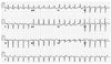
A 36-year-old man with no specific past illnesses was referred for evaluation of his tachycardia and dyspnea for a week. After admission, physical examination revealed grade 2/6 systolic murmur at the left sternal borders and decreased breathing sounds with both basal rales. The initial electrocardiogram (ECG) revealed wide QRS tachycardia (Fig. 1), and the chest X-rays showed marked cardiomegaly with pulmonary congestion. A routine blood profile, including myocardial markers and inflammatory markers, did not reveal any specific abnormalities. The wide QRS tachycardia observed in the ECG showed RBBB, left axis deviation and atrioventricular dissociation. Echocardiography showed 25% of left ventricular ejection fraction and enlargement of the left ventricle and left atrium without a regional wall motion abnormality. Intra-venous and oral verapamil was administered under the suspicion of tachycardia-mediated cardiomyopathy resulting from idiopathic posterior fascicular VT. However, continuous telemetry monitoring demonstrated incessant VT without any response to the treatment. Therefore, electrophysiological studies and radiofrequency catheter ablation (RF-CA) were carried out.
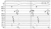
During the electrophysiological recordings, intermittent sinus capture beats were observed, nevertheless, VT continued, and it could not be terminated with 12 mg of adenosine or right ventricle overdrive pacing (pacing cycle length 260-200 ms). Ablation was started from the distal area, where the earliest Purkinje potential was found (Fig. 2). Entrainment at ablation site did not meet the classical criteria, and pace-mapping at ablation site was similar but not exactly matched with clinical VT. It might be due to fusion of pacing beats with incessant VT. If the VT failed to stop within 20 seconds of RF-CA application, the ablation catheter was moved more proximally, and the RF-CA application was restarted. VT was terminated within about four seconds of RF-CA application at the LV mid septal area during 4th RF energy application. Afterwards, a few ventricular beats were observed for 1-2 seconds before disappearing completely. Two more RF-CA applications were delivered at the same location. After the final RF-CA application, VT was not induced either by the programmed stimuli or an isoproterenol infusion of up to 20 µg/min.
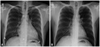
After the procedure, there was no left bundle branch block (LBBB) or intraventricular conduction delay on the ECG. Telemetry monitoring revealed that VT did not return, and that normal sinus rhythm was maintained without a single premature ventricular contraction. The patient's symptoms were improved, and he was discharged two days after the procedure. Follow up echocardiography performed five months after catheter ablation showed 49% of left ventricular ejection fraction and shrinked LV. Chest X-rays performed in an outpatient department six months after discharge showed a decreased cardiac silhouette (Fig. 3) without symptoms. Follow-up observations are ongoing, and neither VT nor any cardiac symptoms have recurred.
DISCUSSION
Because this young patient had pulmonary congestion and cardiomegaly at the time of his visit, myocarditis-induced heart failure with VT was considered also as a possible diagnosis. Ventricular arrhythmias complicating fulminant myocarditis can be initiated and maintained by activity originating from the Purkinje system, and can be suppressed by catheter ablation.3-6 However, a differential diagnosis was made based on the following facts.2-5 First, the initial ECG showed a relatively narrow QRS duration (126 ms), which is not consistent with myocardial inflammation, in which it is usually prolonged to more than 140 ms. Second, the patient's detailed medical history did not indicate any upper respiratory tract infections or flu-like symptoms before the visit to local clinic. Finally, the patient tested negative for all myocardial inflammatory markers that were evaluated in our hospital. In addition, the typical morphology which we observed was compatible with posterior fascicular VT. Therefore, we were able to diagnose the patient with tachycardia-induced cardiomyopathy due to incessant VT originating near or from the Purkinje system. Usually, the known phenomena underlying posterior fascicular VT is macro-reentry sensitive to verapamil. However, this case did not show any response to verapamil, and it did not terminate with programmed electrical stimulation or adenosine. Purkinje fibers can be bystander exit sites, and Purkinje potential is not hallmark of focal Purkinje VT. However, a small number of energy application required to eliminate the VT suggested that the origin of VT was Purkinje fiber.3-6 In view of these observations, the patient was diagnosed as a focal Purkinje VT, due to automaticity rather than triggered activity. Focal Purkinje VT is such a rare disease, so that its clinical and electrophysiological characteristics are not well known. It can appear in two forms: RBBB or LBBB.2,3 It is also known to respond to propranolol, a beta blocker. Additionally, it can be induced possibly by exercise or catecholamines, however, it cannot be induced or terminated by programmed electrical stimulation. Given the fact that Purkinje fibers play a key role in initiating and maintaining the VT and they are located in the subendocardium, RF-CA could be a good treatment for this type of arrhythmia.
 XML Download