Abstract
Purpose
While Mycobacterium kansasii is a common cause of nontuberculous mycobacterial (NTM) lung disease in many developed countries, M. kansasii is infrequently isolated in Korea. We investigated the clinical and radiological features and treatment outcomes of M. kansasii lung disease in Korea retrospectively.
Materials and Methods
We identified 41 patients with M. kansasii lung disease who met the diagnostic criteria for NTM lung disease in two tertiary referral hospitals in Seoul, Korea, between January 1998 and December 2007.
Results
Their median age was 63 years [interquartile range (IQR) 51-75 years] and 33 (81%) were men. Twenty-three patients (56%) were smokers and 13 patients (32%) had previous pulmonary tuberculosis. The most common radiographic findings were nodules (n = 22, 54%) and consolidation (n = 22, 54%). Cavitation was present in 13 patients (32%). Thirty-one patients (76%) were treated with isoniazid, rifampin, and ethambutol. The median treatment duration was 16 months (IQR 9-18 months). The negative conversion rate after 12 months of treatment was 95%.
Mycobacterium kansasii is the second most common nontuberculous mycobacteria (NTM) after the Mycobacterium avium complex in the United States and Japan,1,2 and is the most common cause of NTM lung disease in the United Kingdom and Western Europe.3,4 Infection with M. kansasii probably occurs via an aerosol route. Moreover, unlike other NTM, M. kansasii is much more difficult to recover from natural water supplies.5 Tap water is likely a major reservoir for M. kansasii causing human infection, and thus an association may exist between clinical disease and potable water supplies.
Mycobacterium kansasii usually presents as lung disease that is nearly identical to tuberculosis. Cavitation was found in about 90% of cases in older reports,6-8 while recent survey of 56 adults with M. kansasii lung disease in Israel between 1999 and 2004 noted cavitation in 54% only.9 A variety of radiologic findings have been reported in M. kansasii lung disease, including non-cavitary or nodular/bronchiectatic lesions.10,11
In Korea, M. kansasii is infrequently isolated and a relatively uncommon cause of NTM lung disease.12-15 Although small numbers of cases have been reported,16,17 little is known about the detailed clinical and radiological characteristics or treatment outcomes in Korea. This study investigated the clinical and radiological features and treatment outcomes of M. kansasii lung disease in Korea.
This retrospective study included 41 patients with M. kansasii lung infection who attended two tertiary referral hospitals in Seoul, Korea, between January 1998 and December 2007: Samsung Medical Center and Asan Medical Center. All patients met the diagnostic criteria of NTM lung disease,1 and we reviewed their medical records and radiographs. This study was approved by the institutional review board of Samsung Medical Center and Asan Medical Center, both of which waived the requirement for informed consent of the individual patients due to the retrospective nature of the study.
Mycobacterial stains and cultures were performed using standard methods.18 The results of smear microscopy were reported semiquantitatively. A positive smear was defined as one with > 1 acid-fast bacilli (AFB) per 100 high-power fields. Respiratory specimens were decontaminated using N-acetyl-L-cysteine/2% NaOH, and processed specimens were plated onto 3% Ogawa medium. Inoculated tubes were incubated at 37℃ and then inspected weekly for 8 weeks. To distinguish between M. tuberculosis and NTM, all AFB isolates were assessed according to growth rates, colony morphology, and pigmentation, as well as with a commercial polymerase chain reaction-based assay system (MTB-ID; M&D, Wonju, Korea). NTM species were identified using a polymerase chain reaction and restriction fragment length polymorphism methods based on the rpoB gene, as described previously.14
The medical records of all study patients were reviewed, including information such as age, sex, body mass index (BMI), respiratory and constitutional symptoms, smoking history, underlying illness, pulmonary function tests, tuberculosis history, results for human immunodeficiency virus (HIV) antibody, and treatment history.
Chest radiographs and computed tomography (CT) images were those obtained closest in time to the date of the first positive respiratory culture result. The chest X-ray and CT images were evaluated in terms of the presence of nodules, consolidation, cavitation, and pleural effusion. In addition, hilar or mediastinal lymphadenopathy and bronchiectasis was evaluated on CT.
Nodules (≤ 3 cm in diameter) were considered present when a rounded opacity was observed, either well or poorly defined. Consolidation was defined as a homogeneous increase in pulmonary parenchymal attenuation that obscures the margins of vessels and airway walls. A cavity was regarded as present when a gas-filled space was seen within pulmonary consolidation, a mass, or a nodule. Bronchiectasis was defined as bronchial dilatation relative to the accompanying pulmonary artery, a lack of tapering of bronchi, and bronchi identified within 1 cm of the pleural surface.19
The distribution (upper vs. middle and lower lung zone) and laterality (unilateral vs. bilateral infiltrates) of the lung lesions were also analyzed. Lesions were considered to be in the upper zone of the lung if cephalad to the aortic arch, in the lower zone if caudad to the inferior pulmonary vein, and in the middle zone if between these two zones.20
Sputum conversion was defined as three consecutive negative cultures within 6 months, with the time of conversion defined as the date of the first negative culture. If the patient could not expectorate sputum during the treatment period, the sputum was considered to have converted to negative. Sputum relapse was defined as two consecutive positive cultures after sputum conversion.21
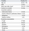
The baseline characteristics of the patients are shown in Table 1. The median age was 63 years [interquartile range (IQR) 51-75 years] and 33 (81%) were men. Twenty-three (56%) patients were current or ex-smokers. None of the patients were HIV positive. Comorbid conditions included previous pulmonary tuberculosis in 13 (32%) patients, chronic obstructive pulmonary disease in 13 (32%), underlying malignancy in nine (22%), and diabetes mellitus in two (5%). The most common presenting symptoms were cough (n = 27, 66%) and sputum (n = 26, 63%). Ten (24%) patients had dyspnea and four (10%) had hemoptysis.
The diagnosis was confirmed by obtaining an adequate number of positive sputum cultures (n = 37, 90%). Patients, who were either unable to produce sputum or had negative sputum cultures, were diagnosed using bronchial aspirates (n = 3, 7%) or lung tissues (n = 1, 2%). Sputum AFB smear examinations were positive in 11 patients (27%). Testing for rifampin susceptibility was performed on M. kansasii isolates recovered from 28 patients and 89% (25/28) were susceptible to rifampin.
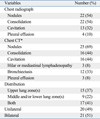
All 41 patients had abnormal chest radiographic findings at the time of diagnosis (Table 2). The most common radiographic findings were nodules (n = 22, 54%) and consolidation (n = 22, 54%). Cavitation was present in 13 patients (32%) and pleural effusion in four (10%). Radiographic abnormalities were confined to the upper lung zones in 15 patients (37%), and 21 patients (51%) had bilateral disease.
A chest CT was available in 36 patients (88%). The most common CT finding was nodules (n = 25, 69%). Consolidation and cavitation were present in 16 patients each (44% each). Combined bronchiectasis was found in 12 patients (33%).
Ten (24%) patients did not receive anti-mycobacterial drugs after the diagnosis of M. kansasii disease. Of these, eight patients had mild symptoms and no significant change on their radiographs during the follow-up period. One patient was lost to the follow-up 1 month after the diagnosis. The upper lobar consolidation resolved spontaneously without antibiotic therapy in one patient (Table 3).
Thirty-one (76%) patients received antibiotic therapy including isoniazid, rifampin, and ethambutol. Of these, 16 patients completed the treatment and were followed for a median of 13 months (IQR 7-28 months) without relapse. Sputum relapse occurred after treatment completion in one patient who had no history of pulmonary tuberculosis and the isolate of M. kansasii was sensitive to rifampin. Four patients remained on the treatment as of the end of May 2009. Nine patients were lost to the follow-up after antibiotic therapy for a median of 6 months (IQR 2-10 months) and two patients died of causes unrelated to M. kansasii lung disease during antibiotic therapy (Table 3). Three patients had rifampin-resistant M. kansasii lung disease. One patient was lost to follow-up after 1.5 months of therapy with isoniazid, rifampin, and ethambutol before drug susceptibility results were available. Treatment regimens were changed to clarithromycin and levofloxacin in two patients after the confirmation of resistance to rifampin. However, one patient was lost to the follow-up and the other patient died of causes unrelated to M. kansasii lung disease during antibiotic therapy.
The median time for negative culture conversion was 1 month (IQR 1-3 months) in patients who achieved negative conversion. For 21 patients who received antibiotic therapy for more than 12 months, the median treatment duration was 16 months (IQR 9-18 months) and the 12-month culture conversion rate was 95% (20/21).
In Korea, M. kansasii constitutes only 2-4% of NTM organisms isolated from clinical specimens,13-15 although the isolation of M. kansasii has increased steadily in recent years.15 In this study, we found that M. kansasii lung disease had diverse radiographic findings. Nodules and consolidation were the most common radiographic features, while cavitation was seen on the chest X-ray in only 32% of patients. A rifampin-containing three-drug regimen was effective, especially in patients treated for more than 12 months.
The radiographic features of M. kansasii lung disease are frequently reported as being indistinguishable from those of tuberculosis.8,22,23 Older studies found cavitation in 75-96% of patients with M. kansasii lung disease,3,8,23-25 while the proportion of cavitary lesions was about 50% in recent studies.9,17 In our series, cavitation was seen in only 32% of patients on chest X-rays. This discrepancy might be explained by recent improvements in the diagnosis and microbiological isolation of the organism.9 Moreover, some studies have reported that patients with M. kansasii lung disease can present with non-cavitary or nodular/bronchiectatic lesions as a major features of M. avium complex lung disease, which is the most common etiology of NTM lung disease.3,10,11
In addition to the low proportion of cavitation, we found that M. kansasii lung disease had diverse radiographic features. Upper lobe lesions were not predominant and pleural effusions were seen in four patients (10%). NTM infection is rarely accompanied by pleural involvement, although a few case reports have described pleural effusion caused by M. kansasii.26-28 Pleural effusion was associated with parenchymal consolidation in three of four patients in our study. On the chest CT, more nodular and cavitary lesions surrounded by consolidation were found in these patients.
Regarding the antibiotic treatment for M. kansasii lung disease, a three-drug combination consisting of isoniazid, rifampin, and ethambutol is recommended and these antibiotics should be maintained for at least 12 months after negative sputum conversion.1 The key to successful therapy for M. kansasii lung disease is to include rifampin in a multidrug regimen. Adding isoniazid and ethambutol to rifampin is essential for preventing the emergence of resistance to rifampin.9
A prospective trial of 28 patients compared treatment for 12 or 18 months with this three-drug regimen.29 Only one relapse occurred; the patient was in the 12-month chemotherapy group. In a series of 56 adults with M. kansasii lung disease in Israel, the mean treatment duration was 21 ± 7 months.9 Another case series of 75 patients with M. kansasii lung disease in Spain showed that a 12-month multidrug treatment regimen, including isoniazid, rifampin, and ethambutol, supplemented with streptomycin during the first 2-3 months, was effective in most cases of M. kansasii lung disease, although five patients (7%) relapsed.30
We found that the rifampin-based multidrug regimen was effective, especially in patients treated for more than 12 months. Those patients had a 95% culture conversion rate after a median treatment duration of 18 months. Relapse occurred in one patient. However, the relapse rate may be an underestimate because the loss to follow-up was high in our study.
In summary, the radiographic characteristics of M. kansasii lung disease in recently diagnosed patients are diverse in Korea. Nodules and consolidation are found more frequently than cavitary lesions. A three-drug combination regimen containing rifampin for more than 12 months is effective in patients with M. kansasii lung disease.
Figures and Tables
ACKNOWLEDGEMENTS
This work was supported by the Korea Science and Engineering Foundation (KOSEF) grant funded by the Korea government (MEST) (R11-2002-103).
References
1. Griffith DE, Aksamit T, Brown-Elliott BA, Catanzaro A, Daley C, Gordin F, et al. An official ATS/IDSA statement: diagnosis, treatment, and prevention of nontuberculous mycobacterial diseases. Am J Respir Crit Care Med. 2007. 175:367–416.
2. Tsukamura M, Kita N, Shimoide H, Arakawa H, Kuze A. Studies on the epidemiology of nontuberculous mycobacteriosis in Japan. Am Rev Respir Dis. 1988. 137:1280–1284.

3. Evans AJ, Crisp AJ, Hubbard RB, Colville A, Evans SA, Johnston ID. Pulmonary Mycobacterium kansasii infection: comparison of radiological appearances with pulmonary tuberculosis. Thorax. 1996. 51:1243–1247.

4. Marras TK, Daley CL. Epidemiology of human pulmonary infection with nontuberculous mycobacteria. Clin Chest Med. 2002. 23:553–567.
5. Falkinham JO 3rd. Nontuberculous mycobacteria in the environment. Clin Chest Med. 2002. 23:529–551.

6. Ahn CH, McLarty JW, Ahn SS, Ahn SI, Hurst GA. Diagnostic criteria for pulmonary disease caused by Mycobacterium kansasii and Mycobacterium intracellulare. Am Rev Respir Dis. 1982. 125:388–391.
7. Johanson WG Jr, Nicholson DP. Pulmonary disease due to Mycobacterium kansasii. An analysis of some factors affecting prognosis. Am Rev Respir Dis. 1969. 99:73–85.
8. Christensen EE, Dietz GW, Ahn CH, Chapman JS, Murry RC, Hurst GA. Radiographic manifestations of pulmonary Mycobacterium kansasii infections. AJR Am J Roentgenol. 1978. 131:985–993.
9. Shitrit D, Baum GL, Priess R, Lavy A, Shitrit AB, Raz M, et al. Pulmonary Mycobacterium kansasii infection in Israel, 1999-2004: clinical features, drug susceptibility, and outcome. Chest. 2006. 129:771–776.

10. Griffith DE, Brown-Elliott BA, Wallace RJ Jr. Thrice-weekly clarithromycin-containing regimen for treatment of Mycobacterium kansasii lung disease: results of a preliminary study. Clin Infect Dis. 2003. 37:1178–1182.

11. Shitrit D, Priess R, Peled N, Bishara G, Shlomi D, Kramer MR. Differentiation of Mycobacterium kansasii infection from Mycobacterium tuberculosis infection: comparison of clinical features, radiological appearance, and outcome. Eur J Clin Microbiol Infect Dis. 2007. 26:679–684.
12. Koh WJ, Kwon OJ, Lee KS. Diagnosis and treatment of nontuberculous mycobacterial pulmonary diseases: a Korean perspective. J Korean Med Sci. 2005. 20:913–925.

13. Lee JY, Choi HJ, Lee HY, Joung EY, Huh JW, Oh YM, et al. Recovery rate and characteristics of nontuberculous mycobacterial isolates in a university hospital in Korea. Tuberc Respir Dis. 2005. 58:385–391.

14. Koh WJ, Kwon OJ, Jeon K, Kim TS, Lee KS, Park YK, et al. Clinical significance of nontuberculous mycobacteria isolated from respiratory specimens in Korea. Chest. 2006. 129:341–348.

15. Ryoo SW, Shin S, Shim MS, Park YS, Lew WJ, Park SN, et al. Spread of nontuberculous mycobacteria from 1993 to 2006 in Koreans. J Clin Lab Anal. 2008. 22:415–420.

16. Koh WJ, Kwon OJ, Suh GY, Chung MP, Kim H, Lee NY, et al. A case report of three patients with nontuberculous mycobacterial pulmonary disease caused by Mycobacterium kansasii. Tuberc Respir Dis. 2003. 54:459–466.
17. Yim JJ, Park YK, Lew WJ, Bai GH, Han SK, Shim YS. Mycobacterium kansasii pulmonary diseases in Korea. J Korean Med Sci. 2005. 20:957–960.
18. Diagnostic Standards and Classification of Tuberculosis in Adults and Children. This official statement of the American Thoracic Society and the Centers for Disease Control and Prevention was adopted by the ATS Board of Directors, July 1999. This statement was endorsed by the Council of the Infectious Disease Society of America, September 1999. Am J Respir Crit Care Med. 2000. 161:1376–1395.
19. Hansell DM, Bankier AA, MacMahon H, McLoud TC, Müller NL, Remy J. Fleischner Society: glossary of terms for thoracic imaging. Radiology. 2008. 246:697–722.
20. Koh WJ, Yu CM, Suh GY, Chung MP, Kim H, Kwon OJ, et al. Pulmonary TB and NTM lung disease: comparison of characteristics in patients with AFB smear-positive sputum. Int J Tuberc Lung Dis. 2006. 10:1001–1007.
21. Kobashi Y, Matsushima T, Oka M. A double-blind randomized study of aminoglycoside infusion with combined therapy for pulmonary Mycobacterium avium complex disease. Respir Med. 2007. 101:130–138.
22. Anderson DH, Grech P, Townshend RH, Jephcott AE. Pulmonary lesions due to opportunist mycobacteria. (Review includes 30 cases of M. kansasii infections). Clin Radiol. 1975. 26:461–469.
23. Banks J, Hunter AM, Campbell IA, Jenkins PA, Smith AP. Pulmonary infection with Mycobacterium kansasii in Wales, 1970-9: review of treatment and response. Thorax. 1983. 38:271–274.

24. Christensen EE, Dietz GW, Ahn CH, Chapman JS, Murry RC, Anderson J, et al. Initial roentgenographic manifestations of pulmonary Mycobacterium tuberculosis, M kansasii, and M intracellularis infections. Chest. 1981. 80:132–136.
25. Ahn CH, Lowell JR, Onstad GD, Shuford EH, Hurst GA. A demographic study of disease due to Mycobacterium kansasii or M intracellulare-avium in Texas. Chest. 1979. 75:120–125.
26. Igari H, Kikuchi N. Nontuberculous Mycobacterium pulmonary infection with pleural effusion caused by Mycobacterium kansasii. Kekkaku. 1993. 68:527–531.
27. Kamiya H, Toyota E, Kobayashi N, Kudo K. A case of pulmonary Mycobacterium kansasii infection complicated with pleural effusion. Kekkaku. 2004. 79:397–400.
28. Olafsson EJ, Naum CC, Sarosi GA, Mastronarde JG. Bilateral pleural effusions and right pneumothorax in a 25-year-old man. Chest. 2004. 126:986–992.
29. Sauret J, Hernández-Flix S, Castro E, Hernández L, Ausina V, Coll P. Treatment of pulmonary disease caused by Mycobacterium kansasii: results of 18 vs 12 months' chemotherapy. Tuber Lung Dis. 1995. 76:104–108.


 PDF
PDF ePub
ePub Citation
 Print
Print


 XML Download