

 PDF
PDF ePub
ePub Citation
 Print
Print


INTRODUCTION
Pericardial effusion is one of the most common cardiac manifestations of human immunodeficiency virus (HIV) infection.1 Pericarditis is found during autopsy in 30% of AIDS patients and pericardial effusions have been detected in about 20% of HIV-infected patients.1,2 In patients with HIV infection, pericardial symptoms and clinical signs may be the first indication of cardiac disease without the presence of other clinical symptoms of HIV infection.3 Therefore, HIV infection should be included in the differential diagnosis of unexplained pericardial effusion or tamponade.4 In this report, we present an exceptionally rare case of cardiac tamponade as the first symptom of HIV infection, which has not been previously reported in Korea.
CASE REPORT
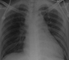
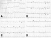
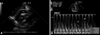
A previously healthy 29-year-old heterosexual man was admitted to the emergency room of our hospital due to a sudden onset of dizziness with presyncope. Dizziness was accompanied by palpitation, nausea, and vomiting. He also complained of a two-week duration of dull, substernal chest discomfort, which was neither pleuritic nor related to exertion, swallowing, or position change. He denied a past medical history of intravenous drug abuse or blood transfusion. A physical examination revealed an acute ill-looking appearance, cachexia, dehydration, and cool extremities. His vital signs were as follows: blood pressure 60/30 mmHg, pulse rate 71 beats/min, respiratory rate 22/min, and body temperature 36.5℃. Initially, he had no peripheral edema and jugular venous pressure was not elevated. No lymphadenopathy or oral thrush was found. Pulsus paradoxus was not measured. A chest examination revealed clear breath sounds. The heart sounds were normal. No pericardial rub was audible. The abdomen was not tender and was without organomegaly or ascites. A chest X-ray demonstrated a slightly enlarged cardiac silhouette with normal lung fields (Fig. 1). A 12-lead electrocardiogram (ECG) revealed a normal sinus rhythm at 71 beats/min and non-specific ST-segment abnormalities (Fig. 2A). Laboratory data showed a hemoglobin level of 13.9 g/dL, a white blood cell count of 8,010/mm3 (50.5% neutrophils, 42.8% lymphocytes), platelet count of 175,000/mm3, creatine kinase-MB isoenzyme 1.7 ng/mL, pro-B-type natriuretic peptide 506.3 pg/mL, creatinine 1.4 mg/dL, alkaline phosphatase 97 IU/L, aminoaspartate transaminase 29 U/L, alanine transaminase 30 U/L, erythrocyte sedimentation rate 1 mm/h, and C-reactive protein 0.4 mg/dL. The arterial blood gas results were pH 7.50 mmHg, PO2 90.9 mmHg, PCO2 20 mmHg, and HCO3 15.7 mmol/L. With vigorous intravenous hydration of 2 liters of normal saline during a 2 hour period, jugular veins became progressively distended and jugular venous pressure was elevated. However, the patient remained hypotensive and oliguria persisted. In view of the hypotension and raised jugular venous pressure, the possibility of cardiac tamponade was considered. A large pericardial effusion with the collapse of the right ventricle during diastole was noted on the transthoracic echocardiography (Fig. 3A). The inferior vena cava was dilated with little or no change in respiration. A pulse-wave Doppler study disclosed exaggerated respiratory variation of the mitral inflow velocities consistent with cardiac tamponade physiology (Fig. 3B). Consequently, a total of 900 mL of hemorrhagic pericardial effusion was aspirated by apical pericardiocentesis. After pericardiocentesis, the blood pressure improved to 110/70 mmHg without inotropics support. Pericardial fluid cytology and cultures for bacteria, mycobacteria, adenovirus, and fungus were all negative. Blood and urine cultures were also sterile. A serial 12-lead ECG during hospitalization revealed diffuse upward concave ST-segment elevation and PR-segment depression, suggestive of acute pericarditis (Fig. 2B). Serological tests using antibodies to cytomegalovirus, herpes simplex virus, Epstein-Barr virus, influenza virus types A and B, adenovirus, Coxsackie virus types B1, B2, B3, B4 and B5, parainfluenza virus types 1, 2, 3, 4, hepatitis A, B and C virus, Legionella spp., Mycoplasma spp., and Cryptococcus spp. showed no evidence of recent infection. However, HIV infection was detected by an enzyme-linked immunosorbent assay and confirmed by Western blot. The CD4 cell count was 168 cells/mm3. Finally, the diagnosis of cardiac tamponade with HIV-associated hemorrhagic pericarditis was made. On the seventh day of hospitalization, the ECG showed nearly complete resolution of the ST-segment elevation, which was followed by T wave inversion (Fig. 2C). The pericardial drain was removed after four days, with the cessation of drainage. An echocardiogram performed on the tenth day of hospitalization showed small amounts of residual pericardial effusion which did not increase. ECG also normalized (Fig. 2D). On the eleventh day of hospitalization, the patient was discharged in stable condition.
DISCUSSION
To our knowledge, this is the first reported case of cardiac tamponade due to HIV-associated pericarditis in Korea. In our patient, pericardial symptoms and clinical signs were the first clinical findings in the course of HIV infection. In such a young patient with minimal coronary risk factors who nevertheless has a history of acute chest pain, ECG findings such as sinus tachycardia, diffuse ST-segment elevation, and low QRS amplitude may suggest acute pericarditis. Echocardiography performed at the bedside may be the most appropriate diagnostic procedure to detect pericardial effusion and to rule out cardiac tamponade in HIV infection.5 The prevalence of cardiac involvement in Acquired Immune Deficiency Syndrome (AIDS) patients has been reported in as many as 45% to 66% of people infected.6 In most patients with HIV, the effusions are small and most patients are asymptomatic.7,8 The reported cases of cardiac tamponade in HIV patients showed hypotension, jugular venous distention, tachycardia, and edema.9 However, our patient initially did not have these clinical signs of cardiac tamponade such as tachycardia, jugular venous distention, or edema. These signs may be absent in patients with "low-pressure tamponade".9 Patients with intravascular fluid depletion due to dehydration, cachexia, and wasting states associated with advanced AIDS may have low pericardial pressure tamponade. Therefore, in patients with pericardial effusion accompanied by clinical signs of hemodynamic embarrassment, cardiac tamponade should be ruled out and pericardiocentesis should be considered, even with the absence of typical clinical signs of cardiac tamponade.10
In conclusion, cardiac tamponade can be the first presentation of HIV infection. It must be considered for the presence of unexplained pericardial effusion or cardiac tamponade.
 XML Download