Abstract
Purpose
The purpose of this study was to study the protective effect and influence of sodium hyaluronate (Na-HA) on mRNA expression of peroxisome proliferators-activated receptor gamma (PPAR-γ) in cartilage of rabbit osteoarthritis (OA) model.
Materials and Methods
Forty eight white rabbits were randomly divided into A, B, and C groups. Group A was normal control group, B and C groups underwent unilateral anterior cruciate ligament transection (ACLT). The rabbits in group B were injected normal saline after ACLT; and Group C received intraarticular1% sodium hyaluronate (HA) injection 5 weeks after surgery, 0.3 mL once a week. At 11th week after surgery, all the rabbits were sacrificed. The cartilage changes on the medial femoral condyles were graded separately. Cartilage sections were stained with safranin-O and HE, and messenger RNA (mRNA) expression of PPAR-γ was detected by using real time polymerase chain reaction (Real Time-PCR).
Osteoarthritis (OA) is the most prevalent type of arthritis that is caused by the breakdown and eventual loss of the cartilage of one or more joints. However, OA develops most frequently in the absence of a known cause. Mechanical, biochemical, and genetic factors are involved in the etiology of OA. In recent years, apoptotic death of articular chondrocytes has been implicated in the pathogenesis of human and animal OA. There are numerous mediators that may induce apoptosis in chondrocytes in vitro, but it is likely that far fewer will function in vivo. Among these, the key players are the free radical nitric oxide (NO),1 Fas and its ligand FasL,2,3 and pro-inflammatory cytokines such as tumor necrosis factor (TNF)-α.4 It is becoming more and more evident that chondrocyte apoptosis plays an important role in cartilage destruction. Recent studies suggest that peroxisome proliferator-activated receptor gamma (PPAR-γ) may be associated with the apoptosis of chondrocyte.5
However, there are only a few reports about the effect of HA on the messenger RNA (mRNA) expression of PPAR-γ, and the mechanisms of therapeutic effect of hyaluronate (HA) on OA remain to be elucidated. In this study, we determined the protective effect of HA on articular cartilage and the expression of PPAR-γ mRNA in experimentally induced OA rabbits.
Forty eight mature New Zealand white rabbits, weighing 2.2-2.8 kg, were used, and randomly divided into three groups. Group A was normal control, and animals in group B and C were anesthetized intravenously with ketamine hydrochloride (1.0 mg/kg), and received unilateral anterior cruciate ligament transection (ACLT). A medial parapatellar incision was made on the skin, and a medial arthrotomy was performed. The patella was dislocated, and the knee was placed in full flexion. The ACL was visualized and transected without destroying the articular cartilage. The knee was irrigated with physiological saline. Then, the capsule and skin were closed, and the animal was individually kept in a cage (60 cm×60 cm×40 cm) without any immobilization and maintained under the same environmental condition.6 On the 5th week after surgery, group B animals were injected with 0.3 mL normal sodium (NS) after ACLT, and animals in group C received 0.3 mL of intraarticular1% sodium HA injection, once a week for 5 weeks. All animals were killed at 11 weeks following surgery.
Evaluation of the cartilage damages was performed by two observers. Qualitative macroscopic evaluation of cartilage injuries was performed by one investigator. Cartilage damages were reported in a semi-quantitative grading table modified from the human literature with a five-grade scale (0 = normal; 1 = surface striations or tears; 2 = bucket handle or transverse tears; 3 = fibrillation of the entire cartilage, protrusion; 4 = loss of cartilage, or ulcer).
Tissues from 8 rabbits of each group were dissected, fixed 48 h in neutral buffered 4% formaldehyde in 0.1 M sodium phosphate buffer (PBS), pH = 7.4, decalcified 3-4 weeks in 10% ethylenediaminetetraacetic acid (EDTA), dehydrated in graded alcohols, and embedded in paraffin. Sections, 5 µm thick, were stained with both hematoxylin and eosin for general morphology, or with 0.1% safranin-O and fast green to demonstrate sulfated proteoglycans (PGs). The area of positively stained safranin-O metachromasia, indicative of PGs, and the total area of the joint cartilage in frontal sections were measured. Each data point represents results obtained from measurements of two sections from each of three individual animals belonging to the same group.
Cartilage tissue from the medial femoral condyle was harvested from the operated knee, and was frozen in liquid nitrogen. The samples were powdered in liquid nitrogen by hand milling. The total RNA was extracted by using the Trizol reagent (Invitrogen Technologies, Carlsbad, CA, USA) according to the manufacturer's protocol. The RNA samples were spectrophotometrically quantified at A260. Then, RNA was reverse-transcribed to cDNA using RT-PCR kit (TaKaRa, Dalian, China) according to protocol. The cDNA was analyzed immediately or stored at -20℃. Quantitative-polymerase chain reaction (PCR) amplification was performed by ABI 7900HT Fast Real-Time PCR System (ABI Company, Forst City, CA, USA) and the SYBR Green I fluorescent dye method was used to quantify cDNA. The sequences of the primers used are shown in Table 1.
The final volume of the RT-PCR reaction was 10 µL and included SYBR Premix Ex Taq 5 µL, primer (10 µmol/L) 0.2 µL each, 50× ROX Reference Dye 0.2 µL, template 1 µL, adjusted to 10 µL with distilled water. PCR cycling conditions consisted of an initial denaturing step for 10 seconds at 95℃, and then 40 cycles of 5 seconds at 95℃, and 30 seconds at 60℃. A stable and reliable standard curve was established using synthesized oligonucleotides resembling complementary DNA (cDNA) fragments in 5-fold decrements as template. The glyceraldehyde 3-phosphate dehydrogenase (GAPDH) from the same sample was used as internal control, and the relative contents of copy numbers of the target gene's mRNA were calculated, through which we could determine the gene expression level and its trend of change. Specificity of each reaction was controlled by melting curve analysis. A negative PCR control containing water instead of cDNA was performed. Real-time PCR was conducted in triplicate in three independent experiments.
The cartilage on condyles of normal rabbits was macroscopically normal. In NS-treated OA rabbits, cartilage lesions of moderate grade and size were present on condyles, Rabbits treated with HA presented a marked reduction in the severity of lesions on the condyles (Table 2).
Cartilage from normal rabbits had a normal histologic appearance, specimens from NS-treated OA rabbits had morphologic changes, including cartilage fibrillation and fissures, and a loss of Safranin 0 staining (Figs. 1 and 2). In the HA-treated OA rabbits, the lesions on the condyles were significantly less severe (p < 0.05) compared with the NS-treated OA rabbits. This difference was largely due to a decrease in severity of structural changes and loss of Safranin 0 staining (Fig. 2) (Table 3).
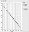
Based on the relationship between the concentration of GAPDH and Ct value, a standard curve with high relativity (r = 0.9949) was obtained (Fig. 3).
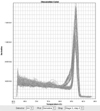
Fig. 4 and 5 show the amplification plot and dissociation curve of PPAR-γ and GAPDH, respectively. The results of PPAR-γ mRNA expression were rectified by the control gene (GAPDH mRNA). Shown in Fig. 6 are the values that represent steady-state levels of PPAR-γ mRNA relative to GAPDH mRNA. PPAR-γ mRNA levels determined by quantitative RT-PCR indicated an approximate increase (1.5-fold) in the cartilage of NS-treated group compared with the normal group (p < 0.05). There was no significant different in the PPAR-γ mRNA expression between HA-treated group and normal group (p > 0.05).
An imbalance between synthetic and catabolic processes affecting components of the extracellular matrix can result in destruction of articular cartilage. Because articular chondrocytes are the only cells residing in cartilage tissue, these resident cells are highly specialized and solely responsible for the maintenance and turnover of extracellular matrix macromolecules including type II collagen, aggregating proteoglycans and noncollagenous proteins.7 Therefore, so their survival is essential for maintaining proper architecture of the articular cartilage. In OA cartilage, hypocellularity and matrix degeneration are believed to contribute to joint degradation.8,9 Numerous studies have demonstrated significant involvement of apoptosis of chondrocyte in the pathologic degradation of the cartilage matrix. It is becoming more and more evident that apoptosis of chondrocytes plays an important role in cartilage destruction. However, in cartilage, there are no macrophages. In those conditions, secondary necrosis would inevitably result in uncontrolled release of lysosomal enzymes from apoptotic bodies causing serious damages to the extracellular matrix.10 There are numerous mediators that potently induce apoptosis in chondrocyte cultures, although the mechanisms involved are not yet fully elucidated. These mediators include nitric oxide,1 Fas and its ligand FasL,2,3 and Bcl-2/Bax family members.11 One member of the prostaglandins (PGs) family, PG E2, has been reported to induce bovine chondrocyte apoptosis.12
However, the chondrocyte apoptosis of OA could not entirely be attributed to these mediators. Nevertheless, many recent investigations have been focused on the possible role of PPAR-γ which is a transcriptional factor that belongs to the nuclear receptor superfamily.13,14 PPAR-γ mRNA expression level in chondrocytes was up-regulated by the proinflammatory cytokines IL-1β and TNF-1α. Specifically, the two synthetic PPAR-γ agonists, ciglitazone and pioglitazone, induced chondrocyte apoptosis.5 Therefore, PPAR-γ could be a potential target to modulate the effects of cytokines on cartilage.15 Furthermore, activation of PPAR-γ by ciglitazone in growth plate chondrocytes inhibits T3-induced terminal differentiation and promotes apoptosis through increased levels of caspase 3/7 activity and decreased expression of the anti-apoptotic protein Bcl-2,16 and Yoon, et al.17 reported that the PPAR-γ agonist, 15-deoxy-Δ12,14-prostaglandinJ2 (15d-PGJ2), causes articular chondrocytes dedifferentiation and induces COX-2 expression and PGE2 production of chondrocytes.
In the present studies, we demonstrated that PPAR-γ is synthesized by chondrocytes of both NOR and OA articular cartilage in rabbit. Real Time PCR analyze showed that NOR and OA cartilage express the PPAR-γ mRNA, and that there is a significant increase in cartilage sample of OA. And activation of PPAR-γ in these cells may result in activation of a pathological form of apoptosis.16 Intra-articular injection of hyaluronan (Na-HA) has widely been used in the treatment of OA. Na-HA is one of the important components of cartilage matrix, and has a protective effect on the progression of OA. Injection of exogenous HA can inhibit prostaglandin E2 (PGE2) synthesis, induced by interleukin-1 (IL-1),18,19 and protect against proteoglycan depletion and cytotoxicity induced by oxygen-derived free radicals, IL-1, and mononuclear cell-conditioned medium, as well as other alterations.20-22 Na-HA is also reported to inhibit the expression of matrix metalloproteinase (MMP-1) and MMP-3 induced by interleukin-1β.23 In our study, the lesions on the condyles in HA treated group were significantly less severe compared with the NS-treated OA rabbits, and the severity of structural changes and loss of Safranin 0 staining were decreased.
In conclusion, our present studies demonstrated that PPAR-γ is involved in the progression of OA, and that the activation of PPAR-γ in chondrocytes may result in activation of a pathological form of apoptosis. Exogenous Na-HA could protect cartilage degeneration, at last partly, through inhibition of PPAR-γ mRNA synthesized by chondrocytes of OA articular cartilage in rabbit. However, we could not detect the isoforms of PPAR-γ, and also did not determine the influence of Na-HA on them. The mechanism needs to be further investigated, and PPAR-γ, may be the novel therapeutic target in OA.
Figures and Tables
 | Fig. 1Representative sections of cartilage from the femoral condyles (HE staning). (A) Cartilage of normal showing no evident changes. (B) NS group showing loss of height of articular cartilage, increased flaking of cartilage, clefts deep to the radial zone, loss of coumnization of cellular distribution, and increased areas of hypocellularity. (C) HA could protect articular cartilage from loss of height, and decrease the flaking of cartilage. NS, normal sodium treated group; HA, sodium hyaluronate treated group. |
 | Fig. 2Representative sections of cartilage from the femoral condyles (Safranin-O staining). (A) Cartilage of normal showing an intact profile, and there is no evident loss of proteoglycan (PGs). (B) NS group showing severe loss of PGs, and clefts deep to the radial zone. (C) HA could inhitit the loss of PGs in cartilage matrix. |
 | Fig. 3Stand curve of GAPDH in fluorescent quantitative PCR. PCR, polymerase chain reaction; GAPDH, glyceraldehyde 3-phosphate dehydrogenase. |
 | Fig. 4Amplification plot of PPAR-γ and GAPDH. PPAR-γ, peroxisome proliferators-activated receptor gamma; GAPDH, glyceraldehyde 3-phosphate dehydrogenase. |
 | Fig. 5Dissociation curve of PPAR-γ and GAPDH. PPAR-γ, peroxisome proliferators-activated receptor gamma; GAPDH, glyceraldehyde 3-phosphate dehydrogenase. |
 | Fig. 6Real Time PCR for the relative expression of PPAR-γ mRNA in cartilage. There was significant difference between NS and NOR, HA cartilage (p < 0.05), and no difference between NOR and HA cartilage (p > 0.05). PCR, polymerase chain reaction; PPAR-γ, peroxisome proliferators-activated receptor gamma; mRNA, messenger RNA; NS, normal sodium treated group; NOR, normal control group; HA, sodium hyaluronate treated group. |
References
1. Studer R, Jaffurs D, Stefanovic-Racic M, Robbins PD, Evans CH. Nitric oxide in osteoarthritis. Osteoarthritis Cartilage. 1999. 7:377–379.

2. Hashimoto S, Setareh M, Ochs RL, Lotz M. Fas/Fas ligand expression and induction of apoptosis in chondrocytes. Arthritis Rheum. 1997. 40:1749–1755.

3. Kühn K, Hashimoto S, Lotz M. Cell density modulates apoptosis in human articular chondrocytes. J Cell Physiol. 1999. 180:439–447.

4. Aizawa T, Kon T, Einhorn TA, Gerstenfeld LC. Induction of apoptosis in chondrocytes by tumor necrosis factor-alpha. J Orthop Res. 2001. 19:785–796.

5. Shan ZZ, Masuko-Hongo K, Dai SM, Nakamura H, Kato T, Nishioka K. A Potential role of 15-deoxy (12,14) prostaglandin J2 for induction of human articular chondrocyte apoptosis in arthritis. J Biol Chem. 2004. 279:37939–37950.

6. Liu SQ, Qiu B, Chen LY, Peng H, Du YM. The effects of carboxymethylated chitosan on metalloproteinase-1, -3 and tissue inhibitor of metalloproteinase-1 gene expression in cartilage of experimental osteoarthritis. Rheumatol Int. 2005. 26:52–57.

7. Muir H. The chondrocyte, architect of cartilage. Biomechanics, structure, function and molecular biology of cartilage matrix macromolecules. Bioessays. 1995. 17:1039–1048.

8. Hashimoto S, Ochs RL, Komiya S, Lotz M. Linkage of chondrocyte apoptosis and cartilage degradation in human osteoarthritis. Arthritis Rheum. 1998. 41:1632–1638.

9. Choy EH, Panayi GS. Cytokine pathways and joint inflammation in rheumatoid arthritis. N Engl J Med. 2001. 344:907–916.

10. Roach HI, Aigner T, Kouri JB. Chondroptosis: a variant of apoptotic cell death in chondrocytes? Apoptosis. 2004. 9:265–277.
11. Feng L, Precht P, Balakir R, Horton WE Jr. Evidence of a direct role for Bcl-2 in the regulation of articular chondrocyte apoptosis under the conditions of serum withdrawal and retinoic acid treatment. J Cell Biochem. 1998. 71:302–309.

12. Miwa M, Saura R, Hirata S, Hayashi Y, Mizuno K, Itoh H. Induction of apoptosis in bovine articular chondrocyte by prostaglandin E(2) through cAMP-dependent pathway. Osteoarthritis Cartilage. 2000. 8:17–24.

13. Schoonjans K, Staels B, Auwerx J. The peroxisome proliferator activated receptors (PPARS) and their effects on lipid metabolism and adipocyte differentiation. Biochim Biophys Acta. 1996. 1302:93–109.

14. Carlberg C, Wiesenberg I. The orphan receptor family RZR/ROR, melatonin and 5-lipoxygenase: an unexpected relationship. J Pineal Res. 1995. 18:171–178.

15. Bordji K, Grillasca JP, Gouze JN, Magdalou J, Schohn H, Keller JM, et al. Evidence for the presence of peroxisome proliferator-activated receptor (PPAR) alpha and gamma and retinoid Z receptor in cartilage. PPARgamma activation modulates the effects of interleukin-1beta on rat chondrocytes. J Biol Chem. 2000. 275:12243–12250.

16. Shao YY, Wang L, Hicks DG, Tarr S, Ballock RT. Expression and activation of peroxisome proliferator-activated receptors in growth plate chondrocytes. J Orthop Res. 2005. 23:1139–1145.

17. Yoon EK, Lee WK, Lee JH, Yu SM, Hwang SG, Kim SJ. ERK-1/-2 and p38 kinase oppositely regulate 15-deoxy-delta (12,14)-prostaglandinJ2-Induced PPAR-gamma activation that mediates dedifferentiation but not cyclooxygenase-2 expression in articular chondrocytes. J Korean Med Sci. 2007. 22:1015–1021.

18. Yasui T, Akatsuka M, Tobetto K, Hayaishi M, Ando T. The effect of hyaluronan on interleukin-1 alpha-induced prostaglandin E2 production in human osteoarthritic synovial cells. Agents Actions. 1992. 37:155–156.

19. Tobetto K, Yasui T, Ando T, Hayaishi M, Motohashi N, Shinogi M, et al. Inhibitory effects of hyaluronan on [14C]arachidonic acid release from labeled human synovial fibroblasts. Jpn J Pharmacol. 1992. 60:79–84.

20. Larsen NE, Lombard KM, Parent EG, Balazs EA. Effect of hylan on cartilage and chondrocyte cultures. J Orthop Res. 1992. 10:23–32.

21. Presti D, Scott JE. Hyaluronan-mediated protective effect against cell damage caused by enzymatically produced hydroxyl (OH.) radicals is dependent on hyaluronan molecular mass. Cell Biochem Funct. 1994. 12:281–288.

22. Campo GM, Avenoso A, Campo S, Ferlazzo AM, Altavilla D, Calatroni A. Efficacy of treatment with glycosaminoglycans on experimental collagen-induced arthritis in rats. Arthritis Res Ther. 2003. 5:R122–R131.
23. Sasaki A, Sasaki K, Konttinen YT, Santavirta S, Takahara M, Takei H, et al. Hyaluronate inhibits the interleukin-1beta-induced expression of matrix metalloproteinase (MMP)-1 and MMP-3 in human synovial cells. Tohoku J Exp Med. 2004. 204:99–107.


 PDF
PDF ePub
ePub Citation
 Print
Print


 XML Download