

 PDF
PDF ePub
ePub Citation
 Print
Print


INTRODUCTION
Bone tissue engineering is rapidly developing, thanks to the development of cell biology and material science. The basic premise of tissue engineering is that lost or damaged tissues can be regenerated by three-dimensional, porous, biodegradable scaffolds and healthy progenitor cells which have been grafted to injured areas. Poly-ε-caprolactone (PCL) is a polyester with chemico-physical characteristics that can be made into an appropriate scaffold for bone engineering. Its mechanical properties and tissue stability have been proven through many applications.1,2 Rai. et al. stated that the synergy of PCL that contained bioactive tricalcium phosphate (TCP) and recombinant human bone morphogenetic protein-2 (rhBMP-2) is an effective method of bone regeneration.3
Bone morphogenetic protein (BMP) was discovered by Urist in 1965 as a growth factor that can induce bone formation. Local delivery of growth factors can induce cell proliferation, chemotaxis, differentiation, and matrix synthesis, and thus, exhibits potential for regeneration.4 Although rhBMP can sufficiently induce bone formation by itself, its rapid spread in the grafted site may reduce its osteoinductive effect. Therefore, proper carrier systems are needed for the delivery, maintenance and discharge of BMP to the grafted site.5,6
Cell source is an important element for successful tissue renewal. Mesenchymal stem cells (MSCs) are multipotent cells that can be differentiated into cells that make up bone and cartilage.7,8 In a study, an experiment on the effect of rhBMP-2 on ectopic bone formation with absorbable collagen sponge (ACS) and beta TCP as carriers, Kim, et al.9 found that the carrier for delivering BMP must be scaffolds, which offer bone-forming cells a space for bone formation, and that this space must be maintained for a considerable length of time for sufficient maturing of the new bones.10,11 If mechanical resistance to compressive force transmitted through mucogingival flaps is restricted, particularly for onlay bone graft such as alveolar ridge augmentation, a space for bone formation may be maintained.
This study was designed to investigate the provision of space for bone formation by PCL-TCP scaffolds through appropriate mechanical properties, the survival and proliferation of dMSCs in the scaffolds when a graft material that consists of auto-fibrin glue (AFG), rhBMP-2, dMSCs, and PCL-TCP scaffolds was placed by onlay grafting techniques on the bone surface.
MATERIALS AND METHODS
Animals
This experiment was approved by Animal Care Committee of the College of Medicine, Korea University. Two 1.5 year-old adult beagle dogs, weighing about 12 kg, were used in this study.
Scaffold design and fabrication
PCL-TCP (80 : 20%) scaffolds were purchased from Osteopore International Pte Ltd,Singapore.
The composites of the scaffold manifested a lay-down pattern of 0/60/120°, a porosity of about 70% and volume measuring as 5.0×5.0×8.0 mm.
The scaffolds were soaked for 3 h at 37.1℃ in phosphate-buffered saline (PBS, 137 mM NaCl, 2.7 mM KCl, 10 mM Na2HPO4, 1.8 mM KH2PO4, pH 7.4) prior to rhBMP-2 seeding.
Bone morphogenetic protein
Ten µg of recombinant human BMP-2 (rhBMP-2) (Cowellmedi Co, Busan, Korea) was used at concentrations of 50 µg/mL on each block containing rhBMP-2, and four experimental groups were made (Table 1). Ten µL of rhBMP-2 was mixed with 190 µL of auto-fibrin glue, which was evenly pipette-loaded on each scaffold
MSCs isolation and cultivation
dMSCs were isolated from 10 mL of the canine marrow aspirates using a modification of techniques previously established for human MSCs (hMSCs) and incubated three weeks before grafting surgery.
Making of auto-fibrin glue
Ten mL of blood collected from the beagle dog was anticoagulated (3.8% sodium citrate) to obtain plasma. After two cycles of thawing and freezing, the plasma was centrifuged at 2,500 r.p.m. for 40 minutes to obtain white sediments, which were used to make solution A. Solution B was prepared by mixing commercial bovine thrombin 100 IU and 2% calcium chloride. Fibrin curd was obtained after 10 to 15 seconds using equal quantities of solutions A and B. To prepare auto serum gel before grafting, solution A was mixed with cells (2×10⁷ cells/mL) stained with PKH26 (Sigma-Aldrich, Inc., St. Louis, MO, USA), and 100 µL each of solutions A and B were injected to the PCL-TCP scaffolds. After this procedures, the BMP-2 was added.
Surgical techniques
Each beagle dog was given the premedication, atropine sulphate (0.05 mg/kg), and anesthesia was induced with Zoletil (5 mg/kg) and isoflurane. The general anesthetic was delivered and monitored under the supervision of an experienced veterinarian.
To expose the scapular bone, the skin, subcutaneous part and muscles around the scapular origin were cut out. An orthopedic bur was used to remove the cortical bone to match the PCL-TCP scaffolds (5.0×5.0×8.0 mm). After placing the grafted material in the defect, the grafted material was fixed by orthodontic mini-implant, Dual-top® (titanium alloy, Jeil Co., Seoul, Korea).
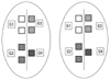
Wound closure was carried out in layers (subcutaneous, muscle, and skin) using a silk suture that ensured complete coverage of the scapular bone. The graft materials were placed and fixed separately in BMP groups (G3, G4) and non-BMP groups (G1, G2) around the scapular origin. (Fig. 1.)
Staining tissue samples
At 2 and 4 weeks after implantation, each dog was anesthetized, followed by euthanasia via perfusion with formalin through the left ventricle of the heart.
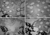
The sampled tissues were divided into two parts. One half was stained with hematoxylin/eosin (H-E), and the other half was checked with a fluorescence microscope for the survival and proliferation of the dMSCs cells that had been injected. The survival of the dMSCs that had been stained by PKH26 was assessed histologically. Von-Kossa staining was used, depending on the degree of matrix formation of the tissues. The tissues were observed at magnification ×40 and ×100.
RESULTS
The surgical sites healed well; and there were no complications or inflammation.
Removal resistance
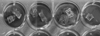
Non-BMP groups (G1 and G2) were easily removed from the scapular bone bed after the mini-implant for fixation was removed at both time points. BMP groups (G3 and G4) exhibited some resistance at week 2, but separated at week 4 when the graft materials adhered completely to the scapular bone while a part of the graft materials remained in the scapular bone (Fig. 2). This remaining graft material was harvested and stained.
Histological observation
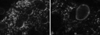
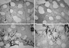
Histologically, new bone was found to grow in the scaffolds near the scapular bone at two weeks (Figs. 3 and 5). The tissues collected at weeks 2 and 4 showed that the PCL-20% TCP scaffolds were clinically observed to maintain their shape without deformation which might possibly be due to the pressure of soft tissues such as surrounding muscles. Histological observation also revealed that the injected cells were proliferating well in the G2, G3, and G4 scaffolds (Figs. 3 and 4).
When compared to G1 and G2, this phenomenon can be seen as a bone formation accelerating effect of rhBMP-2 on the host bone. The survival and proliferation of dMSCs that had been fluorescent-stained could be observed in the G2, G3 and G4 tissues at weeks 2 and 4. More dMSCs were observed at week 4 than at week 2. However, while survival and proliferation of the grafted cells were observed at weeks 2 and 4 after tissue collection in G2, which consisted of dMSCs graft materials, little sign of bone formation was observed in H-E stained or Von-Kossa stained samples. More bone formation was observed in the graft materials at week 4 than at week 2 in G3 and G4, however, less bone formation was observed in G1 and G2 (Figs. 5 and 6) . Calcium deposition was observed in the bone matrix by Von-Kossa stains of G3 and G4 tissues.
DISCUSSION
The main purpose of this pilot study was to find a means of accelerating bone formation, to obtain the following basic data for application in the facial bone, and to answer the following questions.
The PCL-20% TCP scaffold used in this experiment was reported to be a effective material in bone tissue ingrowth,12,13 and showed osteoblast proliferation, differentiation, and mineralized tissue formation. Furthermore, the degradation behavior and bioactive nature14 of this scaffold and its effect as a delivery system for BMP-2 and15 PRP16 have recently been reported. This scaffold has sufficient physical properties to withstand both wound contraction force and load bearing applications, unlike ACS, hyaluronic acid based hydrogel, or polylactic acid which are currently used in bone tissue engineering. This scaffold can prevent the use of titanium reinforced e-polytetrafluoroethylene (e-PTFE) for vertical ridge augmentation in the oral cavity, as well as secondary removal or complications following augmentation.17 Rah concluded that porous scaffold was a safe and effective bone substitute for contouring the facial skeleton.18 Slow degradation provides space for bone formation by ensuring that bone forming cells and growth of the new bone in scaffolds occur over a considerable length of time. The PCL scaffolds in this study worked as we expected and it seemed to withstand the forces.
The bone marrow MSCs are ideal cell sources for tissue engineering of the maxillofacial region for a few reasons. They pose no ethical or legal problems, are easy to get and use, have a wide-ranging self-proliferation abilities, can be easily differentiated to a desired cell type, and present almost no possibility of immune response or tumorigenesis.19 Whether the cells used in this experiment were actual stem cells or not could not be assured, because dMSCs were obtained by an ordinary method and not confirmed by specific markers. The lack of bone formation observed in both G1 and G2 may be due to the short period of observation and there exists the possibility that stem cells differentiated to cells other than osteoblasts. BMPs play an important role in bone formation and skeletal development.20 BMP is a differentiation factor, and promotes the flow of MSCs into a fractured area and differentiation of bone and cartilage forming cells.21 BMPs stimulate angiogenic factors such as vascular endothelial growth and fibroblast growth and promote the recruitment and activation of endothelial cells, which are required for new vascular formation.22 In this study, 10 µg of rhBMP-2 was grafted together with dMSCs using the auto-fibrin as a carrier in G3 and G4. Unlike in G1 and G2, resistance was felt in G3 at week 2 after tissue collection, and a mass similar to bone was found below the graft material. At week 4, the scaffolds were completely adhered to the scapular bone, and a part of the scaffolds remained in the scapular bone after they were collected. However, a little bone formation could be observed in the scaffolds. The short period of observation and the fact that it was unclear if the grafted cells were dMSCs may account for this. Also, we need a control of scaffold without MSCs.
Fibrin glue is a biological adhesive that originates from blood and acts as a barrier to homeostasis. It is known to play an important role in the formation and infiltration of new capillaries while acting as a scaffold that offers a stable fibrin network for the growth of fibroblasts.23,24 But, this study was designed to evaluate auto fibrin's disadvantage as well, because it is reported27 that fibrin glue prevents bone ingrowth through controlling bone morphogenetic protein diffusion and bone morphogenetic protein-stimulated bone growth. This effect may not be beneficial for bone graft in oral cavity. Moreover, commercial fibrin glues generally use fibrinogen extracted from the blood of people, which carries with risks of potential diseases such as viral hepatitis or AIDS and complications due to immunorejection.
Therefore, auto fibrin glue is usually recommended over commercial fibrin glue.25,26 In addition, fibrin glue improves the stability and cell-attachment of delivering cells in PCL-TCP scaffolds and the loading efficiency of rhBMP-2, and delays rhBMP-2 release. In the present study, we used autologous fibrin as the delivery system for auto-MSCs and rhBMP-2 and found that autologous fibrin could be used as an appropriate carrier for dMSCs and rhBMP-2. Furthermore, there was difference between MSCs alone and MSCs with osteogenic media. We found the survival and proliferation of the grafted dMSCs without inflammation by immunorejection, and better bone formation in BMP groups than in non-BMP groups dMSCs: At weeks 2 and 4 after grafting, PCL-TCP scaffolds by AFG had more proliferation at week 4 than at week 2.
In conclusion,
PCL-TCP can be used as a scaffold to provide sufficient space for bone formation in the case of in vivo onlay graft. However, more healing time is necessary to determine the effect of MSCs in scaffolds on bone formation.
Clinically, we found that PCL-TCP + AFG + dMSC composites loaded with rhBMP-2 is the best combination to form new bone.
rhBMP-2 can accelerate bone ingrowth with PCL-TCP as a scaffold grafting to bone defects. But, MSCs do not affect bone growth without rhBMP-2.


 XML Download