

 PDF
PDF ePub
ePub Citation
 Print
Print


INTRODUCTION
The contrast on radiological images is based on the differences in X-ray absorption by different parts of the specimen, however, absorption is not the only type of interaction between X-rays and matter. The interaction between electromagnetic waves, such as X-rays and condensed matter, is described by the complex refractive index, which is composed of the imaginary part corresponding to absorption and conventional refractive index.1 Specifically, refractive index describes phenomena like refraction, diffraction and interference; in general, effects related to the phase of the X-ray waves. To use these interaction mechanisms related to refractive index, an X-ray source with reasonably high coherence is required. A typical synchrotron light source provides a narrow and collimated beam. The three major techniques have been employed for evaluating the effectiveness of phase contrast techniques: refraction enhanced imaging (REI), diffraction enhanced imaging (DEI), and X-ray interferometry.2,3 The simplest method is REI: the edges between different regions of an object with different refractive indices deviate slightly from a well-collimated X-ray beam. This produces a sharp enhancement of the edge in the image.
The large difference in the X-ray absorption of air and soft tissue makes the lungs an ideal candidate for conventional X-ray radiology. The same is true for phase contrast imaging because the difference of refractive index between air and soft tissue is also large.3 Some studies on small animals have recently been conducted.4-7 In one study,8 the tomography of mouse and rabbit lungs was shown by the edge-enhancement effect.
The purpose of this study is to evaluate the feasibility of phase contrast X-ray microtomography and microradiography, using a polychromatic synchrotron X-ray, for the analysis of the lung microstructure.
MATERIALS AND METHODS
All procedures in this study were conducted under the approval of the animal research committee at the institution, where the study was conducted, and performed according to institutional guidelines under sterile conditions at room temperature, unless noted otherwise.
Animal models for microtomography
Normal mice were sacrificed and some of their lungs were prepared by the following method for lung fixation-inflation which has been used at the Upstate Medical Center in Syracuse, NY.9 The resulting specimens were dry, yet spongy in texture. The specimens were cut into a small cubical mass (2×2×2 mm).
Animal models for microradiography
Remaining lungs of normal mice were removed immediately, and they were then fixed with 10% formalin. After specimens were embedded in paraffin block, all tissues were cut into 10 µm sections. Theses sections of specimen were mounted on slide glasses and then deparaffinized for phase contrast microradiography.
Phase contrast X-ray experiments
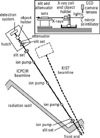
The phase contrast microradiography measurements were performed on a 7B2 beamline at Pohang Light Source (PLS), Korea (Fig. 1). The X-rays were emitted by an electron storage ring with electron energy of 2.5 GeV, and a typical beam current of 150 mA went through two beryllium windows and then reached the specimen. Polychromatic synchrotron X-rays were used with an energy range of 4-15 keV. A set of shutters made of silicon slabs with different thickness was used as an attenuator to control the total X-ray flux and the photon energy distribution. The size of the beam spot was 5×5 mm. The lung tissue samples were placed -200 mm from the scintillator to optimize detection of the phase contrast effects. After passing through the specimen, the X-rays reached a CdWO4 scintillator crystal, producing a visible image. The image produced by the scintillator is reflected by a mirror and then magnified by an interchangeable optical lens system with magnification from 3 to 50X. The magnified image is then detected by a CCD camera. The number of pixels ranges up to 1600×1200 with 14 bit gray scale.
We fixed the inflated lung samples on a rotating holder for microtomography. Two hundred snap shot images at different angle were obtained during 180° degree rotation, and tomographic reconstruction was done.
We fixed the thin lung samples on tilting holder for microradiography. Region of interest was determined by real time fluoroscopic preview. Snapshot microradiographic images were obtained using 400-1000 µm of field of view and 10 ms of exposure time.
Comparison with optical microscopic images
The 10 µm thickness sections of lung samples were examined by not only phase contrast microradiography, but also optical microscopy for 1 : 1 correlation with phase contrast microradiographic images and optical microscopic images. No additional staining of lung samples was performed for optical microscopy.
RESULTS
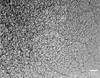
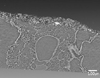
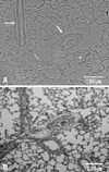
On snapshot phase contrast X-ray images of the inflated lung samples, the overall alveolar structures and small airways were observed as an overlap pattern (Fig. 2). The individual alveolar structures and airways of mouse lung were clearly defined on 3-D reconstruction of phase contrast X-ray microtomography (Fig. 3)
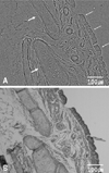
Phase contrast microradiographs of the thin lung samples showed very fine microstructure of the mouse lung. Bronchial cartilage and respiratory epitheliums were clearly defined (Fig. 4). Cilia of pseudostratified columnar epitheliums were also visualized. Individual lung parenchymal structures such as alveoli, bronchiole and arteries were also demonstrated at a micron level of spatial resolution (Fig. 5). Muscular arteries, in which the muscle is enclosed by an internal and an external elastic larmina, were clearly defined. Epithelial layers of bronchioles were seen as folded patterns with underlying interrupted smooth muscle. All of these phase contrast microradiographs correlated with optical microscopic images.
DISCUSSION
In the present study, the REI method was used. DEI and X-ray interferometry generally requires highly monochromatic plane-wave radiation and sophisticated X-ray optics, therefore their use is greatly restricted. REI is a simplified scheme based on an X-ray source having high spatial coherence and can work with polychromatic photons, however, it differs from conventional X-ray radiology only in that a gap is introduced between the object and the detector to allow distance for the refracted rays to diverge from the non-deviated ones. In general, the refraction mechanism has many advantages over the diffraction mechanism. The images are simpler and easier to interpret, and the edge enhancement that is due to a pair of fringes is easier to detect even with limited detector resolution. The imaging technique used in this study is the simplest method that uses polychromatic synchrotron X-ray with several inexpensive pieces of equipment, including scintilator, magnification lens and digital CCD camera. The other experimental conditions, for example, on the source geometry are more relaxed in the case of refraction, and this makes experimental implementation much easier. The 7B2 beamline at PLS eliminates the monochromator. The benefit is a huge increase in the flux delivered to the object, in comparison with monochromatic beamlines. As a result, images can be taken in real time with a speed already exceeding 1 image per millisecond and with excellent lateral resolution (less than 0.7 micron). Furthermore, higher resolution imaging is possible if a higher magnification lens is used, although the field of view would be narrowed. In addition to the elimination of the monochromator, the other important technical features of the beamline are elimination of the hard X-ray optical components (except for windows), the large beam size suitable for imaging with a large field of view, the flexibility, easy, and safe operation and the rapidity for changing experiments. Furthermore, the contrast mechanism is so effective that the radiation dose can be reduced by using attenuators/filters without seriously endangering the image quality. These features mean that this method could act as microradiography for viewing the fine internal structures of biologic subjects.
In several studies that have used DEI, the lung airways and lobe boundaries of small animals have been highlighted, and the inflated lung tissue was seen as a speckled intensity pattern.4,5 Lewis et al.6 demonstrated that phase contrast X-ray imaging using DEI was capable of dynamically imaging the lungs. Refraction enhanced tomography of mouse and rabbit lungs using monochromatic X-ray showed the images of cross sectional images of mouse and rabbit lungs at a spatial resolution of about 40 and 150 µm spatial resolutions.8 In REI with using polychromatic X-rays,10,11 the aerated lung tissue structure of small animals was seen as a speckled pattern. Jheon et al.7 observed terminal respiratory unit microstructures of rat lung ex vivo, using monochromatic X-ray REI. In this study, overall alveolar structure was identified as an overlap pattern on snapshot phase contrast X-ray images of inflated mouse lung sample. Furthermore, microstructures of individual alveoli and airways were clearly seen on phase contrast X-ray microtomography. The phase contrast microradiography of thin lung sample demonstrated the fine pulmonary microstructure such as alveoli, bronchioles, and small vessels with micron level of spatial resolution. The appearance of lung parenchyparenchyma at the structural level of alveoli was also investigated by using absorption-contrast based micro-CT,12,13 however, complicated sample preparation such as silver nitrate staining or infusion of barium sulfate-gelatine-thymol mixture were needed. In the present study we demonstrated very high resolution and contrast to detect microstructure details of mouse lung without complicated sample preparation. However, the present study was performed on ex-vivo animal lung samples to show the microstructure of lung without overlapping. The larger field of view and faster imaging are absolutely needed for the study of non-fixed sample or in vivo microscopic imaging.
Phase contrast microradiography can reveal the microstructure of lung with micron level of resolution. Refraction enhanced imaging is the simplest method among the different techniques for obtaining contrast, based on refractive index rather than on the absorption coefficient. In comparison with diffraction method, REI can work by using polychromatic synchrotron X-ray. This makes experimental implementation much easier and induces huge increase in the flux delivered to the object, resulting in real time imaging with excellent lateral resolution. The images of REI method are simpler and easier to interpret. And complicated sample preparation is not needed. However, the radiation problem exists, although it may be improved by using hard X-ray even in imaging of soft biologic tissue and reducing total X-ray flux. With all these facts in mind, phase contrast microradiography, based on refraction enhanced imaging by using polychromatic synchrotron X-ray, may be considered as a future possibility and its simplicity could be beneficial for its application to the microradiographic imaging.
 XML Download