

 PDF
PDF ePub
ePub Citation
 Print
Print


INTRODUCTION
Osteoporosis affects most commonly postmenopausal women, placing them at a significant risk for fractures. It is well known that osteoporotic fractures frequently occur at the skeletal sites including the spine, hip, and distal radius. Thus, evolving a strategy for the prevention of these osteoporotic fractures would appear to be important in postmenopausal women with osteoporosis.
Up to now, numerous preclinical studies on postmenopausal osteoporosis have shown the influence of an ovariectomy (OVX) on the skeleton in animals and the effect of intervention on the skeleton in OVX animals. It has been suggested that OVX nonhuman primates are a good model for postmenopausal osteoporosis because their trabecular and cortical bone remodeling processes, monthly menstrual cycles, and reproductive hormone patterns are similar to those of humans.1-5 In particular, Jerome et al.5 have shown that OVX cynomolgus monkeys are an excellent model for studying the basic mechanism of postmenopausal osteoporosis and for the development of suitable therapeutic regimens. Peak bone mass is reached in female cynomolgus monkeys at 9 years of age,6 2-3 years after growth plate closure, and animals older than this age would be an ideal for studies designed to model skeletally mature women.7 The lumbar spine has been most frequently studied in cynomolgus monkeys by measuring bone mineral density (BMD) using dual energy X-ray absorptiometry (DXA).8 However, little data exist on the influence of OVX on the 3 clinically important skeletal sites such as the lumbar vertebra, femoral neck, and distal radius in mature cynomolgus monkeys.
Because bone loss at the skeletal site rich in trabecular bone after OVX appears to be more pronounced than that at the skeletal site rich in cortical bone in cynomolgus monkeys,9 a statistically significant loss of trabecular bone mass could be detected at the above described 3 sites even with a smaller sample size. From the point of research and development (R&D) view, it is important to establish the influence of OVX on the clinically important skeletal sites in mature monkeys. The purpose of the present study was to examine the influence of OVX on bone turnover and trabecular bone mass at the 3 clinically important skeletal sites in mature cynomolgus monkeys.
MATERIALS AND METHODS
Treatment of animals
The study was carried out at the Animal Facility of the Simian Conservation Breeding & Research Center (SICONBREC), Inc., Philippines, and the animals were maintained according to the National Institutes of Health (NIH) Guidelines for Care and Use of Laboratory Animals. All the animal protocols were approved by the Laboratory Animal Care Committee of SICONBREC Inc., Philippines.
Six wild-caught female cynomolgus monkeys which had been used for breeding at SICONBREC (Macaca Fascicularis, conventional grade) were housed individually in bracket cages (500 mm in width; 500 mm in depth; 750 mm in height) and used for the study after quarantine and acclimation. Because nonhuman primates have menstrual cycles until near the end of their maximum life span,10 all of them had regular menstruation. According to dentition, their estimated ages ranged from 17 to 21 years. The animals were housed under local vivarium conditions (temperature 26 ± 4℃, humidity 75 ± 25% and 12 h on/off light cycle), and were fed a 100 g Standard Monkey Breeder Pellets containing 0.93% calcium and 0.77% phosphorus (Republic Flour Mills Corp., Makati City, Philippines) once a day and given in-house pumped water ad libitum.
After allowing 1-month's adaptation to the new environment, the monkeys were randomized into 2 groups of 3 monkeys each by the stratified weight method: the OVX group and the sham-operation (Sham) group. After intramuscular anesthesia with a mixed solution of ketamine hydrochloride and xylazine (Troy Laboratories PTY Limited, NSW, Australia) at the dose of 10 mg/kg each, bilateral OVX and a sham-operation were performed for the OVX and Sham groups, respectively. The body weight of the monkeys was monitored weekly and the experimental period was 16 months (64 weeks).
The following parameters were mainly evaluated in the present study; serum and urinary bone turnover markers, lumbar BMD and bone mineral content (BMC) in vivo by DXA, trabecular volumetric BMD (vBMD) of the lumbar vertebra, femoral neck, and distal radius by peripheral quantitative computed tomography (pQCT), trabecular bone mass and dynamics of the lumbar vertebra, femoral neck, and distal radius by static and dynamic bone histomorphometry, and bone strength of the lumbar vertebra and femoral neck.
Biochemical analysis of serum and urine
Serum and urine samples were collected in the morning after fasting from every animal at the baseline and 16, 32, 48, 53, and 64 weeks after the start of the experiment. In particular, urine excreted for 24 hours was collected using metabolic cages. Serum and urine samples were stored at -70℃, and then used for the measurements of the following biochemical markers.
Serum and urinary levels of calcium and phosphorus were measured by the o-cresolphthalein complexone (OCPC) and Fiske-Subbarow methods, respectively using an auto analyzer 7060E (Hitachi Lt., Tokyo, Japan). Serum levels of alkaline phosphatase (ALP) and creatinine and urinary levels of creatinine were measured by the enzyme method using the Auto analyzer 7060E. Serum levels of bone-specific ALP were measured with an enzyme-linked immunosorbent assay (ELISA) method using Osteolinks BAP (Sumitomo Biomedical Co., Ltd., Osaka, Japan). Serum and urinary levels of cross-linked N-terminal telopeptides of type I collagen (NTX) were measured with an ELISA using Osteomark NTX for serum and urine, respectively (Mochida Pharmaceutical Co., Ltd., Tokyo, Japan). Urinary levels of cross-linked C-terminal telopeptides of type I collagen (CTX) were measured with an ELISA using Furelisa β crosslaps (Fujirebio Inc., Tokyo, Japan). Serum levels of intact parathyroid hormone (iPTH) were measured with an ELISA method using I-PTH ELISA (Diagnostic Systems Laboratories Inc., Texas, USA).
Lumbar BMD and BMC measurement in vivo by DXA
DXA scanning of the lumbar spine was performed in vivo at the baseline and 16, 32, 53, and 62 weeks after the start of the experiment. The procedure used for scanning was based on the method previously described.9 After anesthesia with an intramuscular injection of a mixed solution of ketamine hydrochloride and xylazine, the lumbar spine (L1-L7) was scanned by DXA using a DPX-alpha (Lunar Ltd., Madison, WI, USA), with the animals supine on a Styrofoam board together with tap water 10 cm deep. The instrument was set up in a pediatric anteroposterior spine mode (setting: 8 cm scan width, 6 cm/s scan speed, and 0.6 mm×0.6 mm pixel size). The BMD and BMC of the lumbar spine (L4-L6) were determined.
Preparation of bone specimens and measurement of uterus, liver and kidney weight
All the monkeys were labeled with 4 mg/kg of calcein (Dojin Chemical Laboratories Co., Ltd., Kumamoto, Japan) injected intravenously 22 days and 9 days before they were sacrificed. At 64 weeks after the start of the experiment, the animals were anesthetized by intramuscular injection of a mixed solution of ketamine hydrochloride and xylazine, and sacrificed by exsanguination. The uterus, liver, and bilateral kidneys were excised from every animal, and their wet weight was measured. The 3rd, 4th and 5th lumbar spines, bilateral femurs, tibiae and forearm bones were also excised from every animal. Immediately, the length of the left femur and tibia was measured with dial calipers. The 4th lumbar vertebra, left femur, and right forearm bone were stored at -20℃, and then processed for pQCT analysis of trabecular bone of the lumbar vertebra, femoral neck, and distal radius. The 5th lumbar vertebra was stored at -20℃ and then processed for biomechanical testing of the lumbar vertebra. The left femur was also processed for biomechanical testing of the femoral neck immediately after pQCT analysis. The 3rd lumbar vertebra, right femur, and left forearm bone were fixed in 70% cold ethanol, and then processed for bone histomorphometric analysis of trabecular bone of the lumbar vertebra, femoral neck, and distal radius.
Bone histomorphometric analysis of trabecular bone of the lumbar vertebra, femoral neck, and distal radius
The bones fixed in 70% cold ethanol were cut using an Isomet saw (Buehler, Lake Bluff, IL, USA) to obtain specimens of the lumbar vertebra, femoral neck, and distal radius. The bone specimens were stained with Villanueva Osteochrome Bone Stain (Polyscience, Warrington, PA, USA) for 5 days. The specimens were then dehydrated sequentially in ascending concentrations of ethanol (70%, 95%, and 100%) and xylene and then embedded in methyl methacrylate (EM Science, Gibbstown, NJ, USA) at 4℃ by the method of Erben.11 The frontal-sections of the femoral neck, the sagittal sections of the center of the lumbar vertebra, and the frontal sections of the distal radius were cut into 5-µm slices using a microtome (Leica RM2155; Leica Inc., Nussloch, Germany), transferred onto chromium-gelatin-coated slides, dried overnight under pressure at 42℃, and coverslipped with Eukitt mounting medium (Calibrated Instruments, Hawthorne, NY, USA) for static and dynamic histomorphometric analysis of trabecular bone.
A digitizing morphometric system was used to measure bone histomorphometric parameters. The system consisted of an epifluorescence microscope (Olympus BH-2), a color video camera, and a digitizing pad (Numonics 2206) coupled to an IBM computer, and a morphometry program (Osteo-Metrics, Atlanta, GA, USA). The measured parameters for trabecular bone included total tissue volume (TV), bone volume (BV), bone surface (BS), eroded surface (ES), single- and double-labeled surfaces (sLS and dLS, respectively), and interlabel width. These data were used to calculate percent trabecular bone volume (BV/TV), trabecular number (TbN), trabecular thickness (Tb Th), trabecular separation (Tb Sp), ES/BS, mineralizing surface (MS)/BS [(sLS/2+dLS)/BS], mineral apposition rate (MAR), bone formation rate (BFR)/BS, and BFR/BV in accordance with the standard nomenclature proposed by Parfitt et al.12 In the present study, the region of trabecular bone measured was 1.55-5.95 mm distal from the center of growth plate, which consists of secondary spongiosa.
pQCT analysis of trabecular bone of the lumbar vertebra, femoral neck, and distal radius
The lumbar vertebra, femoral neck, and distal radius were scanned with pQCT (XCT Research SA+; Stratec Medizintechnic GmbH, Pforzheim, Germany) in 50% ethanol/saline. The bones were placed inside a glass tube and scanned at a slice thickness of 0.77 mm and voxel size of 0.4 mm. The middle parts of the lumbar vertebra and femoral neck were scanned. For the distal radius, the skeletal site distant from the distal joint by a length corresponding to 4% of the bone length was scanned. The scan line was adjusted using the scout view of the pQCT system. For analysis, a threshold of 395 mg/cm3 at contour mode 2 was used to separate the bone areas from the marrow regions. To separate the cortical areas from the trabecular areas, we used a constant threshold of 690 mg/cm3. Total and trabecular vBMD and area were measured.
Biomechanical testing of the lumbar vertebra and femoral neck
The mechanical strength of the lumbar vertebra and femoral neck was evaluated by the compression test using a materials-testing machine (MZ-500S; Maruto, Co., Ltd., Tokyo, Japan). The femurs were held in the upright position on the loading platform and a 500 kgf load was given by the rectangular parallelepiped crosshead to the center of the femoral head at a rate of 30 mm/min. For the lumbar vertebra, the intervertebral disc was removed to expose the trabecular bone area which was compressed to break over 3 mm or more. A 500 kgf load was given by the rectangular parallelezpiped crosshead to the center of the lumbar vertebra at a rate of 10 mm/min. The specimens were tested in a saline bath at 37℃ Each specimen was submerged in the saline bath for about 3 minutes before the testing, to allow temperature equilibration. Load-displacement curves were recorded, and the parameters analyzed were maximum load, stiffness, and breaking energy.
Statistical analysis
The data were expressed as means ± standard deviation in tables and means ± standard error in figures. Data comparisons between the 2 groups were performed with an unpaired t-test. Longitudinal changes in lumbar BMD and BMC and biochemical markers were examined with an one-way analysis of variance (ANOVA) with repeated measurements. Differences in longitudinal changes in these parameters between the 2 groups were examined with a two-way ANOVA with repeated measurements. All the statistical analyses were performed using the Stat View J-5.0 program (SAS Institute Inc., NC, USA) on a Macintosh computer. A significance level of p < 0.05 was used for all the comparisons.
RESULTS
Baseline characteristics of the animals
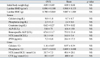
Table 1 shows the baseline characteristics of the animals. There were no significant differences in initial body weight, lumbar BMD and BMC, serum and urinary levels of calcium, phosphorus, and creatinine, and serum and urinary levels of bone turnover markers between the 2 groups.
Changes in body weight
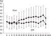
Fig. 1 shows longitudinal changes in body weight. Body weight significantly increased in the Sham group, but did not significantly change in the OVX group (one-way ANOVA with repeated measurements, Table 2). However, there was no significant difference in changes in body weight between the 2 groups (two-way ANOVA with repeated measurements, Table 2).
Changes in lumbar BMD and BMC
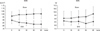
Fig. 2 shows longitudinal changes in the lumbar BMD and BMC. Lumbar BMD significantly decreased in the OVX group, but did not significantly change in the Sham group (one-way ANOVA with repeated measurements, Table 2). Lumbar BMC significantly increased in the Sham group, but did not significantly change in the OVX group (one-way ANOVA with repeated measurements, Table 2). There was a significant difference in changes in the lumbar BMD and BMC between the 2 groups (two-way ANOVA with repeated measurements, Table 2). Thus, OVX induced not only relative osteopenia but also absolute osteopenia, as indicated by a reduction in lumbar BMD compared with Sham controls and the baseline, respectively.
Changes in serum and urine biochemical markers
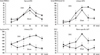
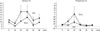
Fig. 3 shows longitudinal changes in the serum and urinary levels of bone turnover markers. Serum and urinary NTX levels significantly increased, while urinary CTX levels did not significantly change in both groups (one-way ANOVA with repeated measurements, Table 2). Bone-specific ALP levels significantly increased in the OVX group, but significantly decreased in the Sham group (one-way ANOVA with repeated measurements, Table 2). There were significant differences in changes in urinary NTX and CTX levels and serum bone-specific ALP levels, but not in those in serum NTX levels, between the 2 groups (two-way ANOVA with repeated measurements, Table 2).
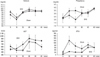
Fig. 4 shows longitudinal changes in the serum levels of calcium, phosphorus, ALP, and iPTH. Serum calcium and phosphorus levels did not significantly change in both groups (one-way ANOVA with repeated measurements, Table 2). Serum ALP and iPTH levels significantly increased in the OVX group, but did not significantly change in the Sham group (one-way ANOVA with repeated measurements, Table 2). There were no significant differences in changes in serum calcium, phosphorus, ALP, and iPTH levels between the 2 groups (two-way ANOVA with repeated measurements, Table 2).
Fig. 5 shows longitudinal changes in the urinary levels of calcium and phosphorus. Urinary calcium/Cr and phosphorus/Cr did not significantly change in both groups (one-way ANOVA with repeated measurements, Table 2). There were no significant differences in changes in urinary calcium/Cr and phosphorus/Cr between the 2 groups (two-way ANOVA with repeated measurements, Table 2).
Final body weight, femoral and tibial length, and uterus, liver, and kidney weight at necropsy
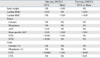
Table 3 shows final body weight, femoral and tibial length, and uterus, liver, and kidney weight at necropsy. There were no significant differences in final body weight, femoral and tibial length, or liver and kidney weight. However, the uterus weight was significantly lower in the OVX group than in the Sham group, suggesting successful surgery in the OVX group.
Bone histomorphometric analysis of trabecular bone of the lumbar vertebra, femoral neck, and distal radius
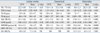
Table 4 shows the results of static and dynamic bone histomorphometric analysis of trabecular bone of the lumbar vertebra, femoral neck, and distal radius. BV/TV of the lumbar vertebra, femoral neck, and distal radius was significantly lower in the OVX group than in the Sham group, which was associated with increased BFR/BV. However, the response of trabecular bone structural parameters and bone formation and erosion parameters differed among the skeletal sites.
pQCT analysis of trabecular bone of the lumbar vertebra, femoral neck, and distal radius
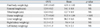
Table 5 shows the results of pQCT analysis of trabecular bone of the lumbar vertebra, femoral neck, and distal radius. Trabecular vBMD of the lumbar vertebra, femoral neck, and distal radius was lower in the OVX group than in the Sham group. In particular, the total area of the femoral neck was greater in the OVX group than in the Sham group, suggesting cortical expansion in the femoral neck caused by OVX.
Biomechanical analysis of the lumbar vertebra and femoral neck
Table 6 shows the results of biomechanical testing of the lumbar vertebra and femoral neck. There were no significant differences in the maximum load, stiffness, and breaking energy of the lumbar vertebra and femoral neck between the 2 groups. The lack of any significant differences in the parameters might be attributable to the low statistical power due to the small sample size.
DISCUSSION
The present study clearly demonstrated that OVX in mature cynomolgus monkeys, aged 17-21 years, increased bone turnover markers such as serum bone-specific ALP and urinary NTX and CTX, and induced trabecular bone loss associated with increased BFR/BV compared with Sham controls at the lumbar vertebra, femoral neck, and distal radius. OVX induced not only relative osteopenia but also absolute osteopenia as indicated by a reduction in lumbar BMD compared with Sham controls and the baseline, respectively, confirming that mature cynomolgus monkeys could be utilized for preclinical studies of postmenopausal osteoporosis to examine the effects of interventions on bone turnover and trabecular bone mass at the 3 clinically important skeletal sites. Thus, we support the concept that nonhuman primates are a good model for postmenopausal osteoporosis based on densitometric, histomorphometric and serum bone marker analysis.7
Concern has been expressed that the OVX monkey model does not produce consistent, statistically significant osteopenia and bone fragility, and frequently exhibits a "failure to gain" bone, rather than the clear bone loss expected of a model for postmenopausal osteoporosis.8 Relative osteopenia compared with intact animals is observed in younger OVX cynomolgus monkeys who were fed a normal calcium diet, whereas absolute osteopenia develops in OVX cynomolgus monkeys at age > 9 years and concomitantly fed a low calcium diet.13 In the present study, however, OVX in cynomolgus monkeys, aged 17-21 years, induced absolute osteopenia under the condition of a normal calcium diet (0.93%), as indicated by a reduction in lumbar BMD from the baseline. Thus, not only the dietary calcium content but also ages of the animals might be important to develop absolute osteopenia in OVX cynomolgus monkeys.
Urinary levels of NTX and CTX and serum levels of bone-specific ALP levels increased after OVX in the present study. Previous studies have shown that OVX in cynomolgus monkeys increased urinary levels of deoxypyridinoline and serum levels of osteocalcin, ALP, bone-specific ALP, and tartrate-resistant acid phosphatase.5,9,14-16 However, recent studies demonstrated OVX-induced elevations in urinary levels of CTX in addition to the above markers in cynomolgus monkeys.17,18 Serum and urinary levels of bone turnover markers were shown to increase within a few months, peak at 6-12 months, and remain elevated for 18 months or longer.8 All of these results indicate that OVX rapidly increases the serum and urinary levels of bone turnover markers and maintains this state for a couple of years in cynomolgus monkeys, consistent with our results (Fig. 3). However, the present study confirmed changes in newer bone markers including urinary NTX and CTX in OVX cynomolgus monkeys.
In the present study, trabecular vBMD and trabecular bone mass (BV/TV) of the lumbar vertebra, femoral neck, and distal radius were similarly lower in the OVX group than in the Sham group. Trabecular bone loss compared with Sham controls at the 3 skeletal sites was clearly associated with an increased bone formation rate (BFR/BV). This result is consistent with those of the study conducted by Jerome et al.5,16 They reported that vertebral trabecular bone loss was associated with dramatically increased bone formation rates, primarily caused by higher activation frequency of basic multicellular units of bone in cynomolgus monkeys (4-14 years of age),5 and that OVX increased the bone formation rates in trabecular bone obtained by iliac biopsy in cynomolgus monkeys (5-8 years of age).16 Thus, increased bone formation rates appear to be observed at multiple trabecular bone sites with bone histomorphometry.8
Because of the small sample size, we could not detect any significant changes after OVX in bone strength of the lumbar vertebra and femoral neck, as observed in cynomolgus monkeys, aged 11-15 years.14 Furthermore, we could not find any significant changes after OVX in cortical bone mass and strength of the femoral and tibial diaphysis, evaluated by pQCT analysis and biomechanical testing (three-point bending test), respectively (data not shown). No significant changes in cortical bone mass as well as cortical porosity were detected at the femoral neck and distal radius, even though we performed histomorphometry of cortical bone (data not shown). Jerome et al.13 recommended a group size of 25 for studies in cynomolgus monkeys. Thus, further studies are needed to clarify the influence of OVX on bone strength and cortical bone mass in mature cynomolgus monkeys. However, the statistically significant changes in bone turnover and trabecular bone mass at the 3 clinically important skeletal sites are worth reporting in the present study, as well as absolute osteopenia as indicated by a longitudinal decrease in lumbar BMD from the baseline even with a small sample size in mature cynomolgus monkeys.
In conclusion, the present study clearly demonstrated that OVX in mature cynomolgus monkeys, aged 17-21 years, increased bone turnover markers and induced trabecular bone loss compared with Sham controls at the lumbar vertebra, femoral neck, and distal radius. OVX also induced absolute osteopenia as indicated by a reduction in lumbar BMD from the baseline. Thus, the results strongly indicate that mature cynomolgus monkeys could be utilized for preclinical studies of postmenopausal osteoporosis to examine the effects of interventions on bone turnover and trabecular bone mass at the three clinically important skeletal sites.


 XML Download