INTRODUCTION
The potent anti-inflammatory agents, glucocorticoids (GCs) are frequently used for the treatment of inflammatory diseases such as arthritis, pulmonary diseases, and skin diseases. It is well known that high-dose or long-term use of GCs results in several adverse side effects including osteoporosis.1 Epidemiological studies have demonstrated the high incidence of fractures at the spine (the skeletal site rich in trabecular bone), followed by fractures at the hip (the skeletal site rich in both trabecular and cortical bone).2 This is probably because GC-induced osteoporosis is more evident in trabecular bone than in cortical bone.3 Thus, the primary strategy for GC-induced osteoporosis seems to be the prevention of vertebral fractures.
Both vitamin K2 and risedronate are widely used in the treatment of GC-induced osteoporosis in Japan,4 because both drugs have been reported to be effective in reducing incidence of vertebral fractures.5-7 Risedronate decreases bone turnover, increases bone mineral density, and prevents vertebral fractures in patients treated with GCs.5,6 An iliac biopsy study clearly demonstrated that risedronate prevented the deterioration of trabecular bone architecture, reduced the degree of mineralization, and preserved elastic modulus within the trabeculae in patients treated with GCs.8 However, there are no data to show the effect of vitamin K2 on bone formation and resorption and trabecular bone in patients treated with GCs.
Several preclinical studies using GC-treated rats have demonstrated that risedronate suppresses bone turnover and increases trabecular bone mass,9 while vitamin K2 attenuates reductions in periosteal bone formation and cortical bone mass as well as trabecular bone mass evaluated by micro computed tomographic analysis.10 However, the effects of vitamin K2 on bone mass and bone formation and resorption in trabecular bone remain uncertain. The purpose of the present study was to compare the effect of vitamin K2 and risedronate on trabecular bone in GC-treated rats.
MATERIALS AND METHODS
Treatment of animals
Fifty Sprague-Dawley female rats, 3 months of age, were purchased from Hilltop Lab. Animals, Inc. (Scottdale, PA, USA). The animals were housed under local vivarium conditions (temperature 23.8℃ and 12-h on/off light cycle), and were fed a pelleted standard chow diet containing 1.36% calcium and 2,400 IU/kg vitamin D (Rodent Diet 8604, Harlan Teklad, Madison, WI, USA), with free access to water. Following a 1-week adaptation to the new environment, the rats were randomized by the stratified weight method into 5 groups of 10 rats each according to the following treatment schedule: age-matched control, CG administration, and GC administration with concomitant administration of vitamin K2, risedronate, or vitamin K2 + risedronate. Five-hundred mg of methylprednisolone sodium succinate (Pharmacia & Upjohn Company, Kalamazoo, MI, USA) was reconstituted with 15 mL of bacteriostatic water and then subcutaneously administered as the GC at a dose of 5.0 mg/kg body weight three times a week. Vitamin K2 (menatetrenone, Eisai Co., Ltd., Tokyo, Japan) was suspended in 0.1 mL of 1,2-propanediol and glycerol solution at a dosage of 30 mg/kg body weight and administered by gavage into deep mouth three times a week. Risedronate (Eisai Co., Ltd., Tokyo, Japan) was dissolved in 0.1 mL of PBS solution at a dosage of 10 µg/kg body weight, and then subcutaneously administered 5 times a week. The doses of vitamin K2 and risedronate were considered to be effective in rats, in accordance with previously published data.9,11,12 Two rats in the GC + vitamin K2 + risedronate group were deleted from the study because of a failure of GC injections during the experiment in 1 rat and a failure of making bone sections for bone histomorphometry in the other. The body weight of the rats was monitored weekly, and the total experimental period was 8 weeks. The study was carried out at Winthrop-University Hospital, and the animals were maintained according to the National Institutes of Health (NIH) Guidelines for Care and Use of Laboratory Animals. All animal experimental protocols were approved by the Laboratory Animal Care Committee of Winthrop-University Hospital.
Preparation of specimens
All the rats were labeled with 10 mg/kg of calcein (Sigma Chemical, St. Louis, MO, USA) injected intramuscularly 10 days and 3 days before they were sacrificed. The animals were anesthetized with intraperitoneally injected ketamine at 80 mg/kg, together with xylazine at 12 mg/kg, and sacrificed by exsanguination. The left femur and right tibia were collected from every animal. The femurs were stored in a freezer (-70℃) and processed later for the measurements of the femoral length and bone mineral density (BMD) as described below. The right tibiae were used for bone histomorphometric analysis of the tibial proximal metaphysis; the bones were fixed overnight in 40% cold ethanol, and then cut into three parts using an Isomet saw (Buehler, Lake Bluff, IL, USA). The proximal tibial metaphyses were stained with Villanueva Osteochrome Bone Stain (Polyscience, Warrington, PA, USA) for 5 days. The specimens were then dehydrated sequentially in ascending concentrations of ethanol (70%, 95%, and 100%) and xylene and then embedded in methyl methacrylate (EM Science, Gibbstown, NJ, USA) at 4℃, in accordance with the method of Erben.13 Frontal sections of the proximal tibial metaphysis were cut at 5-µm thickness using a microtome (Leica RM2155; Leica Inc., Nussloch, Germany), transferred onto chromiumgelatin-coated slides, dried overnight under pressure at 42℃, and coverslipped with Eukitt mounting medium (Calibrated Instruments, Hawthorne, NY, USA) for static and dynamic histomorphometric analyses.
Bone histomorphometric analysis of the tibial proximal metaphysis
A digitizing morphometric system was used to measure bone histomorphometric parameters. The system consisted of an epifluorescence microscope (Nikon E-400, OsteoMetrics, Atlanta, GA, USA), an Osteomeasure High Resolution Color Subsystem (OsteoMetrics, Atlanta, GA, USA) coupled to an IBM computer, and a morphometry program (OsteoMetrics, Atlanta, GA, USA). The parameters measured for trabecular bone included total tissue volume (TV), bone volume (BV), bone surface (BS), eroded surface (ES), single- and double-labeled surfaces (sLS and dLS, respectively), and interlabel width. These data were used to calculate percent trabecular bone volume (BV/TV), trabecular number (Tb N), trabecular thickness (Tb Th), trabecular separation (Tb Sp), ES/BS, mineralizing surface (MS)/BS [(sLS/2+dLS)/BS], mineral apposition rate (MAR), bone formation rate (BFR)/BS, and BFR/BV, in accordance with the standard nomenclature proposed by Parfitt et al.14 In the present study, the region of trabecular bone measured was 1-4 mm distal to the lower margin of the growth plate in the proximal tibial metaphysis, which consists of secondary spongiosa. In addition to measurement of the above parameters, interlabel width beneath the growth plate was used to calculate longitudinal growth rate (LGR).
Measurements of femoral length and BMD
The length of the whole left femur was measured with a dial caliper. The BMD of the whole left femur was determined by dual energy X-ray absorptiometry (DXA) using a Hologic QDR-4500 Plus (Hologic Inc., Bedford, MA, USA). The instrument was adapted for an ultra-resolution mode, with line spacing of 0.0254 cm, resolution of 0.0127 cm, and collimation of 0.9 cm diameter. The bone was placed in a Petri dish, and to simulate soft-tissue density, tap water was poured around the bone to a depth of 1 cm. The bone mineral content and bone area were measured, and the BMD of that area was calculated by dividing bone mineral content by bone area. The coefficient of variation of these measurements at our laboratory was less than 1.0%.15
Statistical analysis
All the data were expressed as means and standard deviation (SD). Mann-Whitney U-test was used to compare the data between the age-matched control group and other groups. Two-way factorial analysis of variance (ANOVA) was used to examine the effect of vitamin K2 and risedronate and their combination effect, using the data from the GC, GC + vitamin K2, GC + risedronate, and GC + vitamin K2 + risedronate groups. All statistical analyses were performed using the Stat View J-5.0 program on a Macintosh computer. A significance level of p < 0.05 was used for all the comparisons.
RESULTS
Effect of GC administration
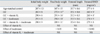
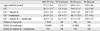
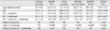
GC induced reductions in body weight and femoral BMD (Table 1). GC also induced reductions in trabecular BV/TV, Tb N and Tb Th and increase in Tb Sp (Table 2), as a result of decreased bone formation (MS/BS, MAR, BFR/BS, BFR/BV) and increased bone erosion (ES/BS) (Table 3). GC reduced LGR/day (Table 3), but did not affect femoral length (Table 1).
Effect of vitamin K2 administration on GC-treated rats
Vitamin K2 did not affect body weight and femoral BMD (Table 1). However, vitamin K2 attenuated GC-induced reduction in trabecular BV/TV (Table 2) by preventing GC-induced decrease in bone formation (MS/BS) and subsequently reducing GC-induced increase in bone erosion (ES/BS) (Table 3). Vitamin K2 did not affect either LGR/day (Table 3) or femoral length (Table 1).
Effect of risedronate administration on GC-treated rats
Risedronate attenuated GC-induced reduction in body weight and prevented GC-induced reduction in femoral BMD (Table 1). Risedronate also prevented GC-induced reductions in trabecular BV/TV and Tb Th (Table 2) by preventing GC-induced increase in bone erosion (ES/BS) although it also suppressed bone formation (MS/BS, MAR, BFR/BS, BFR/BV) (Table 3). Risedronate did not affect either LGR/day (Table 3) or femoral length (Table 1).
Effect of combined administration of vitamin K2 and risedronate on GC-treated rats
There was no combination effect of vitamin K2 and risedronate on body weight and femoral length and BMD (Table 1) as well as trabecular bone structural parameters (Table 2). However, vitamin K2 mildly attenuated the suppression of bone formation (MS/BS) and bone erosion (ES/BS) caused by risedronate when administered in combination (Table 3).
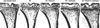
Images of the proximal tibial metaphysis
Fig. 1 shows the images of the proximal tibial metaphysis, confirming the results of bone histomorphometric analysis regarding trabecular BV/TV.
DISCUSSION
The present study found the differential effect of vitamin K2 and risedronate on trabecular bone in GC-treated rats. Vitamin K2 attenuated GC-induced trabecular bone loss by preventing GC-induced decrease in bone formation (MS/BS) and subsequently reducing GC-induced increase in bone erosion (ES/BS). Risedronate prevented GC-induced trabecular bone loss by preventing GC-induced increase in bone erosion (ES/BS) although it also suppressed bone formation (MS/BS, MAR, BFR/BS, BFR/BV). Vitamin K2 mildly attenuated suppression of bone formation (MS/BS) and bone erosion (ES/BS) caused by risedronate without affecting trabecular bone mass when administered in combination.
It has been argued that rat is a poor model of GC-induced osteoporosis, because GC inhibits bone resorption by osteoclasts, resulting in a protective effect on the skeleton in mature rats.16 However, GC administration has been shown to induce losses of trabecular BV/TV, Tb N, and Tb Th in young rats.10,17-20 Therefore, the clearly observed trabecular bone loss in GC-treated young rats in the previous studies could be due, at least in part, to growth inhibition. However, GC treatment is required even in children with nephritis, systemic lupus erythematosus, asthma, juvenile idiopathic or rheumatic arthritis, lymphoblastic leukemia, and other diseases. Therefore, it is important to study the effects of drugs on GC-induced bone loss in young rats. In the present study, GC administration induced trabecular bone loss in young rats compared with age-matched controls.
GC-induced trabecular bone loss has been associated with decreased bone formation and increased bone resorption, however the key histological feature of GC-induced trabecular bone loss has been shown to be reduction in the thickness of trabecular bone, reflecting suppressed bone formation21. In the present study, GC-induced trabecular BV/TV loss compared with age-matched controls was associated with decreases in Tb Th and Tb N and increase in Tb Sp because bone formation was decreased and bone erosion was increased by GC administration. Because bone erosion reflects the coupling of bone formation to bone resorption, both decreased bone formation and increased bone resorption could account for increased bone erosion.
Vitamin K2 is known to have an anabolic action on bone; regulation of bone formation by vitamin K2 may involve γ-carboxylation of osteocalcin and/or may be mediated by the steroid and xenobiotic receptor.22-26 In the present study, vitamin K2 attenuated GC-induced trabecular bone loss by preventing GC-induced decrease in bone formation (MS/BS) and subsequently reducing GC-induced increase in bone erosion. These results suggest at least the mild anabolic effect of vitamin K2, because bone erosion reflects the coupling of bone formation to bone resorption. However, the effects of vitamin K2 on trabecular bone mass and bone metabolism seem to be modest.
The bisphosphonates inhibit osteoclast-mediated bone resorption, and loss of osteoclast function and apoptosis are the consequence of loss of function of one or more important signaling proteins. In particular, nitrogen-containing bisphosphonates, such as alendronate and risedronate, are not metabolized but can inhibit enzymes of the mevalonate pathway, thereby preventing the biosynthesis of isoprenoid compounds, which are essential for the post-translational modification of small GTPases.27 In the present study, risedronate prevented GC-induced trabecular bone loss by preventing GC-induced increase in bone erosion although it also suppressed bone formation (mineralizing surface, mineral apposition rate, and bone formation rate). Thus, risedronate ameliorated the imbalance of bone formation and resorption. These results are consistent with those of previous studies.9 However, oversuppression of bone formation as well as bone resorption is concerned.
Because vitamin K2 and risedronate have different actions on bone formation and resorption, the beneficial effects of combination of the 2 agents on trabecular bone could be expected. A few studies showed the effects of combined treatment with bisphosphonates and vitamin K2 in other models of osteoporosis.28-31 Ito et al.29 demonstrated additive effect of risedronate and vitamin K2 in preventing deterioration of the trabecular bone architecture in ovariectomized rats, whereas Otomo et al.30 showed no significant additive effect of risedronate and vitamin K2 on bone turnover, bone mass/density, bone structure, mineral properties (mineral-to-matrix ratio), and bone strength evidenced by no significant effect of vitamin K2 on bone parameters in ovariectomized rats. Furthermore, Iwasaki et al.31 reported that concomitant use of vitamin K2 with incadronate ameliorated suppression of bone formation and more effectively prevented cancellous bone loss in tail-suspended rats. In the present study, risedronate strongly suppressed bone formation and erosion, and vitamin K2 mildly attenuated the suppression of bone formation (MS/BS) and bone erosion caused by risedronate without affecting trabecular bone mass when administered in combination. Bone strength reflects both bone mass and bone quality, and bone quality is determined by the bone architecture, turnover, damage accumulation, mineralization, and matrix.32 Thus, strong suppression of bone formation as well as bone resorption caused by risedronate might possibly lead to deterioration of bone quality. Vitamin K2 might have a mild anabolic effect, possibly playing a role in attenuating deterioration of bone quality in rats treated with GC and risedronate.
In conclusion, the present study showed differential effects of vitamin K2 and risedronate on trabecular bone in GC-treated rats. Vitamin K2 attenuated GC-induced trabecular bone loss via a mild anabolic effect. Risedronate prevented GC-induced trabecular bone loss via a strong antiresorptive effect. These results suggest that risedronate should be a first line drug, while vitamin K2 remains to be a second line drug in preventing vertebral fractures in patients treated with GCs. Vitamin K2 might play a role in improving bone turnover possibly counteracting by risedronate-induced oversuppression of bone turnover when administered in combination.