

 PDF
PDF ePub
ePub Citation
 Print
Print


INTRODUCTION
Understanding genes that function in the pathogenesis of human disease is essential for developing effective diagnoses and therapies, and animal models are indispensable tools for studying gene function. N-ethyl-N-nitrosourea (ENU) is a powerful spermatogonial mutagen that induces one point mutation per million base pairs in laboratory mice.1 Several research centers are conducting large-scale screening programs for producing novel ENU-induced mutants that model human disease.2 The point mutagenicity induced by ENU potentially allows for the identification of multiple allelic variants at each gene locus that lead to complete loss of gene function (null allele), partial loss of function (hypomorphic), gain of function (hypermorphic), dominant negative function (antimorphic), or novel gain of function (neomorphic).3 Screening for ENU mutagenesis is phenotype-driven, and is a powerful way to identify gene function due to the absence of a prior assumptions.
The vertebrate limb is an ideal model system for studying signaling mechanisms and pattern formation.4,5 Signaling by the polarizing region expressing Shh establishes both the number and characteristics of the digits that can be temporally uncoupled.6-9
Hox genes are divided into 4 linkage groups (called Hoxa, -b, -c, and d) that are essential for the development of limb extremities. Hoxd12 differentially affects the paraxial and postaxial chondrogenic branching in the limb, and regulates Shh in a positive feedback loop.10 Moreover, studies of the Hoxd12 regulatory elements have revealed global and local control elements of the Hoxd complex.11 Hoxd expression data suggest that the most anterior digit in a bird wing is homologous to digit 1 (and not digit 2) in other amniotes.12,13
In this study, we carried out both genome-wide screening and point mutant screening to identify the affected gene. A new ENU-generated mouse mutant exhibited microdactyly on the fore and hind limbs as a recessive trait, however appeared normal otherwise. This study fell short in establishing a strong correlation between the point mutation identified and the microdactyly phenotype observed. Positional cloning allowed the point mutated gene in Hoxd12 to be identified. We also found that Lmx1b and Fgf4 were significantly up-regulated in their mutant mouse line. The present observation would be of potential interest for the understanding of the function of Hox genes in digit development.
MATERIALS AND METHODS
All experiments were performed according to the guidelines of the Yonsei University, College of Dentistry, Intramural Animal Use and Care Committee.
Animals
Mice were obtained from Charles River, Japan. All mice were maintained in the barrier system under specific-pathogen-free conditions with regulated light (07:00 - 19:00 hours), temperature (23 ± 1 ℃), humidity (50 ± 5%), and ventilation (10 - 12 times per hour). The care and treatment of the animals was in accordance with the Guide for the Care and Use of Laboratory Animals (NIH). All animal experiments were approved by the Institutional Animal Care and Use Committee of the Korea Research Institute of Chemical Technology. All experiments were carried out in accordance with the Guidelines for Animal Experimentation. The BALB/cJ male mice were intraperitoneally injected twice with ENU (100 mg/kg), 2 weeks apart. Using a 3-generation breeding scheme, an ENU induced microdactyly was established as a mutant strain.
Skeletal staining
For skeletal analyses, mice were collected at 8 weeks. The mutant and wild type (BALB/cJ) mice were processed and stained with Alcian Blue-Alizarin Red Skeletal Staining.
Genetic mapping
To genetically map the limb mutant locus, intraspecific progenies were generated. A mutant (Mt) mouse was mated with C57BL/6 (B6) mouse. The F1 progeny of the (Mt × B6) mating were crossed with [(F1 × Mt) F2] and produced Mt mouse. Twenty one Mt progeny were used for mapping. The genetic markers were obtained from Mouse Genome Informatics (Blake et al. 2003). Two polymorphic microsatellite markers (D2Mit329 and D2Mit285) were used for linkage analysis using standard techniques. Briefly, genomic DNA was extracted from tails according to the manufacturer's protocols (QIAamp Tissue Kit; Qiagen, Inc., Santa Clarita, CA, USA). Amplifications by polymerase chain reaction (PCR) were performed according to standard protocols. Primers were purchased from Bioneer, Inc. (Daejeon, Korea). Genetic linkage was assessed by segregation analysis. The statistical significance of the recombination was determined by × 2 analysis.14
Mutation analysis
Genomic DNA was prepared from the liver of wild-type (WT) and homozygous mutant mice by the standard protocol and used as the template for PCR. Primer pairs were generated to GenBank (www.ncbi.nlm.nih.gov). PCR was performed in a 20 µL reaction mixture containing 25 ng of template DNA, 1× Taq buffer, 1.5 mM MgCl2, 200 µM dNTPs, 0.2 µM each of primer, and 1 U of Taq polymerase (Superbio, Suwon, Korea). PCR products were purified with a Qiagen purification kit (Qiagen, Inc., Valencia, CA, USA) and sequenced using the ABI Prism BigDye Terminator Cycle Sequencing Ready Reaction Kit version 1.1 and a sequencer, ABI Model 3100 (Applied Biosystems, Foster City, CA, USA). The PCR product was digested, and DNA fragments were separated on a 12% polyacrylamide gel with 1 × TBE buffer.
Quantitative real-time PCR (RT-qPCR)
RNA was extracted from the skin of WT and homozygous mutant mice using a total RNA isolation procedure according to the manufacturer's recommendations and further purified using the WelPrep™ Total RNA isolation reagent (WelGENE, Daegu, Korea). For cDNA synthesis, reverse transcription of RNA was performed using the Revert Aid™ M-MiLV Reverse Transcriptase (Fermentas, Burlington, Ontario, Canada). Real-time PCR was performed using a Thermal Cycler Dice™ Real Time System (Takara, Shiga, Japan) according to the manufacturer's instructions. Reverse transcriptase and buffer were from SYBR Premix EX Taq™ (Perfect Real Time, Takara, Shiga, Japan). The PCR for each sample was performed in triplicate, and the amounts of the PCR products were normalized using GAPDH as an internal control. The data were analyzed with the Thermal Cycler Dice™ Real Time System analysis software (Takara TP800, Shiga, Japan). To determine the relative level of gene expression, we used the comparative ΔΔCt method, previously described by Livak and Schmittgen. The Student t-test was employed to determine significant changes at the 95% confidence level (*p < 0.01).
RESULTS
ENU-induced microdactyly mice
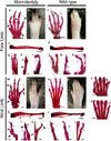
Recessive mutagenesis screening produced ENU-induced microdactyly mice at the third generation that could be distinguished from their normal littermates at birth. Both wild-type (WT) and mutant mice had five digits on the fore and hind limbs, and our mutant mice did not show the syndactyly or polydactyly phenotype (Fig. 1).
WT and heterozygous mice appeared identical at birth, showing normal growth and limb development. In contrast, however, homozygous mutants induced by ENU were characterized by small limbs and digits at birth, whereas other organs such as kidney and lung appeared to be normal (data not shown). Skeletal analysis of microdactyly mice showed additional abnormalities in the radius (R), ulna (U), tibia (T), and fibula (F) (Figs. 1e, f, q, and r). Microdactyly mice had widely expanded digits, and digit I was shorter than those in WT mice (Figs. 1a-d, i, and j). Only digit IV was unaffected on the fore limb (Figs. 1a-d), and the tip of digit I was missing in microdactyly mice (Figs. 1a, g, and j). All the phalanges of digit V were shorter in microdactyly mice than in WT mice (Figs. 1a, b, h, and k), especially for the metacarpal bone (first phalange: microdactyly, 0.1 cm; WT, 0.2 cm, Table 1) and medial phalanx (third phalange: microdactyly, 0.1-cm; WT, 0.2 cm, Table 1), which were half the length of those in WT mice (Figs. 1a, b, i, and l). In addition, both the radius and ulna were smaller and misshapen in microdactyly mice (Figs. 1e and f); specifically, the bones were thinner and the interosseous space of fore limb was wider than in WT mice, although the total length (1.3 cm, Table 1) was the same (Figs. 1e and f).
The microdactyly hind limb had abnormal digits at birth, with the limbs also being shorter than in WT mice (Figs. 1m-p). Although no phalanges were missing, the abnormalities were more significant than in the fore limbs (Figs. 1a-d and m-p). Digits I, II, and V were more curved than normal digits (Figs. 1m-x). Moreover, all the first phalanges were shorter and thicker, and all the joints were thicker in microdactyly mice than in WT mice (Figs. 1m, o, and s-z). The metatarsal bone of digit I was shorter than normal, and was not connected properly to the end of the metatarsal bone. The digital phalanx (tip) was normal (Figs. 1m, o, s, and v). Similar to digit I, digit II of the metatarsal bone was also shorter, thicker, and curved, and also showed a hind limb abnormality (Figs. 1m, o, t, w, y, and z). The proximal phalanx of digit II was thinner in microdactyly mice than in WT mice, and the middle phalanx was bent proximally (Figs. 1m and o). Digit V of microdactyly mice was also curved proximally, and therefore the space between digits IV and V was larger than in WT mice (Figs. 1m-p, v, and x-z). Microdactyly mice exhibited abnormalities in the tibia and fibula of the hind limb, with the interosseous space being narrower than normal (Figs. 1q and r).
Point mutation of Hoxd12
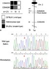
To identify the mutated gene, mutant (BALB/cJ) mice were crossed with C57BL/6 mice, and the (mutant × B6) F1 was crossed with (F1 × F1) F2 to produce ENU-induced microdactyly mice. Twenty one microdactyly offspring were genotyped for linkage analysis. Among the loci associated with the microdactyly phenotype, we chose the Hoxd locus as the candidate mutant gene, based on the phenotypic similarity of mutant mice to ENU-induced microdactyly mice. The genotypes of the F2 mice are summarized in Fig. 2a. The order of the markers was centromere-D2Mit329 and D2Mit 285 (Fig. 2a), and the Hoxd cluster was located between D2Mit329 and D2Mit 285 on chromosome 2. Therefore, to identify the point mutation, we directly sequenced the Hoxd cluster in ENU-induced microdactyly mice (Fig. 2b). The sequences were determined using BLAST searches, and weidentified a G-to-T transition at nucleotide 1359 that cosegregated with the mutant phenotype. This mutation had alanine changed to serine at the residue 453 in Hoxd12 (Fig. 2c).
Alteration patterns of significant molecules in microdactyly mice
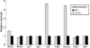
After identifying the mutated gene, we used quantitative real-time PCR to explore the functional consequences of the Hoxd12 point mutation on limb development. These analyses were performed on 8-week-old mice. The gene-specific primers used are listed in Table 2. Compared to WT mice, the mRNA expression of Hoxd12 in ENU-mutated microdactyly mice was not altered (data not shown), whereas that of Bmp2 was moderately increased, Fgf4 and Lmx1b were dramatically increased, and Bmp4, En1, Fgf8, Ptch1, and Shh were slightly decreased (Fig. 3). Expres sion levels of Gli3 and Shh were not changed in point mutated Hoxd12 mice (Fig. 3).
DISCUSSION
ENU mutagenesis is an effective strategy for developing mice models of human disease by inducing mutations that lead to unique phenotypes for gene-targeted strategies. Moreover, mutant phenotypes associated with point mutations might be more appropriate as models of human inherited disorders, since these are predominantly associated with changes to single base pairs.15 Furthermore, mutagenesis screening represents an alternate source of mutant phenotypes for investigations.16
In limbs, Hoxd12 transcripts are absent from the intermediate segments, the presumptive zeugopods in both fore and hind limbs, with especially Hoxd12 being unaffected both in the proximal/posterior domain and in the future digit area.17 In our ENU screening, we identified the phenotype, resulting from a point mutation in the Hoxd12 gene. Targeted mutagenesis of individual and multiple 5' Hoxd genes alter the size, shape, and number of bones, and delay chondrification and ossification.18,19 Thus, these genes determine region-specific growth and differentiation in limb skeletal elements, acting in a combinatorial fashion during both early and late developmental stages.20 The ENU-induced Hoxd12 mutation produces a distinct autosomal recessive phenotype of short digits and limbs, and crosstalk between factors such as Hoxd12, Bmps, Fgfs, and Shh during limb development is critical to correct digit formation.4,5,8,21,22 Our mouse model could help understand the specific roles of Hoxd12. We identified the Hoxd12 point mutation involving G-to-T mutation at nucleotide 453, resulting in a change from alanine to serine (Fig. 2). Our analysis revealed that the function of the mutated Hoxd12 protein at the molecular level might affect protein structure and DNA binding, which would be altered by Hodx12 translational events such as changes in the transcription rate or mRNA stability.
There are striking similarities between microdactyly and Hoxd12-knock-out mice. Digits II and V were short in Hoxd12-knock-out mice, in which exon 2 was deleted.23 Digit II was strongly affected and the wrist carpal bone showed a subtle, characteristic indentation at its distal border. In addition, digit V of these mice was also twisted proximally, as in microdactyly mice.23 Moreover, Hoxd12 misexpression caused a posterior transformation, duplications of the distal anterior skeletal elements, and selective reductions in specific proximal limb elements.24 The Hoxd proteins also reversed the Gli3-repressor function by acting downstream of Shh, thereby promoting digit formation.25
Fgf4 influences digit morphogenesis, which correlates with the widespread changes in gene expression, also affecting the posterior members of the Hoxd.26 Hoxd12 and Hoxd13 were consistently induced in the anterior-distal limb mesenchyme, accompanying mirror-image duplication of the digit pattern.27 In order to elucidate Hoxd12 function, we examined factors involved in digit formation, which included Bmp2, Bmp4, En1, Gli3, Fgf4, Fgf8, Lmx1b, and Ptch1.9,28,29 Only Fgf4 and Lmx1b were found to be significantly increased in microdactyly mice (Fig. 3). Fgf4 is the earliest member of the fibroblast growth factor (FGF) family identified, and is expressed during embryogenesis where it plays an essential role in post implantation development, limb growth, and patterning.30 In addition, increased FGF signaling occurs when the Fgf4 gain-of-function allele is activated in a WT limb bud. Excess Fgf4 causes supernumerary posterior digit (postaxial polydactyly) formation and cutaneous syndactyly between all the digits.31 These observations underscore the importance of controlling FGF expression during normal limb development. Lmx1b is involved in dorsal-ventral patterning and may play additional roles in anterior-posterior patterning and growth.28
In the resesent study, the mutation appeared to be more severe than the full knockout reported previously by two groups. We described the analysis of ENU-mutated mice showing a digit phenotype and the identification of a point mutation within the Hoxd12 gene, causing a serine to alanine substitution. Moreover, we observed perturbation of the expression of some genes in mutant mice that play relevant roles in limb development. It is not clear how these abnormalities are related to the increases of Fgf4 and Lmx1b expressions in the mutant mouse. One possibility is that a point mutation rather than the entire deletion of Hoxd12, such as knockout and transgenic mice, causes the abnormal limb phenotype. This discrepancy could reflect the differential phenotypic effects of changes on single amino acids and be useful in phenotype screening to detect mutants. The precise nature of the spectrum of differences obviously requires further investigation.
 XML Download