

 PDF
PDF ePub
ePub Citation
 Print
Print


INTRODUCTION
Larynx squamous cell carcinomas (LSCCs) account for the vast majority of the head and neck squamous cell carcinomas (HNSCCs), with an occurrence rate of 0.4% in men and 2.2% in women. As the clinical outcomes vary greatly among LSCCs, therapeutic decisions can not be made solely on stage and histology. The current genetic findings in these tumors have been observed to be important in their tumorigenesis and therapeutic decisions. The certain chromosome changes have been identified as being shared among HNSCCs, and possible distinct pathways of tumorigenesis should be taken into account, as they are a heterogeneous group of tumors.1 Of the limited number of cytogenetic studies on these types of tumor, the most frequently chromosome aberrations involve 1p, 3p, 3q, 5q, 7p, 9p, 9q, 11p, 11q, 13q, 14q, 15q, 17p and 18q.2-5 Advanced molecular cytogenetic techniques (FISH and CGH) applied on fixed materials have allowed a better understanding of the genetic changes in HNSCCs.6-15 Gains in 3q, 5p, 8q and 11q13, and losses in 3p and 9p appeared as consistent genetic changes, with other certain chromosomal changes occurring in larynx tumors.10 Molecular studies revealed allelic imbalances in chromosome regions with known oncogenes and tumor suppressor genes, and occur most frequently at 3p, 5q, 9p, 9q, 11p, 11q, 13q, 17p and 18q.16-18 Cytogenetic, molecular cytogenetic and molecular genetic studies are being performed to identify the genes responsible for larynx tumorigenesis.
Here, the CGH technique was used to analyze unbalanced chromosomal changes in fifteen cases with a larynx squamous cell carcinoma, and previously reported chromosomal alterations were found as well as those being reported for the first time.
MATERIALS AND METHODS
Paraffin-embedded archival tumor tissues of 15 randomly selected patients with LSCC (13 male and 2 female) were studied. The patients' ages varied between 40 and 72, all tumors were primary and none of the patients had received any therapy prior to sampling. Clinical staging was performed according to the UICC TNM Staging System (Table 1). Two patients had distant metastases, 4 of the tumors had glottic, 10 were supraglottic and 1 transglottic locations. Hematoxylin-eosin stained tissue sections (5 mm) were prepared from each tumor and the histological diagnosis was confirmed in all cases. For the CGH, genomic DNA was extracted by proteinase K digestion and phenol-chloroform extraction. Tumor DNA was labeled with spectrumGreen-dUTP (Vysis Inc. IL, USA). Reference DNA was prepared from peripheral blood lymphocytes of healthy female and male donors, and labeled with spectrumRed-dUTP (Vysis Inc. IL, USA). Both reference and tumor DNAs were labeled using degenerate oligonucleotide primed-PCR (DOP-PCR) using the method described by Telenius et al.19 The DOP-PCR product was run on a 1.2% agarose gel, with a proper marker DNA, to obtain a 400-2,000 bp probe fragment size of uniform and intense hybridization. The CGH was performed, but with modifications to the standard procedures.7,20,21 Briefly, labeled tumor and reference DNAs (500 ng) were mixed with unlabeled Cot-1 DNA (50 μg) in 15 mL of hybridization buffer and hybridized to normal metaphase preparations for 2 days at 37℃ in a moist chamber. After hybridization, the slides were washed according to previously described protocols, and the chromosomes counterstained with 4'-6-diamidino 2-phenylindole (DAPI) in an antifade solution.7,20,21 The hybridization was analyzed using the PSI (Perceptive Scientific International LTD. UK) digital image software Mac Probe version 4.0, installed on a Zeiss Axiophot-2 epifluorecence microscope equipped with a cooled CCD camera (Photometrics LTD, Tucson, AZ, USA) and a filter system (LEP, Hawthorne, NY, USA) consisting of a 6 position computerized Ludl filter wheel. Three fluorochrome (DAPI, Spectrum Green, and SpectrumRed) images were properly registered and processed with the PSI workstation using the Mac Probe Program Version 4.0 software for pseudocolor display which were used to visualize the color changes along the metaphase chromosomes.
For each case, ten metaphases were analyzed for the chromosomal locations of the DNA sequence gains and losses. These regions were determined using green-to-red fluorescence intensity ratio profiles. Defining the gains and losses of DNA sequence copy numbers in the tumors were based on comparison of normal DNAs labeled with two different colors. The decision limits of the green-to-red ratios were < 0.8 for a loss of DNA copy number and > 1.50 for a gain. Control labeling and hybridizations were performed using nick translation (Boehringer Mannheim) and cross red/green labeling of the normal and tumor DNA probes, respectively. Heterochromatic regions of chromosomes 1, 9, 16, 19, Y, and acrocentrics 13, 14 and 15 were excluded from the analysis. Processing and evaluation were carried out as described previously.7,20,21
RESULTS
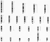
DNA copy number changes were observed in 10 of the 15 samples (66.6 %), the details of which are displayed in Table 1 and Fig. 1. The majority of the chromosomal alterations detected were gains. Three cases revealed polyploidies, and gains were detected in various chromosome regions in 6 patients. One patient displayed a whole chromosome gain of chromosome 1 along with losses within 15q and 22q. Some patients had common regions of DNA amplification (Table 1). The gains and losses of whole and partial chromosomes for each case are shown in Fig. 1.
The minimal common regions of gains were determined in the following chromosomal regions: 3p25-pter. 4p16, 5p15.2-pter, 6q26-qter, 8p23.2-pter, 9p23-pter, 14q31-qter, 17p13.2, 18p11.31-pter and Xq27qter. Three of the four samples with glottic localizations were found to have a normal CGH profile, whereas one had different gains in various chromosomal regions. One sample that had a transglottic localization was also found to have a normal profile. Losses, including the regions 15q26.1-qter and 22q13-qter, were determined in the same patient, who was also the youngest (40 years-old) of the 15 cases.
DISCUSSION
Cytogenetic techniques are accepted as having certain limitations in detecting genomic changes in solid tumors. They require cell cultures, which are technically difficult for such tumors, with the risk of observing chromosomal alterations not belonging to the original tumor. With respect to this, molecular cytogenetic techniques have many advantages over cytogenetic methods in the detection of chromosomal alterations. Comparing our results with cytogenetic and molecular cytogenetic findings on LSCCs revealed some similar and some different results, some of which corresponded to regions with known cancer-related genes, as reported in various molecular genetic studies.22,23 Polyploidies were identified in 3 cases using the CGH technique. Polyploidy was also reported cytogenetically in short term cultures from biopsy specimens,24 cell lines25 and in tumor cells by flow cytometry.26 Aneuploidy, another sign of genomic instability in tumors, was observed in some of the patients in this study along with other chromosome changes, in the form of whole chromosome gains (Table 1). Amplification of the 5p15.2-pter, 9p23-pter and Xq27-qter regions were observed as common findings in two of fifteen cases. 9p23-pter and Xq27-qter amplifications have not previously been reported to occur in LSCCs, while amplification of a region including 5p12.2-pter was reported in a CGH study in 2 out of 38 cases of primary LSCCs.9 The 5p15.2-pter region lies within 5p14-pter, amplification of which has previously been reported in 2 out of 23 having primary larynx tumors with metastases to adjacent lymph nodes.25 However, two of our three cases with a polyploidy profile had metastases, whereas two with 5p15.2-pter had no distant metastases. With an increased number of cases, the relation between these findings and metastases may be revealed. These regions may gain significance if recurrently reported as putative oncogene-harboring regions in future studies. Some of the amplified regions found in our study, all of which were detected in 1 out of 15 cases, had been previously reported by other researchers. 7q21.3 and 13q33 lie within the 7q21-q22 and 13q32-qter regions, respectively, the amplifications of which have been reported by other groups in SCCs of the head and neck were detected in 6 out of 43 and 1 out of 13 cases studied, respectively.27-29 Gain of 13q33 has been previously reported as an amplification of 13q32-34, which harbors the gene coding for FGF14 (Fibroblast Growth Factor-14). Amplification in the 12p12-pter and 14q32 regions were detected in our study, as well as in 4 out of 43 and 1 out of 30 cases, respectively, in the studies of Bockmuhl, Brzoska and Speicher.27-29 There are no oncogenes in these regions known to be activated by amplification. The 18p11.31-pter region is included within 18p, which is known to harbor the YES1 oncogene, and was reported, by Hermsen et al, to be amplified.10 Overexpression of the latter two regions may play a role in the tumorigenesis of LSCCs. Some of the regions detected as amplified in our study have not been previously reported: 2p25-p26, 8p23.2-pter, 10q26-qter, 14q32, Xq27-qter, 3p25-pter, 4p15.3-pter, 17p13.2, 6q26-qter, 2q27.2, 3p24.3, 4p16, 6p24-pter, 8q23.1-pter, 17p13.1-pter, 6q22.1-qter, 11p15.4 and 14q31-qter. This suggests that these regions may harbor oncogenes which potentially play an important role in tumorigenesis and progression of LSCCs. Losses of 15q26.1-qter and 22q13-qter, which occurred in only 1 patient in our study, have not frequently been reported in the literature.9,10 The FES protooncogene was localized to the 15q26.1 chromosome region, and the NBK/BIK gene, a proapoptotic BCL-2 family member which is considered a putative tumor suppressor gene, was mapped to the 22q13 region.30 The losses occurring in the youngest patient (40 years of age) were noteworthy, suggesting a possible role in the earlier stages of LSCC development. These findings may be rendered significant if observed in more than one case when a greater number of cases are studied.
Five of our 15 cases were found to have normal CGH profiles. The cell tumorigenesis and tumor progression of these cases may have been due to point mutations in proto-oncogene(s) and balanced chromosomal exchanges unable to be detected by CGH and may also have an effect on these genes. Although it is well recognized that each tumor may have its unique genomic alterations, studies aiming to reveal common recurrent genetic changes in laryngeal cancers, as in any type of tumor, will contribute to the collection of useful data, which eventually can be used for the correlation of genomic abnormalities with clinical outcome.
 XML Download