

 PDF
PDF ePub
ePub Citation
 Print
Print


INTRODUCTION
Age-related maculopathy (ARM) is the main cause of adult blindness in developed countries.1 Although there is considerable information on the nature and prevalence of visual impairment and blindness in Western countries, only a few population-based articles have appeared, describing the prevalence of ARM and the association of ARM with various patient characteristics in different racial and ethnic groups in Western countries.2-5 Only limited population-based data on the prevalence, characteristics, and risk factors for ARM in Asian populations are available.6-13
Studies from Western countries implicate lifestyle choices, nutritional parameters, and genetic risk factors in the pathogenesis of ARM.14 Known risk factors for developing ARM are age,14-18 smoking, 14-17,19-21 and hypertension.22-24 Hyperlipidemia and fat intake are related to the presence of exudative ARM,24 and obesity25-28 and atherosclerosis24 contribute to ARM development in some studies. Asian lifestyles and nutritional choices are different, however, from those of Western populations. The purpose of this report is to provide an estimate of the prevalence of ARM in a large survey sample, and to evaluate possible risk factors for ARM in the South Korean population.
PATIENTS AND METHODS
The Yonsei Eye Study was carried out in Seoul, which is the largest city in South Korea. The Medical Ethics Committee of the Yonsei University medical center approved the study protocol, and all participants were well-briefed on the purposes of the study before giving written informed consent in accordance with the tenets of the Declaration of Helsinki. Baseline interviews and ophthalmic examinations, with concurrent general medical examinations, took place between January 2006 and December 2006. Of 13,016 participants who came in for a health check-up during that period, 9,530 subjects (73.2%) were ≥ 40 years old.
All examinations were carried out at the Yonsei Medical Examination Center in Seoul, Korea. After measuring visual acuities and intraocular pressures (using a Type WT-200 instrument; Canon Inc, Tokyo, Japan), digital photographs of the maculae and optic discs were taken using a digital fundus camera (model TRC.NW 100; Topcon, Tokyo, Japan). Medical examinations were performed, which included blood analyses, urine analyses, and electrocardiograms. Additionally, the education level, income status, smoking habits, and illness history of each subject were assessed by questionnaire. Subjects were asked to report existing or previous diagnoses of diabetes mellitus, hypertension, cardiac disease, or hepatic disease.
Ophthalmic examination
For the assessment of ARM, the Wisconsin Age-Related Maculopathy Grading system was used, as described previously in detail.29,30 Fundus photographs were examined by ophthalmologists with special training in retinal diseases (MIR, JHK). Intergrader and intragrader agreement was assessed using the quadratic weighted kappa statistic on a random subset of 30 subjects. There was excellent inter-and intraobserver agreement (κ = 0.82) for the grading of fundus photographs. Senior investigators also reviewed all photographs graded as late ARM (ARM 4). Grading of the fundus photographs was based on the staging systems developed by the investigators of the Rotterdam Eye Study31: ARM 0, no feature characteristic of early ARM, or hard drusen only; ARM 1, either soft drusen or pigmentary irregularities only; ARM 2, either soft, indistinct (≥ 125 µm) or reticular drusen only, or soft, distinct, drusen with pigmentary irregularities; ARM 3, soft, indistinct, or reticular drusen with pigmentary irregularities; and ARM 4, neovascular ARM or geographic atrophy. Neovascular ARM was considered to be present if any of the following lesions were found: serous or hemorrhagic retinal pigment epithelial detachment, subretinal neovascular membrane, or subretinal hemorrhage. Geographic atrophy was considered to be present if preretinal fibrous scarring or retinal pigment atrophy with visible choroidal vessels was found. We defined ARM stage 2 and 3 as early ARM and stage 4 as late ARM. Subjects were excluded from the study whose fundi could not be graded because of poor photograph quality or dense ocular media.
Data analysis
For smoking analysis, subjects were categorized as non-smokers (those who had never smoked), current smokers, and prior smokers (those who had smoked earlier but were not smoking for 6 months at the time of the study). Education level was graded on the basis of final education level attained, as follows: 0, no school education; 1, graduated from elementary school; 2, graduated from middle school; 3, graduated from high school; 4, bachelors degree from college and, 5; masters degree from college. Monthly income was divided into 6 categories: 1, monthly income less than 1.5 million won; 2, monthly income between 1.5 million and 2.5 million won; 3, monthly income between 2.5 million and 3.5 million won; 4, monthly income between 3.5 million and 4.5 million won; 5, monthly income between 5.5 million won and 6.5 million won, and; 6, monthly income more than 6.5 million won. Hypertension and hepatitis were considered to be present when subjects selfreported these conditions, and diabetes was considered to be present when subjects self-reported the condition and the self-report was confirmed by a measurement of fasting blood glucose level > 126 mg/dL.
Statistical analyses
Student t-test was used for analyses of continuous variables, and the Pearson χ2 test was also employed for nominal variables. Age-adjusted logistic regression analysis was performed to determine risk factors for early and late ARM, using odds ratio calculations, and estimations of the 95% confidence intervals. We then developed multivariable models using the following procedure. The initial specification included independent variables consistently identified in previous research as risk factors for early and late ARM, or independent variables identified in univariate analyses as related to outcomes (p > 0.10). We then sequentially removed non-significant (p > 0.15) covariates, making it certain that these eliminations did not change estimated coefficients of the exposure variable by more than 10%.32 Age and gender were adjusted regardless of association. Because our models might have been overspecified, given the limited number of ARM in the study group, we also constructed reduced form models by selecting covariates, based on their statistical significance in the adjusted models. SPSS version 12 (SPSS Inc, Chicago, IL, USA) was used to perform statistical analyses. A two sided p value < 0.05 was considered to be statistically significant.
RESULTS
Study population
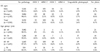
A total of 13,016 potential participants (7,109 men and 5,770 women), who underwent examinations at the Yonsei Medical Examination Center between January 1, 2006 and December 31, 2006, were identified. Of the participants, 9,530 (73.2%) were aged ≥ 40 years, and underwent full clinical examinations. Of these, 9,468 (99.3%) completed all study procedures, including dilated fundus photography. Fundus images were not available for 62 (0.7%) participants because subjects refused to be photographed, and poor-quality, unusable photographs were obtained from 151 (1.6%) subjects because of corneal opacities, phthisis bulbi, or small pupils. Table 1 shows the prevalence of ARM by gender and age. Percentages of the population in various age groups were 42.3% in the group aged 40 - 49 years, 36.0% in the group aged 50 - 59 years, 17.3% in the group aged 60 - 69 years, and 4.5% in the group aged ≥ 70 years. ARM was seen in 235 (2.46%) subjects. Estimated prevalence of early ARM (ARM 2 and ARM 3) and late ARM (ARM 4) increased from 0% and 0.1%, respectively, in the 40 - 49 year age group, to 2.0% and 0.1% in the group aged 50 - 59 years, to 6.7% and 0.2% in the group aged 60 - 69 years, and to 8.0% and 2.4% in subjects aged ≥ 70 years.
Mean age of the subjects with ARM was 63.17 ± 7.45 years[mean ± standard deviation (SD)]. One hundred and twenty-six (53.6%) ARM patients were male and 109 (46.4%) were female, thus males showing a slight predominance. Early ARM and late ARM were present in 2.3% (215 subjects) and 0.2% (20 subjects) of participants, respectively. Among these, ARM was bilateral in 123 subjects (52.3%) and unilateral in 112 subjects (47.7%). Among bilateral subjects, geographic atrophy was found in both eyes in a single subject, and 2 subjects each showed geographic atrophy in 1 eye and multiple drusens in the other eye. The remaining subjects showed multiple drusens in both eyes with or without pigment abnormalities. Among the unilateral ARM subjects, 17 (15.2%) eyes showed late ARM (neovascular AMD in 5 subjects and geographic atrophy in 12 subjects), and remaining 95 eyes showed early ARM. Among ARM subjects, there were no differences in distributions of ARM features between males and females (ARM 2, ARM 3, and ARM 4) (Pearson chi-square test, p = 0.991), or late ARM features (Pearson chi-square test, p = 0.897). Bilateral ARM was more prevalent in females (Pearson chi-square test, p = 0.011).
Neovascular AMD was found in 5 subjects. Among these, 2 patients showed subretinal hemorrhages with exudates, a single subject showed serous pigment epithelial detachment with hyperpigmentation, and 2 subjects had large subretinal hemorrhages with orange nodules.
Risk factors for ARM
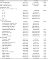
When Student t-test and Pearson χ2 test were used for analyses of variables, the following risk factors for early and late ARM (ARM 2, 3 and 4) were subjected to age-adjusted univariate analysis: gender, socioeconomic status (monthly income and education level), smoking history, self-reported diagnosis of hypertension, self-reported diagnosis of cerebral vascular accident, self-reported diagnosis of hepatitis, and body mass index. Serum levels of cholesterol, triglycerides (TG), high-density lipoprotein (HDL), low-density lipoprotein (LDL), lactate dehydrogenase (LDH) and C-reactive protein (CRP) were measured. In addition, serum hepatitis B surface antigen status (HBsAg), hepatitis B core antibody level (HBcAb), hepatitis B surface antibody level (HBsAb), and hepatitis C antibody level (HCVAb) were also assessed.
Subjects with ARM were significantly older (p < 0.001), and monthly income and final education level were lower than subjects without ARM. Also, while 27% of the ARM subjects had hypertension, only 14.1% of the subjects without ARM had hypertension history (p < 0.001). As for blood sample analysis, significantly different platelet, LDH, and TG levels were found with ARM subjects (Table 2).
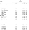
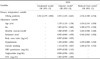
Therefore, estimated prevalence of ARM was strongly age-related (logistic regression; p < 0.001; OR 1.134; 95% CI; 1.114 - 1.154), as expected. The results of age-adjusted logistic regression analyses of risk factors for ARM are listed in Table 3. After adjusting for age, hepatitis B infection (positive status for HBsAg and HBcAb) and serum triglyceride levels and HDL levels remained as significant risk factors for ARM. Multivariate regression analysis indicated that serum HBsAg-positive status was significant and associated with a > 2-fold increase in the risk of ARM, relative to normal macular subjects, in both the adjusted (OR 2.55 ; 95% CI 1.476 - 4.421) and reduced (OR 2.736; 95% CI 1.588 - 4.714) analyses (Table 4).
DISCUSSION
We estimated the prevalence and risk factors for ARM in Seoul, Korea. In our study population, 2.46% of participants had signs of early or late ARM in one or both eyes, suggesting that ARM is relatively rare among adult South Koreans in Seoul. A total of 2.26% of the study subjects showed early ARM, defined as soft indistinct drusen with or without pigmentation abnormalities. Late ARM (defined as neovascular AMD or geographic atrophy) was seen in 0.2% of the study population. Among those with late ARM, neovascular AMD in at least one eye was seen in 5 subjects and geographic atrophy in 15 subjects.
Although direct comparison is difficult due to the fact that our studies showed estimate prevalence of ARM, these estimates of early and late ARM are lower than the incidence rates observed in Western countries. In the Beaver Dam study,33 the prevalence of indistinct soft drusen was 8.4% and patients with either pure geographic atrophy or exudative macular degeneration constituted 0.6% of the study population. The Framingham Eye Study34 showed an incidence of 1.5% of exudative macular degeneration in people ≥ 52 years of age. In a population-based prevalence study (the Beijing Eye Study), Li et al.13 found that the overall prevalence rates of early and late ARM were 1.4% and 0.2%, respectively, and these data are similar to our results. The prevalence of late ARM in the black population was 0.59% in a Barbados study,4 and was 0.5% in Hispanic individuals aged ≥ 50 years in Arizona.35
The prevalence of early ARM obtained in the present study on South Koreans living in the Seoul area was slightly higher (2.46% vs. 1.4%) than in the Chinese population group of the Beijing Eye Study,13 nevertheless, the two studies yielded similar estimates of the prevalence of late ARM (0.2% vs. 0.2%). The prevalence rates of early ARM in the age groups of 40 - 49 years, 50 - 59 years, 60 - 69 years, and ≥ 70 years were 1.54%, 2.74%, 3.41%, and 6.31%, respectively, in the Beijing study. In the same study, prevalence rates of late ARM were 0.14%, 0.52%, 0.14% and 1.11%, respectively, in these age groups. These figures are similar to our data (Table 1). The slight between-study difference in prevalence of early ARM might have been due to a population difference, geographic difference (rural vs. urban), ethnic differences between Chinese and Koreans, differences in dietary or nutritional habits, environmental parameters, and difference in the study design.
Several studies14-19,22,23 have identified risk factors for ARM, and both population-based studies and case control studies have been carried out. Refractive error,36-38 iris color,39,40 hypertension and cardiovascular disease,23,24,28,41 smoking,20,41-44 and alcohol consumption,44-46 are known risk factors for ARM. In our study, age was significantly associated with estimated early and late ARM prevalence, as expected. When adjustments were made for subject age in univariate analysis, seropositivity for HBsAg and HBcAb, indicating (past or present) infections with hepatitis B, serum levels of HDL and TG all remained significantly associated with ARM (Table 3).
Two previous studies47,48 have demonstrated statistically insignificant, but inverse associations between education levels attained and ARM. Although crude analysis showed significant relationships between education level and monthly income and ARM (Table 2), no significant inverse association was noted after age adjustment (p = 0.582, OR 0.970, 95% CI 0.868 - 1.083 for education, p = 0.910, OR 0.995, 95% CI 0.917 - 1.081 for monthly income).
Also, although smoking has been reported to be one of risk factors for ARM, our study showed no definite association between smoking and ARM (p = 0.866, OR 1.034, 95% CI 0.700 - 1.528). This might have been due to relatively small number of late ARM in our study, which is more likely related with smoking.20,42
One of the most distinct risk factors for ARM found in the present study is the association between hepatitis B and ARM in South Korea. Even after adjusted multivariate analysis for socioeconomic factors such as education level and monthly income in this study, HBsAg (p < 0.001; OR 2.736; 95% CI 1.588 - 4.714) and anti HBc antibody (p < 0.05; OR 1.475; 95% CI 1.092 - 1.992) were significantly associated with ARM, (Tables 3 and 4). The statistical significance of both HBs antigen and anti HBc antibody with ARM suggest that there is less opportunity than that this association is by chance alone (1/20 × 1/20 = 1/400). The reason why there has been no epidemiologic study about Hepatitis B and ARM was that Hepatitis viral marker has not been included as covariates in previous epidemiologic studies. Since Hepatitis B infection is highly prevalent in South Korea, viral markers were checked in most examinees. Although the prevalence of hepatitis B surface antigenemia has declined because of an effective vaccination program, HBV remains endemic in South Korea. Seropositivity for HBsAg was estimated at 7 - 9% in the 1980s.49 It was also reported in 1998 Korean National Health and Nutrition survey that prevalence of HBsAg was 5.1% in males and 4.1% in females, with low prevalence rates in individuals under 20 years old.50,51 There is a biologic plausibility underlying this association. First, HBs Ag is found in subretinal fluids with increased detection rate of antibodies to S-antigen in healthy virus carriers, increasing risk for uveoretinal pathology.52,53 This inflammatory process may induce drusen formation. Secondly, molecular mimicry between retinal S-antigen and Hepatits S antigen54,55 can induce cross reactivity which can predispose to uveoretinal inflammation. Furthermore, as viral hepatitis is associated with decreased level of complement C3, C4 and complement factor H related protein 1,56 this may activate the alternative complement pathway, thereby increasing the risk of drusen formation.57
Associations between ARM and hypertension, cardiovascular disease,23,24,28 serum lipid level,14,41 obesity,24,27 cardiovascular biomarkers,28,58-59 and BMI26 have previously been documented. In our subjects, no significant associations between ARM and hypertension, serum cholesterol level, or cardiovascular biomarker CRP were found after age-adjustment. The serum level of TG was inversely associated with the prevalence of ARM, whereas the serum level of HDL showed a significant positive association. However, the association between low TG level, high HDL level and ARM adjusted for age was not maintained in the adjusted model (Table 4), which was adjusted for age, HBs Ab positivity, gender and hypertension. This could be explained by the positive confounding by one or more of the above risk factors for ARM, although there are some reports to support the association between low TG, high HLD and ARM.28,57-59
Strength of this study is the relatively large number of subjects examined. The protocol was standardized, thereby providing an accurate estimate of the prevalence, and risk factors for ARM in Seoul, Korea. Also, the strength of this study is that socioeconomic factors, cardiovascular risk factors, and hepatitis viral markers were considered for analysis. A limiting factor of the present study is that this is a cross-sectional design which cannot assess the causality. Therefore, prospective data are needed to confirm the issue of ARM causality. Although this data may not represent the general population of South Korea, subjects were those who visited the health examination center for regular check-ups, but not for the treatment of confirmed disease, which may lower the selection bias compared with previous hospital- based studies. Moreover, since there was only small number of subjects with ARM, the power of the study to identify all significant factors may be decreased. Therefore, the results of the present study may be taken as practical evidence for those associations that were statistically significant, but not as a proof for those associations which did not show statistical significance. Also, subjects, in whom a dense cataract or hazy cornea media were considered to be reasons for visual impairment, might additionally have had ARM. This fact might artificially reduce the prevalence figures for ARM.
In conclusion, the single most important risk factor for ARM in our study, besides age, is seropositivity for HBsAg. However, factors such as current smoking, history of previous smoking, and the level of smoking, in addition to self-reported diagnoses of systemic diseases such as diabetes mellitus and cerebrovascular condition which had previously been reported as risk factors for ARM, did not show statistically significant influences on the prevalence of ARM in this study.
 XML Download