

 PDF
PDF ePub
ePub Citation
 Print
Print


INTRODUCTION
Asthma is a chronic inflammatory airway disease that can be severe and sometimes fatal.1 It has been estimated that 5 to 10 percent of patients with asthma have severe disease that is not effectively controlled by typical therapies, and these patients are at high risk of asthma-related death.1,2 Severe asthma has several distinct characteristics compared to mild-to-moderate asthma, such as female dominance, less allergic sensitization, and history of more frequent aspirin-hypersensitivity.2 However, there is still little information on the pathophysiological mechanism responsible for severe asthma.1-3
It has repeatedly been suggested that autoimmune mechanism might be involved in the pathogenesis of asthma.4-6 The autoimmune hypothesis of asthma is based on the presence of autoantibodies to the target tissue of inflammation (lung and bronchial mucosa),6-8 common genetic association, frequent coexistence with autoimmune diseases,9,10 and response to medications used for systemic autoimmune diseases such as systemic corticosteroid and anti-TNF reagents.11,12 However, a pathogenic role of autoimmune response in the pathogenesis of asthma has not yet been demonstrated.
Interestingly, IgG autoantibody to a 55-kDa protein in endothelial cells and platelets has been reported to be associated with severe asthma.8 Recently, alpha-enolase protein has been identified as a common autoantigen of endothelial cells, bronchial epithelial cells, and platelets, recognized by circulating IgG autoantibodies in patients with severe asthma.13 It has been suggested that alpha-enolase could be the same antigen that has been previously reported as a 55-kDa endothelial autoantigen.8,13
IgM autoantibodies to alpha-enolase have been detected in patients with Behçet's disease14 and IgE autoantibodies to unidentified 55-kDa endothelial cell antigen have previously been detected in patients with severe asthma.8 We hypothesized, therefore, that a preference of specific immunoglobulin isotype of autoantibody response to alpha-enolase protein might be related with different clinical manifestations in the specific diseases with autoimmune response to alpha-enolase protein.
In this study, we analyzed isotype and IgG subclass distributions of autoantibodies response to alpha-enolase protein in serum samples from adult patients with severe asthma and evaluated possible pathogenetic significance of the autoantibody in severe asthma, especially a possibility of IgE-mediated autoallergic reaction or IgG autoantibody-mediated complement activation.
PATIENTS AND METHODS
Subjects
We used serum samples from 10 patients with severe asthma and 7 patients with mild-to-moderate asthma, and 5 healthy controls (Table 1). All patients had typical clinical history of asthma and documented reversibility of FEV1 greater than 12% after inhalation of bronchodilator. All patients with severe asthma had typical clinically characteristics of difficult-to-control by standard medical treatments and required frequent administrations of oral corticosteroids for control of their asthma. They had clinical history of at least 1 severe asthmatic exacerbation requiring an emergency department visit or admission in the last year despite continuous typical therapies as defined previously.13 All patients underwent skin-prick test with 50 common aeroallergens (Bencard Co., Brentford, UK). Atopy was defined when there was a positive skin reaction to any one of the 50 common aeroallergens.13 All subjects gave written informed consent and the institutional review board approved this study.
Production and purification of recombinant human alpha-enolase protein
Recombinant human alpha-enolase protein was produced using Escherischia coli BL21 cells and purified as previously described.14
Immunoblot analysis of IgG, IgA, and IgM autoantibodies to alpha-enolase
Recombinant human alpha-enolase protein was separated by a discontinuous sodium dodecyl sulfate/polyacrylamide gel electrophoresis (SDS-P AGE) as previously described.13 Following electrophoresis, protein in the gel was transferred onto a polyvinylidene difluoride membrane (PVDF; Bio-Rad Laboratories, Hercules, CA, USA). After the transfer, the membrane was incubated for 1 hour with Tris-buffered saline (TBS) containing 0.1% Tween 20, 10% Skim milk, and 10% bovine serum. The membrane strips were incubated for 2 hours with 1 mL of serum samples at a dilution of 1 in 100 (v/v) at room temperature. After washes, the membrane was incubated for 2 hours with alkaline phosphatase-conjugated goat anti-human IgG, anti-human IgA, or anti-human IgM (Sigma Chemical Co., St. Louis, MO, USA) at room temperature. After a final washing, the membrane was stained with a substrate solution (nitro blue tetrazolium/5-bromo-4-chloro-3-indoyl phosphate; Sigma Chemical Co., St. Louis, MO, USA). Goat specific antibody to human alpha-enolase protein (Santa Cruz Biotechnology, Santa Cruz, CA, USA) was used as a positive control and detected by alkaline phosphatase-conjugated anti-goat antibody. Autoantibody was defined as positively detected if a stained density of immunoblot of the serum sample was definitely visible and comparable to density produced by a goat specific antibody to human alpha-enolase.
Immunoblot analysis of IgG subclass autoantibodies to alpha-enolase
For the detection of IgG subclass autoantibodies to alpha-enolase protein, PVDF membrane strips described above were incubated with various dilutions of serum samples (IgG1, 1 : 100 ; IgG2, 1 : 100 ; IgG3, 1 : 10, and IgG4, 1 : 10 dilution) and incubated for 3 hours at room temperature. After washes, monoclonal anti-human IgG subclass antibodies (clone 8c/6-39 for IgG1, HP-6002 for IgG2, HP-6050 for IgG3, HP-6023 for IgG4; Sigma Chemical Co., St. Louis, MO, USA) were diluted at 1 : 1000 and incubated for 2 hours at room temperature. After washes, the membrane was incubated with alkaline phosphatase-conjugated goat anti-mouse IgG (Sigma Chemical Co., St. Louis, MO, USA) for 2 hours at room temperature. After a final washing, the PVDF strips were stained as described above.
Immunoblot analysis of IgE autoantibody to alpha-enolase
Immunoblot analysis of IgE autoantibody to alpha-enolase was carried out according to the method used for the detection of specific IgE antibody to house dust mite allergen.15 The PVDF membrane strips described above which contained recombinant human alpha-enolase protein were incubated for 3 hours with dilution of serum samples (1 : 10 dilution) at room temperature. After three washes, the membrane was incubated for 2 hours with affinity-purified and biotinylated anti-IgE (Vector Laboratories Inc., Burlingame, CA, USA) at room temperature. After washing, the membrane was incubated for 1 hour with alkaline phosphatase conjugated streptavidin (Sigma Chemical Co., St. Louis, MO, USA) at room temperature. After washing, the PVDF strips were stained as described above.
RESULTS
Isotype distribution of autoantibody to alpha-enolase protein
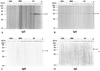
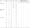
IgG autoantibody to alpha-enolase protein was detected in serum samples from 7 of 10 patients with severe asthma (70%), 1 of 7 patients with mild-to-moderate asthma (14.3%), and 0 of 5 healthy controls (0%) (chi-square test, p < 0.05) (Fig. 1 and Table 1). On the other hand, IgA, IgM, and IgE autoantibodies to alpha-enolase protein could not be detected in all patients with severe asthma on mild-to-moderate asthma, and healthy controls (Fig. 1).
IgG subclass distribution of IgG autoantibody to alpha-enolase protein
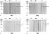
IgG1 autoantibody to alpha-enolase protein was detected in 7 of 10 patients with severe asthma (70%), 1 of 7 patients with mild-to-moderate asthma (14.3%), and none of 5 healthy controls (0%) (chi-square test; p < 0.05) (Fig. 2 and Table 1). IgG2, IgG3, and IgG4 autoantibodies to alpha-enolase protein were detected only in 2 of 10 patients with severe asthma (20%) (Table 1). There were no significant differences in the detection rates of IgG2, IgG3, and IgG4 subclass autoantibodies to alpha-enolase protein among patients with severe asthma on mild-to-moderate asthma, and healthy controls (p > 0.05) (Fig. 2 and Table 1).
The pattern of immunoblot detection of IgG1 autoantibody to alpha-enolase in all subjects tested was exactly similar to the pattern of IgG autoantibody to alpha-enolase (Figs. 1 and 2). Consequently, the IgG1 subclass was the predominant type of autoantibody response to alpha-enolase protein in patients with severe asthma.
DISCUSSION
In this study, we found that IgG antibody was a dominant isotype among the IgG, IgA, IgM, and IgE autoantibody responses to alpha-enolase protein in patients with severe asthma. We also found that IgG1 subclass was the predominant type of IgG subclass autoantibody response to alpha-enolase protein in patients with severe asthma. This finding suggests a possible involvement of IgG1 autoantibody-mediated complement activation in the pathogenesis of severe asthma.
A possible involvement of autoimmune mechanism in the pathogenesis of bronchial asthma has been suggested for a long time (at least more than 40 years).6-8 Previous studies have shown the presence of circulating autoantibodies to the bronchial mucosa tissue, lung tissue, endothelial cells, and epithelial cells in patients with bronchial asthma.4,6-8 The beta-2 adrenergic receptor, vasoactive intestinal peptide, cytokeratin 18 protein, and alpha-enolse protein were identified as autoantigens associated with bronchial asthma.4,7,13,16 Unfortunately, however, autoantibody responses to the above identified autoantigens are not specific to bronchial asthma, and detected in many other chronic inflammatory diseases including systemic vasculitis, inflammatory bowel diseases, endometriosis, autoimmune hepatitis, and rheumatoid arthritis.7,13,16,17 These findings suggest that autoimmune responses observed in the bronchial asthma could be an epiphenomenon which reflects damage of bronchial tissue, resulting from chronic airway inflammation. However, a possibility of pathogenic contribution of autoantibody response by inducing airway inflammation could not be excluded either.
There are several reports on in vitro experiment to study the functional significance of the autoimmune response observed in patients with bronchial asthma.4 One important characteristic of circulating autoantibodies in patients with bronchial asthma was that the autoantibodies were complement-fixing antibodies.18 The presence of antinuclear antibody was significantly associated with elevated serum levels of activated complement in patients with bronchial asthma.19 Circulating concentration of activated complement was also significantly increased in patients with aspirin-sensitive asthma compared to aspirin-tolerant asthma and healthy controls.20 Theoretically, complement fixing autoantibodies to antigens which exist in the bronchial mucosa tissue can activate the complement system, thereby inducing bronchial smooth muscle contraction and mucosal inflammation by generating anaphylactoxin (C3a and C5a).21 A possible involvement of autoantibody-mediated complement activation in the pathogenesis of bronchial asthma can further be supported by the deposition of immunoglobulin and complement and up-regulation of complement receptors on the epithelial cells and endothelial cells in the bronchial mucosa tissue from asthmatic patients who died of severe asthmatic attack.22,23 However, the pathogenetic significance of autoimmune response observed in patients with bronchial asthma should further be tested by establishing an animal model of "autoimmune asthma".
Circulating IgE autoantibody to 55-kDa autoantigen which is commonly expressed in endothelial cells and platelets has previously been detected in 2 among 97 patients with bronchial asthma.8 We have recently identified that an alpha-enolase protein is the autoantigen recognized by circulating autoantibodies from patients with severe asthma and commonly expressed in the endothelial cells, airway epithelial cells, and platelets.13 We also suggested that previously reported 55-kDa endothelial autoantigen could be an alpha-enolase protein.13 In the present study, therefore, we tested a possible existence of IgE autoantibody to human alpha-enolase proteins in the serum samples from patients with severe asthma, however, we could not detect IgE autoantibody in serum samples from 10 patients with severe asthma including 7 patients with severe asthma who had a circulating IgG autoantibody to alpha-enolase protein, suggesting a low possibility of IgE autoantibody-mediated activation of mast cells in these patients with severe asthma. The predominance of IgG1 subclass autoantibody response to alpha-enolase protein observed in patients with severe asthma is also compatible with previous studies which analyzed the IgG subclass distribution of autoantibody response to alpha-enolase protein in patients with primary membranous nephropathy.24 Although IgM autoantibodies to alpha-enolase were detected in patients with Behçet's disease,14 we could not detect IgM autoantibodies to alpha-enolase in patients with severe asthma. These results suggest that a preference of specific immunoglobulin isotype in autoantibody response to alpha-enolase protein might be associated with different clinical manifestations in the specific diseases which have autoimmune response to alpha-enolase protein.
The IgG subclass antibody response was suggested to be regulated by balance of Th1 and Th2 cell response to the antigen.25,26 Generally, IgG1 antibody response has been suggested to be stimulated by the Th1 cells response to the antigen.25,26 Therefore, the predominance of IgG1 subclass autoantibody to alpha-enolase protein in patients with severe asthma suggests that activation of Th1 cells is specific to alpha-enolase protein in patients with severe asthma. The above hypothesis is compatible with previous studies on T cells and cytokines in blood samples and bronchial mucosa tissues of patients with severe asthma.5
In conclusion, IgG1 subclass was the predominant type of autoantibody response to alpha-enolase protein in patients with severe asthma and this finding suggests a possible involvement of IgG1 autoantibody-mediated complement activation in the pathogenesis of severe asthma. However, further studies are needed to determine the functional significance of IgG1 autoantibody response to alpha-enolase protein observed in patients with severe asthma.
 XML Download