

 PDF
PDF ePub
ePub Citation
 Print
Print


INTRODUCTION
Chikungunya (CHIK) fever is a mosquito-borne viral disease of humans, which is characterized by high fever, rash and polyarthralgia by Chikungunya virus (CHIKV) infection.1 CHIKV is transmitted to humans mainly by Aedes (Ae.) aegypti and Ae. albopictus mosquitoes.2 CHIKV, which was first isolated from the serum of a febrile human in Tanganyika (Tanzania) in 1953,3 has caused a number of outbreaks in Africa, India, South East Asia, and Southern Europe.4,5 Recently, a major outbreak occurred in the western part of the Indian Ocean islands, and La Reunion island in 2005 - 2006. In that outbreak, 270,000 cases of CHIKV infection were reported (34% of the population).6 In India in 2006, there was a large outbreak of CHIKV infection with 1.39 million CHIKV reported cases.1,4,7 Increased travel and trade has introduced these vectors to all continents with an increasing risk for globalization of these vector-borne viral diseases.8
The major clinical symptom of CHIKV infection is febrile illness, which is clinically similar to symptoms of Dengue virus infection.9 Both these viral diseases are transmitted by the same species of the mosquitoes Ae. aegypti and Ae. ablopictus, and mixed outbreak of CHIKV with sporadic cases of Dengue virus has been reported in Andhra Pradesh state, India.10 However, unlike Dengue virus infection, CHIKV infection is rarely fatal and usually does not require close clinical supervision. Therefore, the ability to distinguish CHIKV infection from Dengue virus infection would be important to launch control measures, particularly in areas where Dengue virus infection is endemic or epidemic.
CHIKV is an enveloped, positive strand RNA virus, which is an alphavirus of the Togaviridae family.9 The genome of CHIKV consists of a linear, positive-sense, single-stranded RNA of approximately 11.8 kb, and contains structural genes that encode three structural proteins; E1 and E2 of envelope, and nucleocapsid protein.11,12 The CHIKV envelope protein E1 and E2 are components of spikes, which composed of triplets of heterodimer of E1 and E2 glycoproteins, and cover the viral surface in the form of membrane-anchored types. The viral spike proteins facilitate attachment to cell surfaces and viral entry into the cells. The E1 envelope protein is a class II fusion protein that mediates low pH-triggered membrane fusion during virus infection. The E2 envelope protein is a type I transmembrane glycoprotein and has been known to be responsible for receptor biding during the course of alphavirus cycle.13,14
Current main laboratory diagnosis for CHIKV infection is virus isolation, serological tests and molecular method, using reverse transcriptase polymerase chain reaction (RT-PCR). The serological tests include hemagglutination inhibition test (HI test) and ELISA detecting IgM antibodies of CHIKV. HI test is a simple and rapid test, however the results can be difficult to interpret due to cross-reactivity with other viruses.9,15 ELISA is an another popular method to detect viral antigen-specific antibodies because of its high sensitivity and specificity. Presently, the whole virus antigens in crude form have been used as a diagnostic reagent for CHIKV diagnosis. Therefore, CHIKV-specific antigen is urgently needed as a diagnostic reagent for CHIK fever.
In this study, we expressed the CHIKV envelope proteins, E1 and E2, in the baculovirus expression system, and evaluated the seroreactivity of the recombinant envelope proteins as a diagnostic reagent for CHIKV infection using ELISA.
MATERIALS AND METHODS
Sera panel
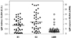
The evaluation panel for CHIKV was purchased from Laboratoire Marcel Merieux (Lyon, France), consisting of 40 positive and 20 negative serum samples, based on the anti-CHIKV IgM antibody titer by IgM capture ELISA (cut off value, A450 = 0.15, Fig. 3). As a negative control, 20 normal serum samples were collected from healthy Koreans who have never traveled to endemic or epidemic areas of CHIKV or Dengue virus. To check the cross-reactivity with Dengue virus infection, twenty Dengue fever-positive serum samples were kindly provided from Arboviruses Laboratory, National Institute of Hygiene and Epidemiology, Hanoi, Vietnam.
Construction of baculovirus transfer vector containing CHIKV E1 and E2 envelope proteins
In order to clone the CHIKV envelope protein genes, E1 and E2, CHIKV (strain TSI-GSD-218) was propagated in C6/36 cells, and CHIKV genomic total RNA was extracted from CHIKV-infected C6/36 cells using an RNeasy mini kit (Qiagen Inc., Valencia, CA, USA). The cDNA synthesis and amplification of the envelope genes were performed using RT-PreMix (Bioneer Inc., Seoul, Korea). The CHIKV envelope protein genes, E1 and E2 were amplified using the following primers encoding appropriate restriction enzyme sites: E1 (sense, 5'-GGATCCCCGAACACGGTGGGAGTACCG-3'; antisense, 5'-AAGCTTTCCCGTGATCTTCTGCACCCAT-3') and E2 (sense, 5'-G GATCCCCATACTTAGCTCACTGTCCCG-3'; antisense, 5'-AAGCTTAGTCATAGTGGGGTACAGCTCA-3'). The PCR-amplified CHIKV envelope protein genes containing enzyme sites were ligated with pCR2.1 Topo vector (Invitrogen, Carlsbad, CA, USA), and then the CHIKV envelope protein genes were subcloned into the baculovirus vector pFastBac HT (Invitrogen, Carlsbad, CA, USA). The resulting plasmid pFast HT-CHIKV-E1/-E2 were used to generate recombinant viruses.
Generation of recombinant baculovirus
Recombinant viruses were generated as described.16,17 Briefly, Sf900 II SFM cells (Sf9, Invitrogen, Carlsbad, CA, USA) were transfected with the recombinant transfer vector pFastBac HT-CHIKV-E1/-E2 to generate recombinant baculovirus. As a control, pFastBac HT-CAT vector was transfected into cells. Liposome-mediated gene transfer was employed with Cellfectin (Invitrogen, Carlsbad, CA, USA). A few plaques were picked, and recombinant viruses were purified by plaque assay and confirmed by PCR amplification and Western blot analysis.
Expression analysis and immunoblotting
Sf9 cells were infected with recombinant baculoviruses at a multiplicity of infection of 5 : 1 PFU: cell and incubated at 27℃. Three days post-infection, cells were harvested and whole-cell lysates were analyzed by a discontinuous sodium dedecyl sulfate polyacrylamide gel electrophoresis (SDS-PAGE) system, and the gels were stained with Coomassie blue. Proteins resolved by SDS-PAGE were electrophoretically transferred onto a nitrocellulose membrane for Western blot analysis.18 The membrane was incubated in PBS containing 2% skim milk and then reacted with 1 : 100 dilution of CHIKV IgM positive serum, which was prepared by pooling 40 CHIKV IgM positive sera. After 1 hour, the membrane was washed several times and subsequently treated with horseradish peroxidase (HRP) conjugated goat anti-human IgM antibodies (Santa Cruz Biotechnology, Santa Cruz, CA, USA) at a 1 : 2,000 dilution for 1 hour at room temperature. Protein binding was detected using the Amersham Biosciences ImmunoBlot System (Amersham Pharmacia Biotech, Stockholm, Sweden).
Affinity purification of the recombinant CHIKV envelope proteins
A high-yield, homogenous preparation of CHIKV envelope proteins, E1 and E2, were obtained by using the nikel-nitrilotriacetic acid (Ni-NTA) resin, according to the standard procedures described by the manufacturer (Clonteck, Mountain View, CA, USA). Briefly, recombinant baculovirus-infected Sf9 cell lysates were pelleted, and the supernatants were added to the equilibrated Ni-NTA agarose in a 1 : 10 volume ratio. The bead slurry was then washed with 10 volumes of 50 mM sodium phosphate, 300 mM NaCl, 10% glycerol, and 20 mM imidazole (pH 8.0). The CHIKV envelope proteins were then eluted with 300 or 500 mM imidazole in 50 mM sodium phosphate, 300 mM NaCl, and 10% glycerol (pH 6.0).
Indircet anti-CHIKV IgM ELISA
Enzyme-linked immunosorbent assays (ELISA) were performed to evaluate the reactivity of recombinant CHIKV envelope proteins, E1 and E2 to anti-CHIKV IgM antibodies as described before.20,21 Ninety-six well enzyme immunoassay (EIA) plates (Costar, Cambridge, MA, USA) were coated overnight with serial dilutions of recombinant CHIKV envelope proteins, E1 or E2 (1, 2, 4, 8 µg/mL) in a volume 100 µL of polycarbonate buffer (pH 9.2) at 4℃. Plates were then washed and blocked with PBS (pH 7.4) containing 1% BSA for 1 hour at 37℃. Serum samples were diluted in PBS containing 1% BSA (1 : 100) and were then added to the wells and incubator for 1 hour at 37℃. The bound antibodies were detected with horseradish peroxidase-conjugated mouse anti-human IgM antibody (1 : 3,000 dilution, Standard Diagnostics, Yongin, Korea). The plates were washed at least four times with PBS containing 0.05% Tween 20 between each step. An enzyme substrate TMB (Sigma Chemical Co., St Louis, MO, USA) was added to the wells and incubated for 30 min. The reaction was stopped by adding 1.5 N H2SO4, and the absorbance was read at 450 nm using an automatic ELISA reader (Molecular Devices, Biotek Instruments, Hyland Park, VA, USA). The absorbance ratio of anti-CHIKV positive to negative serum to the recombinant envelope protein (P/N ratio) was calculated as follows; A450 of positive serum/A450 of negative serum.
RESULTS
Cloning the CHIKV envelope protein genes, E1 and E2, and construction of recombinant baculovirus
The CHIKV envelope protein genes, E1 and E2, were amplified and the amplified envelope genes were ligated with pCR 2.1 Topo vector PCR. The sequences of CHIKV envelope protein genes, E1 and E2, were confirmed (data not shown). The each envelope protein gene was subcloned into the baculovirus vector pFastBac HT from pCR 2.1 Topo vector after enzyme digestion.
Expression of CHIKV envelope proteins in Sf9 cell cultures
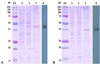
The CHIKV envelope protein gene constructs (pFastBac HT-CHIKV-E1/-E2) were expressed in Sf9 cells. In order to validate the expression of CHIKV envelope proteins, E1 and E2, infected cells were harvested 24, 48, and 72 hours post-infection, whole-cell lysates were analyzed by SDS-PAGE, and the gels were stained with Coomassie blue (Fig. 1). The expression level of recombinant CHIKV envelope proteins peaked at 72 hours post-infection (data not shown). CHIKV E1 and E2 envelope protein with molecular mass of 44 kDa and 40 kDa, respectively, were expressed in Sf9 cells infected with recombinant baculovirus (Fig. 1). These corresponding protein bands of CHIKV E1 and E2 envelope proteins were observed in the cell lysates infected with each recombinant baculovirus and in the purified proteins infected (lanes 2 and 3 in Fig. 1). The each corresponding protein was not present in mock-infected cells (lane 1 in Fig. 1). The identity of the recombinant CHIKV envelope proteins, E1 and E2, was confirmed by Western blot analysis using pooled anti-CHIKV positive serum (lane 4 in Fig. 1).
These results demonstrate that the expression and purification of CHIKV envelope proteins, E1 and E2, was successfully done via the baculovirus expression system.
Titration of recombinant CHIKV envelope protein, E1 and E2, for detection of anti-CHIKV IgM antibodies by indirect ELISA
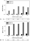
The recombinant baculovirus-expressed CHIKV envelope protein, E1 and E2, were titrated to detect anti-CHIKV IgM antibodies using indirect ELISA. In order to determine the optimal concentration of recombinant CHIKV envelope proteins, each recombinant envelope protein was serially diluted from 0.5 to 8.0 µg/mL (from 50 to 800 ng/well, respectively), and CHIKV envelope proteins, E1 and E2-specific IgM antibodies, were detected using pooled anti-CHIKV positive or negative serum samples. As shown in Fig. 2, as little as 50 ng/well (0.5 µg/mL) of recombinant E1 or E2 protein produced an ELISA signal for pooled anti-CHIKV positive serum. The highest ratio of absorbance value of CHIKV positive to negative serum (P/N ratio) for recombinant E1 protein was 20 at the level of 200 ng/well (2.0 µg/mL) of E1 envelope protein (Fig. 2A). Interestingly, the highest P/N ration for recombinant CHIKV E2 envelope protein was 62.5 at the level of 100 ng/well (1.0 µg/mL) of E2 envelope protein (Fig. 2B), which was considered to be an optimal concentration of the recombinant E1 and E2 envelope protein for evaluation of CHIKV serum samples, because absorbance value was high at the level of 100 ng/well (1.0 µg/mL) of E1 envelope protein even though P/N ratio of E1 protein was the highest, 200 ng/well. In the following experiments, therefore, this quantity of recombinant protein was used.
Evaluation of the recombinant CHIKV envelope protein E1 and E2 using indirect IgM capture ELISA
The recombinant CHIKV envelope proteins, E1 and E2, were evaluated for the detection of anti-CHIKV IgM antibodies using ELISA. Sixty anti-CHIKV serum samples (40 positive and 20 negative) from Laboratoire Marcel Merieux (LMM, Lyon, France) were used to evaluate the recombinant CHIKV envelope proteins as a diagnostic reagent for CHIKV infection. To check the cross-reactivity of the CHIKV envelope proteins with Dengue virus infection, twenty anti-Dengue virus positive serum samples were also included. As a negative control of CHIKV and Dengue virus infection, twenty normal serum samples from healthy Koreans were included.
As shown in Fig. 3, the mean absorbance values for forty anti-CHIKV positive serum samples were approximately 0.88 and 1.23 for the recombinant CHIKV envelope proteins E1 and E2, respectively. The absorbance values of anti-CHIKV positive serums for CHIKV E2 envelope protein were significantly higher than those for CHIKV E1 envelope protein (p < 0.001), and there was significant correlation between absorbance values of anti-CHIKV IgM positive serum samples for recombinant CHIKV E1 and E2 envelope protein (r = 0.8328, p < 0.001). However, there was no significant correlation between the absorbance values of 40 anti-CHIKV IgM positive serum samples for recombinant CHIKV E1 or E2, and absorbance values of 40 anti-CHIKV IgM positive serum samples from Laboratoire Marcel Merieux (Lyon, France).
The reactivity of the recombinant CHIKV E1 and E2 envelope proteins was very low (A450 < 0.2) for twenty anti-CHIKV negative serum samples and twenty normal control serum samples derived from healthy Koreans. The cross-reactivity of the recombinant CHIKV E1 and E2 envelope proteins was very low for all twenty Dengue virus samples (A450 < 0.2).
The sensitivities of the recombinant CHIKV E1 and E2 envelope proteins for anti-CHIKV positive serum samples were 77.5% and 90%, respectively (based on the cut off values, A450 = 0.2) (Table 1). The specificities of the recombinant CHIKV E1 and E2 envelope proteins for anti-CHIKV negative serum samples were 100% (Table 1).
These results indicate that the recombinant CHIKV E1 and E2 envelope proteins were strongly reactive towards anti-CHIKV IgM antibodies, and showed a low cross-reactivity with anti-Dengue virus IgM antibodies.
DISCUSSION
CHIKV and Dengue virus clinically cause similar febrile disease, but the prognosis of Dengue virus infection is more severe. The outbreaks by these viruses infection occur often within the similar geographical area and same vectors, most of which are Ae. aegypti. Thus, the laboratory confirmation between CHIKV and Dengue virus infection is essential in order to launch control measures when an epidemic or endemic outbreak occurs. Current serological laboratory diagnosis for CHIKV is based on HI test, ELIAS, and indirect immunofluorescence test. HI test is a simple and rapid test9,15,22 and ELISA is a popular serological method with high sensitivity and specificity.20,21,23 Currently, indirect immunofluorescence test has been developed as commercial kit.24 However, the whole virus antigens in the crude form were used as a diagnostic reagent for CHIKV in these immunological methods.
Therefore, in this study, CHIKV E1 and E2 envelope proteins were expressed in a baculovirus expression system as CHIKV-specific diagnostic reagents. The seroreactivity of CHIKV E1 and E2 envelope proteins was evaluated using anti-CHIKV positive and negative serum samples, and the cross-reactivity of CHIKV E1 and E2 envelope protein with Dengue virus was evaluated using anti-Dengue virus serum samples. The sensitivities of the recombinant CHIKV E1 and E2 envelope proteins for anti-CHIKV positive serum samples were 77.5% and 90%, respectively (based on the cutoff value A450 < 0.2). The specificities of the recombinant CHIKV E1 and E2 envelope proteins were 100% for anti-CHIKV negative serum samples. The sensitivity of CHIKV E2 envelope protein was higher than that of CHIKV E1 envelope protein, even though there was a highly significant correlation in the seroreactivity between CHIKV E1 and E2 envelope protein for anti-CHIKV positive serum samples (p < 0.001). This difference between CHIKV E1 and E2 envelope protein might be explained by the fact that CHIKV E1 envelope protein is buried almost in the virus structure,25 which may give CHIKV E1 envelope protein less chance to exposure to host immune system. The sensitivity and specificity of the recombinant CHIKV E2 envelope protein to detect anti-CHIKV IgM antibodies using indirect IgM ELISA were similar to the sensitivity (96.9%) and specificity (98.3%) using indirect immunofluorescence test.24 These results indicate that recombinant CHIKV E2 envelope protein is also immunogenic as whole virus antigens which was used as a serodiagnostic reagent in the indirect immunofluorescence test. The absorbance values of anti-CHIKV positive serum samples to CHIKV E1 and E2 envelope proteins appear to be higher than the absorbance values of anti-CHIKV positive serum samples from Laboratoire Marcel Merieux (Lyon, France), even though the direct comparison of these data is not possible because different ELISA methods were used. The cross-reactivity of the recombinant CHIKV E1 and E2 envelope proteins with twenty Dengue positive serum samples was not observed.
In the endemic or epidemic areas of CHIK fever, previous infection of CHIKV could induce seroreactivity with CHIKV antigens, because anti-CHIKV IgM antibodies persist more than one year. Further assessment using the serum samples which are derived from both previous and currently CHIKV-infected people is needed.
In next extensive studies, cross-reactivity of recombinant CHIKV E1 and E2 envelope proteins also needs to be evaluated with a closely related virus, o'nyong-nyong (ONN) virus, which is a member of the genus Alphavirus in the family Togaviridae,26 and cause a febrile symptom similar to CHIKV and Dengue virus infection, even though ONN virus is transmitted by anopheline mosquitoes, typically Anopheles (An.) funestus and An. Gambiae and there is a district phylogenetic difference between ONN virus and CHIKV.27
These results suggest that the recombinant CHIKV E1 and E2 envelope proteins have a potential as diagnostic reagents of serological diagnostic methods such as ELISA, immunochromatographic assay and indirect immunofluorescence because of high levels of sensitivity and specificity towards CHIK fever.

 XML Download