

 PDF
PDF ePub
ePub Citation
 Print
Print


INTRODUCTION
Angioleiomyomas are formed by proliferation of smooth muscle cells in the vascular wall and are usually subcutaneous or appear as skin nodules in the extremities. Angioleiomyomas developing in organs other than the skin are very rare. Mediastinal angioleiomyoma is very rare, and only 3 other cases have been reported to the best of our knowledge. We report a case of symptomatic angioleiomyoma in the left posterior mediastinum treated uneventfully with video-assisted thoracoscopic surgery.
CASE REPORT
A 66-year-old woman presented with left back and flank pain for 6 months. The pain was getting worse thereafter. She had a history of old pulmonary tuberculosis and hysterectomy 26 years ago for an unknown disease at an other hospital and slip-down on the left lower posterior chest wall 3 years ago.
Physical examination was unremarkable. There were no abnormalities noted on both biochemical and hematologic analyses of the patient's blood, except for high lactate dehydrogenase and high eosinophil fraction.
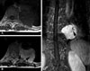
Preoperative frontal and lateral chest radiographs showed a well-circumscribed paraspinal mass with loss of descending thoracic aorta interface, suggesting posterior mediastinal mass. Contrast-enhanced CT scan showed a 4.3 × 5.2 cm mass with relatively homogeneous enhancement in the left paravertebral space. The MRIs are shown in Fig. 1. The mass had a slight extension into the left intervertebral neural foramen.
Considering imaging findings, we suspected a nerve sheath tumor arising from the intercostal or spinal nerve, and planned operation. The patient was positioned with right lateral decubitus. Three ports were introduced for 30° - 10 mm thoracoscope and instruments. The mass was adjacent but apart from intervertebral foramen and broad-based. It was located at the 9th intercostal space. Even though the mass was easy-bleeding, it was uneventfully removed through video-assisted thoracoscopic surgery.
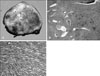
Pathologic examination revealed a well-demarcated tumor mass measuring 4.3 × 3.0 × 3.2 cm. The gross and microscopic findings are shown in Fig. 2. Most cells were spindle shaped and diffusely positive for alpha-smooth muscle actin and h-caldesmon but negative for CD34, S-100 protein, and HMB45. The pathologic diagnosis was angioleiomyoma.
She was discharged 7 days after operation without complications. Her chief complaint, left back and flank pain, disappeared after surgery.
DISCUSSION
Angioleiomyomas are formed by proliferation of smooth muscle cells in the vascular wall. Almost 90% of such tumors are subcutaneous or appear as skin nodules in the extremities, and 70% of extremity angioleiomyomas occur in the lower extremities.1 According to Duhig, 40% of angioleiomyomas appeared in married women between the age of 30 - 60 years.1 Angioleiomyomas developing in organs other than the skin including the liver and kidney have been reported but they are rare.2,3 Mediastinal angioleiomyoma is very rare, and only 3 cases have been reported to the best of our knowledge.4-6 Unlike with peripheral angioleiomyoma, 2 mediastinal angioleiomyomas were found in males and 2 in females including the present case.4-6
To rule out a dumbbell tumor for operative strategy, chest MRI was done. It was tentatively diagnosed as neurogenic tumor because of its close location to the sympathetic trunk and intercostal nerve, and extreme rarity of angioleiomyoma in the posterior mediastinum. Reviewing MRI retrospectively, however, unlike neurogenic tumor, signal void tubular vascular structures within the mass were seen on gadolium-enhanced coronal T1-WI (Fig. 1A). Previously, angioleiomyomas have been reported to have well-defined margins and show homogeneous or heterogeneous enhancement with or without signal void vascular structures on MRI.7 In the present case, MR report and pathologic correlation of angioleiomyoma indicated that interlacing iso-SI areas within the tumor correlated with various amounts of connective tissue and intravascular thrombus. Furthermore, the areas of high SI on T2-WI corresponded to smooth muscle component, and a well-defined peripheral hypo-intense area on T2-WI showed a fibrous capsule.
Regarding the growth of angioleiomyomas, a few theories have been advanced, including estrogen, traumatic, and congestive theory.1 In attempting to explain unusual gender and age distribution, Duhig hypothesized that minor trauma, injury, and irritation or inflammation on dilated vessel for stasis might well produce a mass composed of muscle, collagen, and many vascular channels, especially under the influence of estrogenic hormone. To further support this hypothesis, the close morphologic relationship between angioleiomyomas and dermal hemangimas has been mentioned. Many of the hemangiomas seen in the skin in older patients represent simple reactions to injury.1
In the present case, the trauma 3 years ago seemed to be responsible for the growth of the mass. Compression of the intercostal nerve by the mass seemed to have caused left flank and back pain because the pain disappeared immediately after operation.
Even though it is rare, angioleiomyoma seems to be on the list of differential diagnosis of posterior mediastinal tumor, especially in middle-aged females with trauma history.
 XML Download