

 PDF
PDF ePub
ePub Citation
 Print
Print


INTRODUCTION
Pulmonary sequestration is a rare clinical entity, occurring in fewer than 2% of congenital abnormalities. It has no normal continuity with lung tracheobronchial elements, and arterial blood supply is from the systemic circulation. Thoracic and abdominal aortas are the most common origins for abnormal nutrient arteries. Arterial supply from a coronary artery is extremely rare. We describe a case of pulmonary sequestration with right coronary artery blood supply.
CASE REPORT
A 65-year old female was referred for coronary angiography. She complained of 2-week history of squeezing chest pain at rest. Her previous history was negative for cardiovascular disease. At admission, her blood pressure was 135/80 mmHg; physical examination, laboratory results, electrocardiography, and echocardiography were normal.
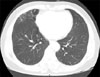
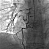
A chest X-ray revealed abnormal shadow in the right lower lobe. Computed tomography of the lesion revealed cystic changes (Fig. 1). Coronary angiography demonstrated normal-caliber coronary arteries without atherosclerosis. A large abnormal artery arose from the proximal right coronary artery, traversed the mediastinum, and fed an area of the right lower lung (Fig. 2); venous drainage was directed into the right lower pulmonary vein. Pulmonary angiography was not performed. The patient was medically managed with a calcium channel blocker and nitrate. She had no further chest pain after discharge.
DISCUSSION
Pulmonary sequestration is an uncommon, often congenital, anomaly that affects both children and adults. It is thought to account for 0.15 to 6.4% of all congenital pulmonary anomalies and is usually located in the lower lobe of the lung.1-3 The sequestrated segment consists of embryonic lung tissue that has no identifiable tracheobronchial communications and receives systemic blood supply. The blood supply derives most commonly from the thoracic or abdominal aorta, but arteries occasionally arise from other sites including the celiac, splenic, intercostal, subclavian, or renal arteries. Pulmonary sequestration supplied by a coronary artery is extremely rare.2-10
Sequestrations are further subdivided into intralobar and extralobar types. Intralobar sequestrations are more common and share the surrounding lung tissue pleura. These sequestrations almost always occur at the left lower lobes. Associated congenital abnormalities are uncommon.
Extralobar types are rare and encapsulated within a distinct pleural envelope separate from the normal lung. Associated congenital anomalies are present in 50% of cases.
Several complications have been reported, including recurrent pulmonary infections, hemoptysis, and heart failure from persistent left-to-right shunting.3,11 The natural history of sequestration supplied by a coronary artery remains unknown. Therefore, in the absence of complications, observation may be an option. Surgical resection is recommended for recurrent pulmonary infections or coronary steal.4
In a patient with sequestration, suspicion of coronary origin is important and requires preoperative angiography before surgical ligation of the feeding artery. Injury or proximal ligation may produce myocardial ischemia, infarction, or death. Although this entity is extremely rare, knowledge of it can prevent inappropriate management and fatal results.
 XML Download