

 PDF
PDF ePub
ePub Citation
 Print
Print


INTRODUCTION
Hemophagocytic lymphohistiocytosis (HLH) has been described in association with a number of viral diseases, including human immunodeficiency virus (HIV) infection.1-3 Most of the HLH cases diagnosed in HIV patients have been identified in patients with advanced stages of HIV infection and were often found in association with concomitant infections or malignant disease. These patients have a reported 21% recovery rate, which suggests that HIV patients with HLH have a poor prognosis.
Acute HIV infection is defined as the period from the initial HIV infection to complete seroconversion. More than 90% of acute infections go undiagnosed and only 50% of acute HIV cases are symptomatic.4 This infection period is associated with high levels of viremia, and the clinical manifestations are variable. HLH caused by acute HIV infection alone has not been reported very frequently. Here, we describe the first case of HLH during the acute stage of HIV infection in a Korean patient. The patient recovered spontaneously without any immunomodulating therapy.
CASE REPORT
A 44-year-old heterosexual man visited our hospital after experiencing 3 weeks of fever, chills, sore throat, generalized malaise, myalgia, and anorexia. This patient had no medical history, except for syphilis, which had been treated 10 years previously. The patient had not been an intravenous drug user and denied having any risk behaviors for HIV infection, except for one incident of sexual intercourse with a man 1 month before presentation. Upon physical examination, the patient appeared acutely ill and had a temperature of 38.7℃, a respiratory rate of 16 breaths/min, and a pulse of 66 beats/min. The patient also had icteric sclera, enlarged tonsils coated with purulent exudates, and multiple, pea-sized, non-tender lymph nodes involving the superficial and deep cervical nodes bilaterally. A non-pruritic morbilliform erythematous rash was present on the trunk and extremities.
Laboratory values were determined as follows: WBC count; 3,900 cells/µL with 24% neutrophils and 50% lymphocytes (4,800 - 10,800 cells/µL; reference values in parentheses), hemoglobin; 13.6 g/dL (12 - 18 g/dL), hematocrit; 40% (37 - 52%), platelet count; 90 × 103/µL (130 - 450 × 103/µL), alanine aminotransferase; 869 U/L (< 37 U/L), aspartate aminotransferase; 494 U/L (< 37 U/L), lactate dehydrogenase; 8,400 U/L (< 37 U/L), alkaline phosphatase; 1,147 U/L (32 - 117 U/L), total bilirubin; 6.6 mg/dL (< 0.6 mg/dL), direct bilirubin; 6.2 mg/dL (< 0.3 mg/dL), total protein; 6.6 g/dL (6 - 8.3 g/dL), albumin; 3.0 g/dL (3.5 - 5 g/dL), total cholesterol; 146 mg/dL (< 200 mg/dL), triglycerides; 480 mg/dL (< 200 mg/dL), LDL-cholesterol; 23 mg/dL (< 120 mg/dL), HDL-cholesterol; 11.6 mg/dL (40 - 80 mg/dL), ferritin; > 30,000 ng/mL (7 - 323 ng/mL), ESR 11; mm/h (< 20 mm/h), CRP; 0.63 mg/dL (< 1 mg/dL), prothrombin time; 10.4 s (< 11.5 s), aPTT; 54.7 s (26 - 41 s), FDP; 20 µg/mL (< 5 µg/mL), D-dimer; 1.7 mg/L (< 0.3 mg/L), and fibrinogen; 178 mg/ dL (200 - 485 mg/dL).
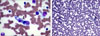
The patient's chest X-ray was unremarkable with no identified focus of infection. Multiple, deep, homogenous enhancements of the lymph nodes were noted in the neck by computed tomography (CT). The abdominal ultrasound and CT demonstrated mild hepatomegaly and acalculous gallbladder thickening with a normal bile duct. A fine needle aspiration biopsy of a cervical lymph node was compatible with a diagnosis of necrotizing lymphadenitis. A bone marrow aspiration and biopsy were also performed. Pathological examination of the bone marrow revealed normocellularity with histiocyte infiltration and hemophagocytosis, but no evidence of malignant cells (Fig. 1).
Blood, throat swab, lymph node, and bone marrow cultures were negative for bacteria, mycobacteria, and fungi. Serological tests were negative for causative pathogens, including immunoglobulin M (IgM) to viral capsid antigens and antibodies of the early antigen of the Epstein-Barr virus, IgM to cytomegalovirus, Herpes simplex virus, measles, rubella, Toxoplasma gondii, hepatitis A virus, the core antigen of the hepatitis B virus (anti-HBc IgM), and antibody to hepatitis C virus. A rheumatological workup was negative for antinuclear antibodies and anti-neutrophil cytoplasmic antibodies. However, the patient's serum was HIV positive when tested by the enzyme-linked immunosorbent assay (ELISA) for HIV. An initial Western blot for HIV revealed antibodies to HIV protein, p24, and gp160 only. Such results were regarded as indeterminate.
The patient was treated with supportive care and all of the aberrant clinical features and abnormal laboratory findings improved gradually. A second Western blot was repeated two weeks after the initial Western blot and indicated clearly positive results with the antibodies to HIV protein, p24, and gp120/gp160. At that time, the patient's peripheral blood CD4 cell count was 157/mm3 (normal 360 - 1,725/mm3) and the serum viral load exceeded 1 × 10⁹ copies/mL.
We diagnosed the patient with acute HIV syndrome presenting with HLH. The patient promptly received highly active antiretroviral therapy (HAART) with zidovudine, lamivudine, and indinavir. No HLH recurrences have occurred since then and the patient has remained well and free of any opportunistic infections for 3 years.
DISCUSSION
Hemophagocytic lymphohistiocytosis is a rare disease resulting from the abnormal proliferation of histiocytes in tissues and organs. According to the HLH-2004 protocol, the diagnosis of HLH is established if five out of eight diagnostic criteria are met. These criteria include five initial criteria [i) fever, ii) splenomegaly, iii) bicytopenia (with at least two of the following: hemoglobin ≤ 9 g/dL, platelets < 100 × 103/µL, and neutrophils < 1.0 × 10³/µL in the peripheral blood), iv) hypertriglyceridemia (≥ 265 mg/dL) or hypofibrinogenemia (≤ 150 mg/dL), and v) hemophagocytosis in the bone marrow, spleen, or lymph nodes without evidence of malignancy] and three new criteria [vi) low or absent NK-cell activity, vii) hyperferritinemia (≥ 500 µg/L), and viii) increased soluble plasma CD25-levels (IL-2Rα chain; ≥ 2,400 U/mL)].5 Our patient had fever, bicytopenia, hypertriglyceridemia, and hyperferritinemia, suggesting a diagnosis of HLH. The presence of hemophagocytosis was subsequently confirmed from bone marrow specimens. Our patient had no significant splenomegaly. NK-cell activity and CD25 levels were not assessed in this patient.
Of the two HLH forms, primary/familial and secondary/reactive, reactive HLH develops secondary to a variety of viral, bacterial, and parasitic infections, as well as to malignancy, various autoimmune diseases, and drug use. To determine the etiology of HLH in our patient, we performed imaging studies and a variety of cultures and serological tests for a number of pathogens and rheumatological markers. Despite an exhaustive workup, the HLH could not be attributed to any associated disorder except for the HIV infection. In this patient, the initial Western blot for HIV did not confirm HIV infection, while a follow-up Western blot 2 weeks later was clearly positive.
Hemophagocytic lymphohistiocytosis has been diagnosed with increasing frequency in patients infected with HIV.2,3 Hemophagocytosis is commonly found, to varying degrees, in the bone marrow of HIV-infected individuals without an underlying diagnosis of HLH, which raises the possibility of a subclinical form of HLH.6 An autopsy study of 56 patients with acquired immunodeficiency syndrome (AIDS) found an approximate 20% prevalence of hemophagocytosis.7 In fact, most of the reported cases of HLH have been diagnosed during the advanced stages of HIV infection, when the patients also suffer from opportunistic infections and malignancies. Recent reports have suggested that HLH is a manifestation of acute HIV infection.8 Although rare, eight cases of HLH during the acute phase of HIV infection have been reported. Our case further confirms that HLH occurs during the seroconversion stage of acute HIV infection.
A recovery rate of 21% (6 out of 28) has been observed in patients with advanced HIV and concomitant HLH with opportunistic infections or malignancies.9 Nearly 50% of the deaths occurred within 1 month after the diagnosis of HLH. However, all nine cases of HLH that were associated with acute HIV infections recovered, including our patient.8,10 Of the eight reported cases with an HLH diagnosis, four patients received immunomodulating agents, such as intravenous immunoglobulin or steroids. However, the benefit of these agents for treating HLH associated with acute HIV infection remains unclear.8 The signs and symptoms of HLH in our patient resolved spontaneously without any immunomodulating therapy. This was similar to the findings of three other reported cases. These findings suggest that HLH associated with acute HIV infection has a favorable prognosis compared to when HLH occurs during the advanced stages of HIV infection.
Genetic defects in patients with primary HLH consist of mutations in the genes required for cytotoxic granule release or function. However, the reason why secondary HLH occurs in response to infectious organisms, malignancy, or autoimmune disease remains unclear.11 The common pathophysiological abnormality in HLH patients is cytokine dysfunction, which results in the uncontrolled accumulation of activated T-lymphocytes and histiocytes in many organs.12 Norris et al. reported that cytokine alteration occurred within 7 days of detectable HIV-1 viremia.13 Although the plasma cytokine levels were not measured in our patient, we surmise that such early cytokine alterations, from a burst of viremia associated with acute HIV infection, were responsible for the HLH in our patient. Even though a patient with advanced HIV infection and HLH, who did not respond to HAART, has also been described,14 Castilletti et al. showed that HAART was very effective in controlling both the acute HIV infection and HLH.10 Therefore, unlike in patients with advanced AIDS, the precipitous decline in plasma viremia during antibody seroconversion may explain the more favorable outcome in patients with acute HIV infection.
Since the recognition of the AIDS epidemic in 1981, the number of patients infected with HIV has increased dramatically worldwide. As the number of newly infected HIV patients has increased recently in Korea, so has the number of cases with acute HIV syndrome.15-17 This paper describes the first report of acute HIV syndrome presenting as virus-associated HLH in Korea. This patient recovered spontaneously, without any immunomodulating therapy. Therefore, acute HIV syndrome should be considered in the differential diagnosis of HLH, especially in patients with any HIV risk factors. In such cases, the appropriate diagnostic evaluation should be performed.
 XML Download