

 PDF
PDF ePub
ePub Citation
 Print
Print


INTRODUCTION
The development of cancer is a complex and multi-step process in which a series of progressive changes culminate in deregulation of all proliferation in nervous cells.1 Alterations of gene expression and several genetic aberrations have been shown to occur during malignant growth, transformation, and progression of glioblastoma.1,2 The glioblastoma is the most common malignant brain tumor in adults.3 Their distinct ability to invade the normal surrounding tissue makes them difficult to completely remove surgically, thus accounting for the extraordinarily high lethality associated with malignant gliomas.4 A variety of genetic alterations in human gliomas comprise signal transduction and cell cycle arrest control of cellular processes.
Current methods for comparing gene-expression profile changes in different tissues or different pathological specimens include mRNA differential display, serial analysis of gene expression (SAGE), cDNA microarray, large-scale cDNA sequencing, expressed sequence tag (EST) database comparison, two-dimensional gel electrophoresis of cellular proteins, subtractive library construction, representational difference analysis (RDA) and suppression subtractive hybridization (SSH).5-8 The mRNA differential display is complicated and labor-intensive, and both SAGE and cDNA array are limited for identifying unknown genes because they formerly require known genetic information. 2,9-11 The chip technology is highly efficient, but also requires formerly known genetic information and expensive equipment for making the analysis. Thus, chip technology is not yet widely available for the searching of unknown genes in glioblastoma study.12-14
Suppression subtractive hybridization (SSH) is an efficient and versatile PCR-based method of identifying rare, tumor-specific transcripts.2,11,13 Since the development of glioblastoma is a complex process involving a large number of genes, we believe that it is important to identify additional new genes, which might be involved in the formation of this tumor. To this end, we employed the SSH technique, which was originally designed to identify genomic differences between two complex genomes. SSH was later adapted to isolation of gene transcripts which are differentially expressed in one sample relative to another, allowing the detection of low-abundance transcripts in a sample. SSH is a powerful technique that enables researchers to compare two populations of mRNA and obtain clones of genes that are expressed in one population but not in the others.15-20
In this report, over-expressed genes involved in glioblastoma were identified by SSH, characterized by RT-PCR and sequenced. The result obtained from SSH screen was confirmed by RTPCR of non-neoplastic brain tissue, human glioma cell lines and other human cancer cell line. To know whether these genes are involved in the cell division, serum deprivation and stimulation were conducted.
MATERIALS AND METHODS
Tissue materials
Through June 2004 to September 2005, glioblastoma patients consented to use of their tissues. We investigated tissues from 15 patients in whom craniotomy and surgical removal were performed due to cerebral glioblastoma. Tumor and non-tumor brain tissues (tumor tissues; N = 15, non-tumor brain tissues; N = 15) were collected during the neurosurgical operations. The patients altogether consisted of 15 patients - 5 females and 10 males, aged 49 to 70 years. Mean age at diagnosis was 55.2 years ± 6.9 (SD). The histological diagnosis was made by two neuropathologists according to World Health Organization (WHO) classification of the astrocytic tumors. Among 15 pairs of tissues (non-tumor brain tissue and glioblastoma tissue), 10 pairs was used for identification of new glioblastoma specific genes and remaining 5 pairs for expression of identified genes.
RNA extraction and SMART™ PCR cDNA synthesis
Total RNA of the non-tumor brain tissue and glioblastoma tissue was prepared using the Trizol reagent according to manufacturer's instructions (Clontech, Mountain View, CA, USA). The concentration and quality of each RNA sample were measured for the synthesis of high quality cDNA by spectrophotometrical determination of a 260/ 280 ratio. About 1 g µ of total RNA samples was treated with DNase I at 37℃ for 15 min and at 65℃ for 10 min, to get rid of genomic DNA contamination. The RNA was dissolved in DEPCtreated water and total RNA was analyzed on 1.2% agarose gel. Total 1 µg RNA was used for SMART cDNA synthesis and was reverse-transcribed in 10 µL mixture with PowerScript™ Reverse transcriptase (BD Biosciences Clontech, Mountain View, CA, USA). The first-strand of cDNA was diluted to a final volume of 40 µL with 1 × TE buffer (10 mM Tris-HCl, pH 8.0, 1 nM EDTA). One µL of the diluted cDNA was used to generate cDNA by long distance PCR with BD Advantage 2 polymerase mix (BD Biosciences Clontech, CA, USA), using PCR primer II A according to the manufacturer' instructions. The final concentration of digested second strand cDNA was 300 µg/ µ.
PCR-based suppression subtraction hybridization (SSH)
SSH was performed with the PCR-Select cDNA Subtraction Kit according to the manufacturer's protocol. Driver ds cDNA was synthesized from 1 µg each of total RNA, using SMART™ PCR cDNA Synthesis Kit user Manual (Clontech, Mountain View, CA, USA). First- and second- strand cDNA synthesis and blunt-ending of DNA ends by T4 DNA polymerase were carried out according to the manufacturer' protocol. The second double-stranded cDNA was digested with 10 units/ul RsaI in a final volume of 50 µL at 37℃ for 3 hr. After extraction and precipitation of digested second strand cDNAs, the pellet was dissolved in 7 µL of sterile H2O, precipitate was washed in 80% ethanol and residual ethanol was evaporated after the supernatant was removed. The final concentration of driver was 300 ≈ng/µL. RsaI digested ds tester cDNA was prepared as described above for the driver. Digested tester cDNA (1 µL) was diluted in 5 µL of H2O. The diluted tester cDNA (2 µL) was then ligated to 2 µL of adaptor land adaptor 2R (10 µLM) in separate ligation reactions in a total volume of 10 µL at 16℃ overnight, using 400 units/µL of T4 DNA ligase in the buffer supplied from the manufacturer. After ligation, 1 µL of 20(EDTA/glycogen was added and the samples were heated at 72℃ for 5min to inactivate the ligase and stored at - 20℃.
cDNA hybridization was used with tester1-1 and tester1-2 that were, mixed with adaptor1 and adaptor 2R respectively. One and half µL of tester1-1 with adaptor1 and tester1-2 with adaptor 2R were, respectively, hybridized with 1.5 µL digested first stranded driver cDNA of brain tumor in 1 µL of 4 × hybridization buffer solution at 68℃for 8 hr. Tester1 - 2 hybridization sample was drawn into the pipette tip. Afterwards, 1 µL of denatured mixture from 1 µL of digested second stranded driver cDNA, 2 µL of H2O, and 1 µL of 4 × hybridization buffer solution at 98℃were drawn into pipette tip with a slight air space below the droplet of the above tester1-2 hybridization sample. The final hybridization was then diluted in 200 µL of dilution buffer (20 mM HEPES/50 mM NaCl/0.2 mM EDTA), heated at 68℃for 7 min and stored at - 20℃ For each subtraction, we performed two PCR amplifications. The primary PCR was conducted in 25 µL. It contained 1 µL of diluted, subtracted cDNA, 1 µL of PCR primer 1 (10 µM), and 23 µL of PCR master mixture prepared by using the 50 (Advantage cDNA polymerase PCR Kit (Clontech, Mountain View, CA, USA). PCR was performed with the following parameters: 75℃for 5 min; 30 cycle at 94℃for 25 sec, 94℃for 10 sec, 64℃for 30 sec, 72℃for 1.5 min. three µL of primary PCR mixture was diluted in 27 µL of water for the nested-PCR reaction for 11 cycle (94℃for 10 sec, 68℃for 30 sec, 72℃for 1.5 min) with the same reagent except the two nested primer1, 2R.
Transformation and sequencing of the subtracted cDNA
The subtracted cDNAs obtained after secondary PCR were cloned with a T/A Cloning Kit (Promega, Madison, WI, USA). The cDNAs were ligated into T/A vectors by incubating 3 µL of the secondary PCR amplification and 1 µL of the vector (50ng/µL) with 3Weiss units of T4 DNA ligase overnight at 4℃. Then, 5 µL of ligated product was transformed into 50 µL of competent JM109 cells for heat shock. Competent JM109 cells transformed by ligated product were grown on LB medium agar plates overnight at 37℃. White colonies containing inserts were obtained, following transformation of competent cells. White colonies were placed into LB medium and shaken overnight at 37℃. The plasmid DNA with inserted fragments was extracted, the extracted DNA was digested with EcoRI restriction enzyme, and the product was analyzed on 1.2% agarose gel. DNA sequencing was performed at Biotechnology Center Macrogen (Seoul, Korea). Nucleic acid homology was searched using the NCBI BLAST program (http://www.ncbi.nih.gov/BLAST).
RT-PCR
For expression of isolated seven genes' mRNA levels on glioblastoma tumor tissues and nontumor brain tissue, RT-PCR was performed using previous primers. mRNA of glioblastoma tissue sample and non-tumor brain tissue which were mentioned above samples was reverse transcribed to cDNA using Superscript™ First-Strand Synthesis (Invitrogen, Carlsbad, CA, USA). RT-PCR was performed on samples with 1 µg of total RNA. The reaction mixture contained 1 µL of SuperScript II reverse transcritase (50 units), 1 µL oligo dT (0.5 µg/µL), 2 µL 10X RT buffer, 2 µL of 0.1 M DTT, and 1 µL each of 10 mM dNTP. The reverse transcription was carried out for 50 min at 42℃. Semi-quantitative RT-PCR was used to assess amounts of mRNA in glioblastoma tissue sample and non-tumor brain tissue. PCR amplification was performed with gene specific primer, and 18srRNA was used as the reference gene using the TaKaRa Ex Taq™. Gene specific primers used in this study were designed from known sequences of the isolated genes, shown in Table 1. Amplification conditions were denaturation at 94℃for 5 min, followed by 35 cycles of denaturation at 94℃for 30 sec, annealing at 55℃for 30 sec and extension at 72℃for 30 sec. A final extension was done at 72℃for 10 min, and the reaction mixtures were then run for 30 min at 100V on 1% agarose/EtBr gel with ethidium bromide staining. Densitometric analyses were performed including standardization from the control (18srRNA) band.
RT-PCR analyses were performed to identify genes in five human glioma cell lines (SF188, SF539, SF126, U87, and U251) (Seoul National University Cancer Research Center, Seoul, Korea). The same methodology described above was used. And also, RT-PCR was also done in other human cancer cell lines; lung (WI-38), colon (CCD-18Co), prostate (RWPE-1), SV40-immortalized cell line (WI-38 VA13), lung carcinoma (NCI-H596), colon carcinoma (KM1214) and prostate carcinoma (DU145). These cell lines were purchased from ATCC (Manassas, VA, USA) and Nikken Cell line bank (Japan).
Serum deprivation and stimulation test
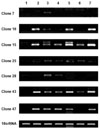
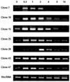
WI-38 (8PDL) cells were routinely maintained in Eagle's minimal essential medium (E-MEM) supplemented with 10% fetal bovine serum and, essential and non-essential amino acids. Cells were seeded at a density of 2 × 10⁶ cells in a 60-mm dish. The cultures were maintained at 37℃in a humidified 5% CO2 atmosphere. They were cultured at a density of 60% cells, washed with PBS, and medium supplemented with 1% FBS was finally added. The cells were washed with PBS after 48 h and supplemented with 10% fresh FBS medium. Cells were then harvested at 0, 0.5, 1, 2, 4, 6, and 16 hr after the 10% FBS treatment. To identify the expression, "clones 7, 10, 15, 25, 28, 43, and 47" mRNA levels were analyzed by RT-PCR. PCR was performed using the TaKaRa Ex Taq™ under the following conditions: first 3 min at 94℃, then 30 sec at 94℃, 30 sec at 55℃, 30 sec at 72℃ for 35cycle, and 10 min at 72℃. After PCR, each of the PCR products was electrophoretically resolved on 1.2% agarose gel and stained with ethidium bromide.
RESULTS
Identification of new over-expressed genes in glioblastoma using SSH
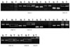
With this experimental design, 130 plasmid clones were obtained. The digested cDNA clones were searched for sequence homologies in the GenBank DNA database by BLAST. DNA sequencing results of the digested 130 clones were found to have high homology to 14 genes known in the public database of the 14 kinds (Table 2). We identified 21 unknown genes, which were not homologous to any of the known genes in the Genbank database (Table 3). Among these 21 genes, we selected 7 genes which were over- expressed in glioblastoma, compared to non-tumor brain tissue (Fig. 1). To be considered over-expressed in glioblastomas, the genes tested had to show a threefold greater expression in tumor samples than in non-tumor brain tissue by semiquantitative RT-PCR. In this study, we considered these genes as candidates for over-expressed genes in glioblastoma.
Different expression of genes in glioblastoma tissues and non-tumor brain tissue
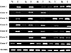
To examine whether these new genes were expressed specifically in glioblastoma tissues, the expression patterns of transcription were examined by RT-PCR analysis for remaining 5 resected pairs of glioblastoma tissues and non-tumor brain tissues. The expressions of clone 25 were found to be relatively over-expressed as compared with non-tumor tissues (Fig. 2). The remaining six clones were not clear.
Expression of genes in human glioma cell lines and other human cancer cell lines
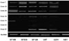
In comparison of five commercial glioma cell lines, the expression profiles of various cell lines could be categorized into two groups (Fig. 3): (1) genes that were over-expressed by all cell lines (clone 7, 25, 28), (2) genes that showed weak expression across the cell lines without any distinct pattern (clone 10, 15, 43, 47).
The expressions of clones 7 and 28 were not specific to other cancer cell lines, except it was weakly expressed in lung cancer cell line. As shown in Fig. 4, the mRNA expressions of clones 10, 15, 25, 43, and 47 were determined to be over- expressed in human lung cancer, prostate cancer cells compared with human non-cancer cell lines.
Serum deprivation and stimulation
As shown in Fig. 5, the time course experiment showed that WI-38 cells starved in serum-free medium did not show mRNA expression at 0 hr. After 10% FBS treatment, however, each clone was found to express the mRNA.
DISCUSSION
Identification of genes that are differentially expressed between brain tumor tissue and normal brain tissue is very important for understanding the molecular basis of these nervous tumors and for defining possible targets for therapeutic intervention.3,21 Human glioblastoma, as with many human cancers, is characterized by genetic and phenotypic heterogeneity despite a fairly uniform appearance by light microscopic findings.1,22 Our study was designed to identify genes with differential expression between glioblastomas cells and non-neoplastic brain cells.
To achieve our goal, we have used the SSH that is suited ideally for the identification of over- expressed genes. When we used the tester (glioblastoma tissue) and driver (non-tumor brain tissue) DNA fragments, the drives cDNA would eliminate the sequences common between the tester and driver cDNA samples during the first and second hybridization step. To determine the efficiency of the SSH, both the subtracted and unsubtracted cDNAs have to be amplified by PCR with GAPDH primers for different cycles: 18, 23, 28, and 33 cycle. The expression of GAPDH was observed in unsubtracted cDNA after 18 cycles and after 33 cycles in subtracted cDNA. In this result, the abundance of GAPDH decreased significantly after subtraction. It was important to confirm that individual clones indeed represent differentially expressed genes. In the present study, we isolated known genes as well as new genes that were over-expressed in glioblastoma tissue as compared to non-tumor brain tissue.
Over 130 libraries were screened after SSH, from which 21 differentially regulated clones were identified. The percentage of up-regulated clones was, therefore, approximately 16%. Of the 21 clones identified, 6 clones were multiple hits, and we selected 7 genes for further evaluation. (Table 3)
In most glioblastoma samples, the expression of mRNA of clone 25 was distinctly higher than that in non-tumor brain tissues (Fig. 2). On the whole, the expressions of six clones except clone 47 were relatively higher than non-tumor tissue. The uniform expression of control 18srRNA indicates equal template loading of the RNA from the tumor specimen.
The expression profiles of the 7 genes in five human glioma cell lines were categorized into two groups. In contrast to expressions of clones 10, 15, 43, and 47, those of clones 7, 25, and 28 were overexpressed by all the glioma cell lines. Especially, the clone 7 and 25 clearly showed over-expressions.
In various human cancer cell lines, the expressions of the clones 7, 25 and 28 were either weak or not detectable, however, those of clones 10, 15, 25, 28, 43, and 47 were relatively over-expressed in other cancer cell lines. Four out of 7 genes were up-regulated by immortalization in lung fibroblasts. Those expressions in immortalized fibroblasts might suggest that the expressions of RT-PCR were due to proliferating state of dividing cells, but not due to quiescent cells. On the other hand, the rest of the genes were not up-regulated in immortalized fibroblasts. The weak expressions of the clones 7 and 28 were observed compared to normal lung line, but expression of these genes in glioma cell line was strong. It is highly likely that some genes of specific cancer would be involved also in other cancer, because of multiplicity of oncogene. In cancer cells, their expression is due to particular genes and shared with other genes in different degrees by in other cancers.
Clones 7 and 28, which were not or weakly expressed in the other cancer cell lines, were expressed in the glioma cell line suggesting that these genes may be related to glioblastoma. Clones 10, 15, 28, 43, and 47 were expressed non-specifically in human glioma cell line and expressed variously in other human cancer cell lines, so these clones can represent genes that are related in a proliferate state regardless of tumor phenotype. In serum deprivation and stimulation, it is said that theses seven genes are related to cell division. But, the expressions of 5 out of 7 genes (clone 10, 15, 25, 43, 47) were increased by serum deprivation and stimulation, indicating some uncertainty of our experiments: It might have been influenced by culture medium condition. Clone 7 needs to be further functional studied.
Our present study is preliminary to derive any definite conclusion. We have cloned one or two over-expressed genes. Its significance and roles are unclear. Further studies are needed to characterize functional significance and roles of these genes in glioblastoma tumorigenesis and progression.
 XML Download