

 PDF
PDF ePub
ePub Citation
 Print
Print


INTRODUCTION
Hypoxia-inducible factor-1 (HIF-1) primarily mediates the hypoxic response. Since discovered in 1992, more than 60 genes have been found to be induced by HIF-1.1 These genes are related with cell survival, cell proliferation, apoptosis and angiogenesis.2 The human HIF-1 is composed of two subunits: HIF-1α and HIF-1β. HIF-1α is undetectable in normoxia condition, but HIF-1β is found under oxygen condition. HIF-1α is located on chromosome 14 (14q21-4) and HIF-1β is on chromosome 1 (1q21). HIF-1α is very labile in normoxia with a half-life of less than 5 minutes. The rapid degradation of HIF-1α is blocked under hypoxic condition.3 Therefore, hypoxia increases the levels of HIF-1α in cells. HIF-1α induction by various stimuli contributes to the cell proliferation and survival.
To investigate the effect of HIF-1α, we used RNA interference technology using small interfering RNA (siRNA). siRNA is one of the latest methods of sequence-specific gene silence. It is a time-effective means to manipulate gene expression and to probe gene functions.4 It was discovered in 1998 and used to silence gene expression. siRNA can chemically be synthesized or expressed from vectors transcribing short double stranded hairpin- like RNAs that are processed into siRNAs inside the cells.5
Therefore, when HIF-1α gene was blocked with HIF-1α-siRNA, we expected the increase of cell apoptosis and sensitivity to chemotherapeutic drug. In the present study, we performed in vitro and in vivo experiments to clarify the effect of HIF-1α on tumor growth using siRNAs and a chemotherapeutic drug.
MATERIALS AND METHODS
Cell culture
MIA PaCa-2 cells were grown in Dulbecco's modified Eagle's Medium (DMEM, Mediatech, Herndon, VA, USA), supplemented with 4 mM glutamine, 1 mM Na-pyruvate, and 10% fetal bovine serum (FBS), at 37℃ in a humidified atmosphere containing 5% CO2. The cells were plated (1.5 × 10⁵ cells/well, 95% confluent) in 6-well plates, one for immunoblotting and one for stable cell line. The following day, confluent cells were transfected by using the lipofectamine™ 2000 transfection reagent (Invitrogen, Carlsbad, CA, USA) following the manufacturer's instructions for siRNA of control and HIF-1α. After 48 hours of incubation, one each well of transfected cells was harvested for immunoblotting. Other each well of transfected cells was selected in DMEM with the addition of G418 sulfate (Geneticin; Mediatech, Herndon, VA, USA). The selection was continued for 3 weeks with 2 mg/mL geneticin.
siRNA preparation
The HIF-1α mRNA specific RNA oligonucleotides with 3'-TTTTT overhangs were constructed. The corresponding RNA oligonucleotides, and the forward and reverse chains of each gene were chemically synthesized. We cloned siRNA via splicing pSilencer 2.1-U6 neo vector plasmid (Ambion Inc., Austin, TX, USA).
Immunoblotting
Cells of one well were washed with ice-cold PBS and were resolved in a mixture of 50 mM HEPES, 50 mM NaCl, 5 mM EDTA, 10 mM Na2P2O7·10H2O, 50 mM NaF, 1 mM NaVO4, 1% Triton X-100, and protease and phosphatase inhibitors (Complete mini, Roche Molecular Biomedical, Indianapolis, IN, USA). The cell lysates were incubated on ice for 30 minutes, centrifuged for 15 minutes at 3000 rpm and 4℃, and the clear solution was sonificated for 15 seconds and then kept frozen (-79℃) until use. Protein concentrations of the resulting solutions were determined using Bio-Rad protein assay kit. The extracts were boiled with sample buffer, and then resolved on SDS-PAGE gels and then transferred to PVDF membrane. After blocking in phosphate saline buffer containing a mixture of 0.2% Tween and 5% dried milk, the blot was probed with a suitable antibody for HIF-1α (BD Biosciences, San Jose, CA, USA). After probing with second antibody, ECL Plus western blotting detection (Amersham Biosciences, Piscataway, NJ, USA) was performed for detection by chemiluminescence and chemifluorescence.
Cell proliferation assay
We used MTS ([3-(4,5-dimethylthiazol-2-yl)-5-(3-carboxymethoxyphenyl)-2-(4-sulfophenyl)-2H-tetr azolium, inner salt]) for cell proliferation assay and crystal violet assay to detect the effect of drug sensitivity. Each stable cell line was plated in two 96 wells plates. And then, we treated the cells with two concentrations (127 µmol/L and 254 µmol/L) of gemcitabine (Eli Lilly Co., Indianapolis, IN, USA). And then one plate was placed under hypoxic condition for 2 days. For hypoxia, cells were incubated at 37℃ in 5% mol/mol carbon dioxide, 1% mol/mol oxygen, and 94% nitrogen atmosphere, a MTS assay were then performed using Celltiter96 aqueous one solution® (Promega Co., Madison, WI, USA) following the manufacturer's instructions. Then, crystal violet assay was performed to confirm MTS assay.
Caspase-3 assay
To examine the basal caspase-3 activities, the cell pellets from each cell line at 80% confluence were collected and lysed with the lysis buffer containing 10 mM Tris-HCl (pH 7.5), 10 mM NaH2PO4/NaHPO4 (pH 7.5), 130 mM NaCl, 1% Triton X-100, and 10 mM Na2P2O7 10 H2O. Caspase activity in the cell lysates was examined according to a standard protocol (Bio-Rad Laboratories, Hercules, CA, USA) using substrates specific for caspase-3-like (Ac-DEVD-AFC), which detects activity of caspase-3. The intensities were measured by a fluorescence microplate reader (Bioteck FL600 Fluorometer, Winooski, VT, USA). RFUs were standardized to RFU/µg or RFU/10 µg protein. For each experiment, control groups with specific caspase inhibitors, including caspase-3 inhibitor (Z-DEVD-CHO; BD PharMingen, San Diego, CA, USA), were included to ensure specificity of the assay.
Animal experiment
Female athymic NCR nu/nu mice (weighing 20 to 23 g; National cancer institute, Frederick, MD, USA) were acclimatized for 2 weeks in a sterile environment. Each cell line (2.5 × 10⁶ cell/mice) was subcutaneously injected into tissue of the back. Both cell lines were injected into same mouse; control siRNA transfected cells into the left back and HIF-1α siRNA transfected cells into the right back. The tumor cells were allowed to grow for 1 week. And mice were randomly assigned to either treatment or the control group. Five mice for each group were assigned. Treatment of mice with gemcitabine was carried out for 21 days by 2 days interval of intraperitoneal injection (120 mg/kg). A mice were daily inspected, and tumor size was measured. Fifty days after tumor injection, the mice were sacrificed for collection of tumors. The collected tumors were frozen and stained with HIF-1α for immunofluorescence. And the remnant tumors were blotted after extraction of proteins. All authors were authorized from Division of Animal Resource, Emory University and this animal experiment was also performed under the permission of Division of Animal Resource, Emory University. Same experiment was performed twice.
RESULT
Down regulation of HIF-1α expressions by siRNA
The expressions of HIF-1α were determined by Western blot analysis. As shown in Fig. 1, HIF-1α siRNA decreased the expression of HIF-1α. The effect of siRNA was amplified in hypoxic conditions.
Differences in inhibition of cell growth by chemotherapeutic drug
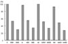
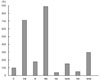
Cell proliferation assay of control and HIF-1α siRNA transfected MIA paca-2 cell lines treated with two concentrations of gemicitabine showed that the growth of HIF-1α siRNA transfected MIA paca-2 cell line was more susceptible to gemicitabine than that of control siRNA transfected MIA paca-2 cell lines. (p = 0.047, z = 1.667) (Fig. 2) We also confirmed the result by crystal violet assay. Caspase-3 assay revealed that caspase-3 activity was more increased in HIF-1α siRNA transfected MIA paca-2 cell line than in control siRNA transfected MIA paca-2 cell lines. And similar results were also obtained in gemicitabine and hypoxia groups (Fig. 3).
Differences in tumor growth of mice experiment
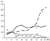
Tumor growth of control and HIF-1α siRNA transfected MIA paca-2 cell lines showed that the growth of HIF-1α siRNA transfected MIA paca-2 cell line was slower than that of control siRNA transfected MIA paca-2 cell lines. Also, tumor growth of control and HIF-1α siRNA transfected MIA paca-2 cell lines treated with gemicitabine showed that the growth of HIF-1α siRNA transfected MIA paca-2 cell line was more susceptible to gemicitabine than that of control siRNA transfected MIA paca-2 cell lines (p = 0.044, z = 1.825)(Fig. 4).
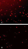
Finally immunofluorescence staining of resected tumor from mice confirmed that the HIF-1α siRNA activity was still effective in mice (Fig. 5).
DISCUSSION
Pancreas cancer remains as an aggressive neoplasm, and survival of patients is still measured in months. It is responsible for almost 6% of all cancer-related deaths.6 Currently, 5-FU and gemicitabine are the main agent of chemotherapy. Gemicitabine is a deoxycytidine analogue with structural and metabolic similarities to cytarabine. Because the reported response rate is less than 30% and the toxic effects may be significant,7 we attempted to find increase the chemosensitivity and the effect of gemicitabine.
HIF-1 primarily mediates the hypoxic response. Since its discovery, more than 60 genes have been found to be induced by HIF-1.1 These genes are associated with cell survival, cell proliferation, energy metabolism, apoptosis and angiogenesis.2 The human HIF-1 is composed of two subunits: HIF-1α and HIF-1β. HIF-1α is undetectable in normoxia condition, but HIF-1β is found under oxygen condition and persistantly expressed. HIF-1α is located on chromosome 14 (14q21-4) whereas HIF-1β is on chromosome 1 (1q21). HIF-1α is very labile in normoxia with a half-life of less than 5 minutes. The rapid degradation of HIF-1α is blocked in hypoxic condition. Therefore, hypoxia increases the levels of HIF-1α in cells. HIF-1α induction by various stimuli contributes to cell proliferation and survival. Therefore, HIF-1 is a pivotal transcriptional factor in the cellular response to hypoxia. In malignant glioma, overexpression of HIF-1α induced by hypoxia is essential event in the activation of glioma cell motility through alteration of invasion-related molecules such as metallloproteinase -2 or 9.8 Also HIF-1 is activated by non-hypoxic stimuli through activation of diacylglycerol-sensitive protein kinase-C pathway.9,10 On the other hand, activation of HIF-1 by non-hypoxic activator seems to be a cell-type specific response. Induction of HIF-1 by growth factors (insulin-like growth factor-2, epidermal growth factor) contributes to autocrine solicitation of proliferation and survival.1 Therefore, the downregulation of HIF-1 in cancer cells could have important effects on tumor growth. Many inhibitors of HIF-1 activity are currently under clinical trials.1
In this study, we tried to knock-down HIF-1α by using siRNA technique, since siRNAs are one of the most recent molecules used to silence gene expression and currently widely used as nucleic acid-based sequence-specific gene silencing molecules.11 siRNAs found in nature are derived from cytoplasmic processing of dsRNA by the RNase-III-type enzyme termed dicer. siRNAs can also be chemically synthesized or expressed by vectors transcribing short double-stranded hairpin-like RNAs that are processed into siRNAs inside the cell.5,12 The HIF-1α siRNA used in the present study was chemically synthesized with RNA oligonucleotides and cloned with splicing vector plasmid.13 Although they did not completely knock-out, we successfully made a new cell lines and confirmed its effectiveness was confirmed by many immunoblotting assays. We confirmed that knock-out of HIF-1α decreased the tumor growth and increased the response rate of gemicitabine in vitro and in vivo studies.
Several cellular steps are required to activate HIF-1, including accumulation of HIF-1α protein, nuclear translocation, formation of HIF-1 complex with HIF-1β protein, and binding to DNA. Among these 4 steps, the accumulation of HIF-1α protein in hypoxia is considered to be a key step for the transcriptional activation of HIF-1.14 Introduction of HIF-1α targeted siRNA results in downregulation of HIF-1α. Therefore, the inhibition of tumor growth results from the decrease of cellular responses to HIF-1α.8
The presence of hypoxic regions in solid tumors is a common phenomenon and results in dramatic alterations in gene expression.15 And hypoxic condition may be more similar to in tumor-in-situ than normoxia. Therefore, we explored whether a hypoxia environment could affect HiF-1α expression and tumor growth. In this study, the effect of HIF-1α siRNA was found to be more remarkable than normoxia under the hypoxic condition.
The next research project should be how to deliver the siRNA to tumor-in-situ like 2ME2, inhibitor of HIF-1 activity.1 Furthermore, the methodology of knock-out of HIF-1α using siRNA can be used in other neoplasms.
Although the gene expression of HIF-1α may be confirmed at protein and RNA levels, the suppression of HIF-1α by the use of siRNA technique results in decrease of cancer cell proliferation and increase of chemo-sensitivity. Consequently, targeting the HIF-1α to inhibit tumor growth may be useful in treatment of some pancreas neoplasms.

 XML Download