

 PDF
PDF ePub
ePub Citation
 Print
Print


INTRODUCTION
Microsatellites are short tandem repeats of DNA bases scattered throughout the genome, and vary in size from tens to hundreds of bases. Microsatellite regions are at particular risk of mutation such as the insertion or deletion of repeating units during DNA replication; this change in length is called MSI.1 In normal tissues, the mismatch repair (MMR) system identifies and excises these abnormalities that occur during DNA replication. Therefore, a defect in the MMR system, including the proteins hMLH1, hMLH3, hMSH2, hMSH3, hMSH6, hPMS1, and hPMS2, results in MSI.2
Because defects in DNA mismatch repair are related mainly to inherited mutations, MSI occurs in about 90% of hereditary non-polyposis colorectal cancer (HNPCC).3 However, MSI has also been observed in a variety of sporadic cancers, including those of the colon, endometrium, pancreas, and bladder.4-7 The potential significance of MSI in some tumors has been emphasized, and MSI has been reported to play roles in tumor diagnosis, prognosis, response to treatment, and tumor biology.8
The incidence of MSI depends on which loci are investigated, because of many microsatellite regions within the human genome. To overcome this confusion, the National Cancer Institute (NCI) introduced guidelines defining MSI in colorectal cancer.8 A panel of five microsatellite loci consisting of two mononucleotide repeats (Bat25 and Bat26) and three dinucleotide repeats (D2S123, D5S346, and D17S250) was established. When these criteria were applied to ovarian carcinoma, the reported frequency of high MSI was 11 - 14%.9,10 Using additional MSI markers, the frequency of MSI in ovarian carcinoma varies from 6 to 37%.10-15 Given these variable results, the prevalence of MSI in ovarian tumors is not yet clear. Therefore, we evaluated the occurrence of MSI in 21 borderline and 25 malignant ovarian tumors using both the five standard MSI markers and nine new MSI markers and determined clinical significance of MSI in sporadic epithelial ovarian tumors.
MATERIALS AND METHODS
Patients and tissue samples
Paired tumor and normal samples were retrieved from 46 patients with ovarian tumors who had been treated surgically at the Department of Obstetrics and Gynecology, Yonsei University Medical Center in Korea. The tumor samples included 21 borderline and 25 malignant ovarian tumors. None of the patients had a personal or family history of ovarian cancer or HNPCC syndrome. The tumors were classified according to the World Health Organization criteria and staged using the International Federation of Gynecology and Obstetrics (FIGO) staging system. Of the 46 ovarian tumors, 10 were serous, 26 mucinous, and 10 endometrioid. Of the 25 malignant ovarian tumors, 9, 3, and 13 were stages I, II, and III, respectively.
DNA isolation and polymerase chain reaction (PCR)
The tumor tissues were snap-frozen in liquid nitrogen and stored at -70℃. Genomic DNA was extracted from the frozen tumor tissues using a DNA extraction kit according to the manufacturer's guidelines. To isolate normal DNA, paraffin-embedded and formalin-fixed blocks were used. Microdissection was performed on some sections of the surgically resected gynecologic organs to confirm the normal tissues. Normal DNA was extracted from normal tissues using a kit according to the manufacturer's guidelines.
Tumor and normal DNA at 14 microsatellite markers were amplified by PCR. The markers used for the investigation included the National Cancer Institute Consensus Panel (BAT25, BAT26, D17S250, D2S123, and D5S346) and 9 other markers: the dinucleotides NME1, D3S1611, D10S197, and D11S904; the trinucleotide androgen receptor (AR); and the tetranucleotides D13S175, DxS981, DXS6800, and DXS6807. PCR was performed in a total volume of 5 µL, containing 0.5 µL of DNA template, 0.475 µL of each primer, and 2.5 µL of PCR MasterMix solution. PCR was carried out in a thermal cycler, beginning with a 15-min denaturing step at 95℃, followed by 30 cycles of 94℃ for 45 s, 55℃ for 45 s, and 72℃ for 45 s, with a final 10-min extension at 72℃.
Microsatellite analysis
After PCR, 0.5 µL of each product was added to 0.5 µL of GS500 size marker and 10 µL of Hi-Di formamide. After 2 min at 95℃, the mixture was immediately immersed in an ice bath. The amplified fragments were separated by denaturing gel electrophoresis using an ABI PRISM 310 Genetic Analyzer (Applied Biosystems, Foster City, CA). The data were analyzed using GeneScan software (Applied Biosystems).
Microsatellite instability was defined as the presence of extra or shifted bands in comparison to the normal control. The tissues were classified as high MSI (MSI-H) if instability was noted in ≥ 30% of the loci investigated, low MSI (MSI-L) if instability was observed in < 30% of the loci investigated, and stable tumors (MSS) if there was no instability.
RESULTS
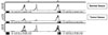
DNA samples from 46 tumors and matched normal samples were analyzed for MSI (Fig. 1). MSI was detected at the BAT25, BAT26, D2S123, NME1, D13S175, and DXS981 loci.
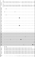
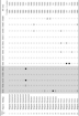
Applying the NCI consensus definition of MSI to the sporadic ovarian tumors, MSI was found in 4 of 46 (8.6%) ovarian tumors, including 2 of 21 (9.5%) borderline ovarian tumors (Table 1) and 2 of 25 (8%) malignant ovarian tumors (Table 2). The MSI loci were BAT25 and BAT26. Whereas all the borderline tumors with MSI were MSI-L tumors, one of the two malignant ovarian tumors with MSI was MSI-H.
When 9 more MSI markers were used to detect MSI in sporadic ovarian tumors, MSI was seen in 7 of 46 (15.2%) ovarian tumors, including 3 of 21 (14.3%) borderline ovarian tumors (Table 1) and 4 of 25 (16%) malignant ovarian tumors (Table 2). However, there was no MSI-H in any of these 7 cases.
To determine clinical significance of MSI in sporadic ovarian tumors, we evaluated the difference in MSI expression according to the histological type and stage. MSI was found in 3 of 10 (30%) serous tumors, 3 of 26 (11%) mucinous tumors, and 1 of 10 (10%) endometrioid tumors. The incidence of MSI was higher in the serous type, although the difference was not statistically significant. According to the stage of malignant ovarian tumors, MSI was observed in 1 of 9 (11%) cases of stage I disease, none of 3 cases of stage II, and 3 of 13 (23%) cases of stage III disease. There was no significant difference in MSI according to the stage of the malignant ovarian tumor.
DISCUSSION
MSI is caused by mutations in the mismatch repair genes. MSI has been implicated in the pathogenesis of colon, endometrial, and gastric carcinomas that occur in the setting of HNPCC syndrome, and also in a subset of sporadic cancers such as upper urinary tract, stomach, colon, and endometrial carcinomas.16,17
In ovarian cancers, the reported incidence of MSI ranges from 0 to 37%, depending on the number and type of markers. Sood et al. first reported the incidence of MSI in ovarian cancer using the NCI criteria.9 Based on the NCI markers, 18% of tumors demonstrated MSI, of which 11% were MSI-H and the remaining 7% were MSI-L. By contrast, we found that only 4 of 46 (8.6%) ovarian tumors had MSI. Similar to our results, Alvi et al. observed MSI in 6.9% of sporadic ovarian cancer by applying the NCI criteria,18 whereas Gras et al. observed MSI in 3.8%,19 suggesting that the NCI standard-based MSI is infrequent in ovarian cancer.
When additional MSI markers such as mono-, di-, and tetranucleotides were added to the standard NCI markers, we found a slight increase in the rate of MSI. Sood et al. also reported an increase in the rate of MSI from 18% to 36% when additional MSI markers were added.9 In other cancer types, increased microsatellite changes at selected tetranucleotides have been reported to be important and are especially frequent in skin (75%), head and neck (56%), lung (51%), and bladder (21 - 40%) cancers.20-23 However, the observed increase of MSI with the additional MSI markers that we used was too weak to decide the usefulness of the additional MSI markers. In addition, the MSI-H observed by using the 5 standard MSI markers was downgraded to MSI-L when all 14 MSI markers were used, suggesting that additional MSI markers might not help detect MSI in sporadic ovarian tumors.
To determine the clinical significance of MSI in ovarian tumors, we evaluated the correlation between MSI-positive tumors and some clinicopathological factors. There was no statistically significant difference in the MSI rates according to histological type or stage. The MSI rate was higher in serous tumors than in endometrioid or mucinous tumors, although it was not statistically significant. Sood et al. also failed to find clinical significance of MSI in ovarian cancers.9 On the other hand, some investigators insist that MSI is more prevalent in the endometrioid type because ovarian endometrioid carcinoma is morphologically and biologically similar to the endometrial endometrioid carcinoma and is the most prevalent type of ovarian carcinoma in patients with HNPCC.19 Nevertheless, Singer et al. suggested that serous ovarian carcinoma had an intermediate frequency of MSI,24 and Tangir et al. postulated that MSI is an important pathogenic mechanism in the development of serous tumors with low malignant potential.13
The lack of MSI in this study could possibly be due to the small number of samples. In addition, the variation in the apparent MSI frequency might have been due to the choice of microsatellites analyzed, analytical methodology, tissue preservation technique, and variable histology of ovarian cancer.
In conclusion, microsatellite instability was infrequent in both borderline and malignant ovarian tumors. Despite increasing the number of MSI markers, MSI was found to be not common in sporadic ovarian tumors. The clinical significance of MSI appears to be weak in patients with sporadic ovarian tumors. Therefore, further study of molecular events that are correlated with ovarian tumors is needed.
 XML Download