

 PDF
PDF ePub
ePub Citation
 Print
Print


INTRODUCTION
PDA is an important problem in preterm infants, particularly in VLBWI, and is with significantly associated with high mortality and morbidity, including bronchopulmonary dysplasia (BPD), retinopathy of prematurity (ROP), intraventricular hemorrhage (IVH), and necrotizing enterocolitis (NEC).1-7 PDA can be closed either pharmacologically with indomethacin or surgically. Indeed, surgical PDA ligation is usually considered after failure or contraindication of indomethacin treatment. Several studies have examined the appropriate time for surgery, influence of indomethacin, or adequate indications of surgery in VLBWI. Some studies reported that early surgical ligation was beneficial compared to delayed surgery,4-6,8,9 and other studies have reported that more courses of indomethacin before surgical ligation leads to increased morbidity.6 However, the optimal timing of PDA ligation in VLBWI remains controversial, and the factors affecting the ultimate outcome of surgical closure are unclear.
Another issue of PDA ligation is the location of the operation. Because of the risks of transferring unstable babies to an operating room, surgery in the NICU bedside is preferred nowadays.3,7,10,11
PDA ligation in our NICU was first performed in 1994. This study evaluated the final outcomes after PDA ligation, such as morbidity, mortality and factors that might affect the final outcomes of surgical ligation and demonstrated the safety of PDA ligation in the NICU.
MATERIALS AND METHODS
Medical records of 94 VLBWI weighing < 1,500 g with PDA who underwent PDA ligation in the NICU at Samsung Medical Center (SMC) from October 1994 to July 2006 were reviewed retrospectively. In all cases, PDA was confirmed using 2-dimensional echocardiography.
Mortality and morbidity of the infants were investigated as the ultimate outcomes. Factors affecting the final outcome were evaluated by dividing the patients into 3 groups according to mortality and major morbidities as follows: (1) the Mo group, infants who expired; (2) Mb group, those who had 1 or more morbidities, including moderate to severe BPD, high grade of ROP, high stage of NEC, and high grade of IVH; and (3) NM group, those who survived without major morbidity.
Factors that might affect the final outcomes were identified by comparing the following variables between the 3 groups: gestational age, birth weight, age and weight at surgery, incidence of RDS, number of courses of indomethacin, perioperative ventilatory parameters and vital signs, the use of dopamine, surgery-related complications, duration of ventilation and total days of oxygen support, and the length of hospital stay. The ventilatory parameters, including the fraction of inspired oxygen (FiO2) and mean airway pressure (MAP) as well as the vital signs, including body temperature, heart rate, and blood pressure, were assessed before the onset of surgery and 48 hours after surgery. Major morbidity was defined as follows: high grade IVH was ≥ grade III; high stage ROP requiring laser therapy; high grade NEC was ≥ grade III; and moderate to severe BPD was defined as an oxygen dependency at the corrected age of 36 weeks according to Walsh et al.12
Surgical ligation of PDA in the NICU
The surgical team performed the ligation on critically ill VLBWI with PDA in the NICU. The experienced surgical team consisted of pediatric cardiothoracic surgeons, circulating nurses, scrub nurses, and a pediatric anesthesiologist. Infants were moved on their overhead radiant warmer next to their incubators for surgery and were then checked for continuous monitoring, including oxygen saturation, heart rate, and blood pressure. General anesthesia was induced with fentanyl, and vecuronium provided curarisation. Infants were positioned in the right lateral decubitus position and all ligations were performed through a left thoracotomy via the 4th or 5th intercostal space intrapleurally. The parietal pleura on the aorta and PDA was divided, and the ductus arteriosus was mobilized and ligated using a 1/0 silk suture with 3 knots or a hemoclip.
Neonatologists remained in continuous attendance of the infant from before surgery until its completion and adequate postoperative stabilization. Anyone entering the NICU were required to wear proper operating attire, including caps, surgical gowns, shoes, and masks.
RESULTS
Outcome of surgery
The postoperative mortality was 1% at 7 days, 13% at 30 days, and 20% at hospital discharge. One (1%) infant who underwent surgery died 3 days later due to underlying sepsis before surgery. There were 20 (21%) final deaths, and the median postnatal age of death was 70 ± 69 days (ranging from 9 to 235 days). The causes of final death were as follows: sepsis (n = 13, 65%), severe BPD and Cor pulmonale (n = 3, 15%), high-grade intracranial hemorrhage (n = 2, 10%), high stage NEC (n = 1, 5%), and ARF (n = 1, 5%). No mortality was directly related to the surgery.
Forty-three (46%) infants had major morbidity. Among those who had more than 1 major morbidity, BPD occurred in 29 (67%) infants. Thirty-six (84%) infants had high stage ROP, 18 (19%) had high grade IVH, and 4 (4%) had high grade NEC. Thirty-one (33%) infants survived to discharge without any major morbidity.
There was no significant difference in the duration of ventilation between the 3 groups (Mb, 50 ± 12 days vs Mo, 50 ± 22 days vs NM, 36 ± 18 days). However, in the Mb group, the number of oxygen support days (Mb, 82 ± 16 days; Mo, 73 ± 70 days; and NM, 59 ± 22 days), and hospital days (Mb, 100 ± 23 days; Mo, 73 ± 70 days; and NM, 62 ± 19 days) was significantly longer than in the Mo and NM groups (p < 0.05).
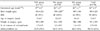
Factors affecting the outcome (Table 1)
In the Mo group, birth weight was significantly lower than in the NM and Mb groups (p < 0.05). However, there was no significant difference in gestational age, weight and age at surgery, incidence of RDS, and number of courses of indomethacin between the 3 groups.
Indications of surgery (Table 2)
Table 2 shows the indications of surgery. There was no significant difference in the indications for surgery between the 3 groups.
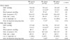
Perioperative conditions (Table 3)
Table 2 shows the ventilatory parameters and vital signs before the onset of surgery and 48 hours after surgery. The preoperative mean FiO2 and mean dopamine dose were significantly higher in the Mo group than in the Mb and NM groups (p < 0.05). In the Mo group, the mean FiO2 and mean dopamine dose 48 hours after surgery were still higher than in the NM group (p < 0.05). Other variables, including MAP, body temperature, blood pressure, heart rate, and number of infants using dopamine, were similar in the 3 groups.
Surgery-related complications
Table 4 shows surgery-related complications. There was no significant difference in the occurrence of surgical complications between the 3 groups. There was minimal or no bleeding within 24 hours postoperatively requiring a blood transfusion, no hypothermia, and no dislodgement of the E-tube and catheter. There were no other surgical complications, such as postoperative wound infections, NEC, phrenic nerve palsy, vocal cord paresis and chylothorax.
All patients received follow-up echocardiography after surgery and there was no residual PDA in any patient.
DISCUSSION
The present results showed low mortality rates of 1% at 7 days, 13% at 30 days, and 20% at hospital discharge after surgical PDA ligation. No mortality was related directly to the procedure but was attributed to the underlying prematurity. This mortality rate is similar to that reported in previous studies,6,13 which showed that PDA ligation performed in the NICU is safe and feasible even in VLBWI.
Because indomethacin inhibits the synthesis of all prostaglandins, a wide variety of adverse effects can be expected, including NEC, intestinal perforation, renal failure, and thrombocytopenia. VLBWI are more susceptible to these complications, and 40% are unlikely to respond to indomethacin therapy.1,5,7,9,14,15 Palder et al.5 reported a higher failure rate of indomethacin in infants < 750 g in weight, suggesting that direct ligation might be the preferable treatment. Little et al.15 revealed that infants weighing < 1,000 g were less likely to respond to indomethacin than infants weighing > 1,000 g. Rudd et al.16 reported that 87% of infants weighing < 1000g at birth were initially closed with indomethacin but 47% of these reopened. Therefore, surgical ligation might be considered in VLBWI who have either a lower probability of PDA closure with indomethacin or a higher risk of developing complications from indomethacin. However, indications, optimal time of surgery, and factors affecting the outcomes of surgical ligation in VLBWI are unclear.
As discussed above, in order to determine the factors affecting the final outcomes after surgery, the infants were divided into 3 groups according to their mortality and major morbidities, and several variables that were expected to affect the outcome of PDA ligation were compared. In the 3 groups, there was no significant difference in gestational age, weight and age at surgery, number of courses of indomethacin, perioperative MAP and vital sign, including blood pressure, pulse rate, and body temperature. This suggests that these factors can lead to a similar final outcome after surgery. However, in the mortality group, birth weight was significantly lower than in the no morbidity and morbidity group but the preoperative and postoperative mean FiO2 and mean dopamine dose was significantly higher than in the NM group. This means that the severity of the preoperative conditions related to underlying prematurity is the most important factor that affect the final outcomes of PDA ligation in VLBWI.
Overall, there was a significant difference in the total oxygen time and hospital days between the 3 groups because in the mortality group, some patients expired due to their underlying severe preoperative condition. In this condition, oxygen support time and hospital days were shorter. Moreover, in the Mb group, infants had major morbidities that required prolonged additive therapy, particularly, BPD, which required prolonged oxygenation support.
It was recently suggested that an early surgical ligation of PDA was preferable for VLBWI. Jaillard et al.8 reported that the ventilatory requirement was lower and nutritional outcome was better when early surgical closure was performed. Lee et al.6 demonstrated delayed surgery and increasing respiratory morbidity if 2 or more courses of indomethacin had been given. But, in the present study, no significant difference was observed in the surgery time and the courses of indomethacin, and there was no significant difference in the surgery time between the 3 groups. Therefore, surgery time may not affect the outcome of PDA ligation.
It was concluded that PDA ligation can be considered at any time in VLBWI when indomethacin either failed to close or was contraindicated because the factors related to surgery, including surgery time, number of courses of indomethacin, preoperative vital signs, and postoperative complications, may not significantly affect the outcomes.
As the design of our study is retrospective, it does not allow us to rule out the comorbidity of indomethacin. The prospective study in the future will be necessary to clarify the above notion.
There are several reports demonstrating that the surgical ligation of PDA in VLBWI operated in the NICU is safe, with a risk for infection similar to the operation room,11 also reducing the risks inherent in patient transport.3 The aim of ligation in the NICU is to eliminate the risks encountered while transferring the most fragile infants to the operating room. The transferal of a sick neonate may be associated with significant morbidity, such as unstable hemodynamics, hypothermia, disruption of the vascular lines, and dislodgement of the endotracheal tube. Furthermore, there may be delays because an operating room may be unavailable when required. In contrast, the advantages of performing surgery at the NICU include elimination of the aforementioned risks involved in transferring the patient and, throughout surgery, the infant can be cared for by the same nurses and doctors who are familiar with the infant, vascular access, and ventilator requirements. Furthermore, vital treatments, such as inhaled nitric oxide, which may have to be discontinued while transferring the neonate to the operating room, can be continued at the NICU without disruption.3,7,10,11 Although an anesthetic machine and gaseous anesthetic agents are generally unavailable in the NICU, the narcotics relaxant technique using intravenous fentanyl used at ligation at NICU is an effective anesthesia for neontates.5 Shew et al.17 reported that the whole-body protein breakdown, protein turnover and skeletal muscle protein breakdown rates improved significantly after surgery with fentanyl in low birth weight infants who had a minimal metabolic reservoir and previously experienced a loss of protein while enduring the physiologic stress of a PDA.
The disadvantage of performing surgery in the NICU includes limited lighting and accessibility, and beds in the ICU are not adjustable for surgery. In addition, the degree of asepsis achieved in the NICU is less effective than in an operating room due to higher levels of traffic in the ICU and lower airflow per minute in the ICU. However, the incidence of complications in several previous reports was quite low despite the marked prematurity of the patients,1,4,11,18 and were not serious complications related to surgery.
This study provides useful data on the outcomes of VLBWI undergoing PDA ligation in the NICU. The retrospective design represents a limitation and warrants future prospective design.
In conclusion, the factors affecting the outcome of surgery in VLBWI are the preoperative conditions related to the underlying prematurity, including birth weight, preoperative FiO2, and mean dopamine dose. In addition, PDA ligation performed in the NICU is a safe and feasible and is not associated with significant complications, even in VLBWI. Therefore, surgical ligation of PDA can be considered at any time for the treatment of VLBWI who have either a lower probability of PDA closure with indomethacin or an increased risk of developing complications from indomethacin.


 XML Download