

 PDF
PDF ePub
ePub Citation
 Print
Print


INTRODUCTION
Catheter ablation has played an increasingly important role in the management of atrial tachycardia and has evolved as first-line therapy in many experienced institutes. This procedure is relatively safe with few complications. However, atrial tachycardia, with its focus near the atrioventricular node (AVN) or near the His bundle, may bear a potential risk of atrioventricular block during the catheter ablation procedure.1-4 In this case, the earliest activation of atrial tachycardia was observed in the non-coronary aortic sinus. After failed radiofrequency energy applications at right His-bundle region, the complete elimination of atrial tachycardia was achieved with an radiofrequency energy application in the non-coronary aortic sinus. This report emphasizes the importance of mapping in the non-coronary aortic sinus in focal atrial tachycardia near the AVN or near the His-bundle.
CASE REPORT
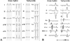
A 55-year-old woman was admitted to our center with recurrent episodes of disabling episodes of palpitation and dizziness for 6 months. The clinical tachycardia presented with abrupt onset and offset and could be terminated by 5 mg of verapamil. She had undergone a left modified radical mastectomy for breast cancer 21 years prior. The patient had been diagnosed with hypertension and diabetes mellitus at that time. A chest radiograph revealed no evidence of cardiomegaly. Echocardiography revealed no evidence of structural heart disease with a normal left ventricular function. The 12-lead electrocardiogram (ECG) at the time of admission demonstrated regular sinus rhythm with a narrow QRS complex and a normal axis (Fig. 1A, left panel). An ECG recorded during an episode of palpitations revealed a narrow QRS complex tachycardia with a cycle length of 480 ms. The P wave polarity preceding the QRS complex was negative in leads V4-6, biphasic (initially negative and a late positive component) in leads V1 and V3, and undetermined in the limb leads (Fig. 1A, right panel).
After giving informed consent, an electrophysiological study was performed without sedation. Three catheters were introduced to the right atrium, the right ventricular apex, and at the His-bundle region via the femoral veins. Also, a 7-Fr multipolar catheter was advanced within the coronary sinus via the left subclavian vein. During initial ventricular pacing, the ventriculoatrial conduction was absent. A tachycardia was induced and terminated reproducibly with incremental atrial pacing and extrastimulation. During the tachycardia, the local electrogram recorded at the His bundle preceded the onset of the surface P wave by 40 ms and the proximal coronary sinus by 15 ms (Fig. 1B).
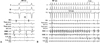
Ten radiofrequency applications were delivered near the His-bundle region where the atrial activation during the tachycardia recorded from the distal electrodes of the ablation catheter, preceded the onset of the surface P wave by 45 ms. The tachycardia was terminated and junctional beats occurred frequently and AV block was observed transiently during applications of radiofrequency energy. However, the tachycardia was induced and sustained after radiofrequency energy deliveries at these sites. Mapping in the left atrium was performed to find the earliest atrial activation site with retrograde transaortic approach. The earliest left atrial activation was located in the anteroseptal region. The earliest left atrial activation was later than that in the right atrium by 5 ms. No His potential was found at this site (Fig. 2A).
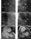
To find the other earliest atrial activation site, attempts were made to map from the aortic sinus of Valsalva. Mapping was performed using a 7 Fr quadripolar catheter with a 4-mm distal electrode (Blazer II, Boston Scientific, Natick, MA, USA). With detailed mapping at the aortic sinus of Valsalva using a retrograde transaortic approach, the earliest atrial activation was found within the non-coronary sinus of Valsalva where the local electrogram during the tachycardia recorded from the distal electrodes of the ablation catheter preceded the onset of the surface P wave by 55 ms (Fig. 2A). After confirming the location of the ostia of both coronary arteries and identifying the contour of the aortic sinus of Valsalva by an injection of a contrast agent, (Figs. 3A, B, C and D), an radiofrequency energy application delivered at that site using a maximum power of 30 W and maximum electrode to tissue interface temperature of 50℃ terminated the tachycardia 5.8 seconds after initiating the application (Fig. 2B). No Junctional beats occurred. No tachycardias were inducible after the catheter ablation. With incremental atrial pacing, the paced cycle length producing AVN Wenckebach block was 280 ms, which was the same as before the ablation procedure. A cardiac MRI was performed to determine the anatomic relationship with the other structures (Figs. 3E and F). The patient was discharged with no need for medications, and she has done well with no recurrence of the tachycardia during a 10-month follow-up.
DISCUSSION
This is a rare case of atrial tachycardia originating from the non-coronary aortic sinus which shows the benefit of mapping in non-coronary aortic cusp in atrial tachycardia near the AVN. The prevalence of atrial tachycardia near the AVN has varied, ranging from 0 - 13% in all patients with atrial tachycardia.1-4 According to the experience from Hamburg, a non-coronary aortic sinus origin was found only in 4.1% with focal atrial tachycardia.6 Atrial tachycardia near AVN may bear a potential risk of atrioventricular block during the catheter ablation procedure. However, elimination of atrial tachycardia was achieved in non-coronary aortic cusp avoiding a potential risk of atrioventricular block.5,6
In this case, VA conduction was absent. The tachycardia was initiated and terminated by incremental atrial pacing and extrastimuli. The tachycardia was reproducibly terminated by a small dose of verapamil without the development of AVN conduction block. On the basis of these findings, the tachycardia mechanism was determined to be due to either micro-re-entry or triggered activity. During the tachycardia, the local electrograms recorded from the non-coronary sinus of Valsalva and near the His-bundle region were earlier than those at other sites. The clinical and electrophysiological characteristics of this focal atrial tachycardia were similar to those described in the patients with focal atrial tachycardia originating from the non-coronary aortic sinus of Valsalva described by Tada et al.5 and Ouyang et al.6
In this case, the radiofrequency energy application near the His-bundle region did not succeed to eliminate tachycardia even with junctional rhythm and transient AV block during the radiofrequency energy application. Finally, the tachycardia was terminated within 5.8 minutes by an radiofrequency energy application in the non-coronary sinus of Valsalva where the earliest atrial activation was recorded. This finding suggests that this tachycardia might have originated closer to the non-coronary sinus of Valsalva than to the His-bundle region in the right atrial anteroseptal region. Of note, no His potential recordings were observed at the successful radiofrequency site. This information may explain why no prolongation of the PR interval or junctional beats occurred during the radiofrequency delivery in this case. Spatially, the aortic root occupies a central location within heart, with the non-coronary sinus of Valsalva anterior and superior to the paraseptal region of the left and right atria close to the superior atrioventricular junction.5 The MRI image 2 days after the ablation in this patient clearly shows that the rightward (anterior) margin of the non-coronary sinus of Valsalva is related to the paraseptal region of the right atrial wall (Figs. 3E and F). There was no damage in aortic valve in MRI and echocardiography after the ablation.
This case strongly suggests that, in patients with focal atrial tachycardia near the AVN or near the His-bundle region, mapping in the non-coronary aortic sinus of Valsalva can improve clinical outcome. This maneuver may avoid any aggressive radiofrequency energy deliveries near the His bundle, with the potential consequence of AV block. Therefore, if the tachycardia mapping were to demonstrate the earliest atrial activation at this region, especially in the patients with a previously failed ablation, delivery through the non-coronary sinus of Valsalva might be safer than a right-sided approach.
 XML Download