Abstract
Purpose
The varied morphology of the umbilical ring and its surrounding structures, such as the ligamentum teres hepatis, and the median and medial umbilical ligaments, has not been thoroughly investigated. Hence, this study was undertaken to clarify the morphologic variations of these structures.
Materials and Methods
The anterior abdominal walls were removed en bloc from 57 adult cadavers and dissected under a surgical microscope.
Results
One case of umbilical hernia was observed, and the remaining 56 umbilical rings were classified into 3 types: oval or round in 33 cases (Type A, 59.0%), obliterated or slitted in 12 cases (Type B, 21.4%), and completely covered by a connecting band between the ligamentum teres hepatis and umbilical ligaments in 11 cases (Type C, 19.6%). The median and medial umbilical ligaments were classified into four types based on their interrelationships. The most common type was the median umbilical ligament terminated by joining one or both medial umbilical ligaments (Type II, 41.1%). The ligamentum teres hepatis frequently ended by dividing into several branches in the area cranial to the umbilical ring, some of which crossed the umbilical ring. The umbilical fascia covered the umbilical ring in 50.0% of cases, and the rest either not covering the ring or not existing.
The umbilical ring (UR) provides a window through which part of the intestine may protrude to cause an umbilical hernia among patients with increased intra-abdominal pressure due to hepatic cirrhosis and ascites,1 and also through which skin infection in the umbilical area can spread, resulting in the development of pyogenic sacroiliitis.2 The morphologic relationship between the UR and the ligamentum teres hepatis (LTH) has been described, 3,4 and a protective function of the ligament against umbilical hernia has also been proposed.3 However, dissections frequently revealed that the relationship between the 2 structures doest support previous descriptions3,4; the LTH terminated in the inferior border of the UR, fused with both sides of the ring, the 2 medial umbilical ligaments (MdU) after bifurcation,3 or had an insertion site at the umbilicus or supra-umbilical region.4 Moreover, the umbilical fascia (UF) does not always cover the UR, and variations of the UR, the median umbilical ligament (MnU), and MdU were found, which have rarely been reported in anatomy textbooks. This study, therefore, aimed to classify variations of the UR, LTH, MnU and MdU, and to clarify the morphologic relationship between the UR and the UF in relation to their preventive role against umbilical hernia.
The anterior abdominal walls were removed en bloc from 57 Korean adult cadavers (33 males, 24 females) with mean age of 68 years (range, 31 to 97 years). One case of umbilical hernia (female, 50 years) was found, and the remaining 56 specimens were dissected under a surgical microscope. The umbilical fascia (UF) was identified through the parietal peritoneum from the inner side. After the peritoneum was resected, LTH, MnU and MdU were exposed and traced up to the umbilical ring. The connections among the ligaments were identified. The fat packing the UR was removed, exposing the ring and the diameters were measured using a digital caliper. Differences in types of UR in relation to age and gender were analyzed statistically by a chi-square test. A p value less than 0.05 was considered to be significant.
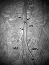
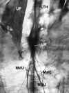
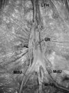
The UR's were classified into three types (Fig. 1); round or oval (Type A, 59.0%) (Fig. 2), obliterated or slitted (Type B, 21.4%) (Fig. 3), or completely covered by a connecting band between the LTH and the umbilical ligaments (Type C, 19.6%) (Fig. 4). The mean diameters of the UR of Type A were 3.4 mm (range, 2.3 - 5.0 mm) and 6.0 × 3.3 mm (range, 2.0 - 9.5 × 1.2 - 6.6 mm) in the round and oval shapes, respectively. The mean length of the UR of Type B was 7.9 mm (range, 1.4 - 10.2 mm) in the slit shape. The UR's were crossed by branches from the LTH or umbilical ligaments in 20 specimens (35.7%) of Type A and 9 (16.1%) of Type B. The types of UR did not differ significantly with either age or gender.
The MnU and MdU terminated in various shapes that were classified into four types (Fig. 5): The MnU ended by fusing with both the MdU's on the inferior border of the UR in Type I (25.0%); The MnU ended by joining 1 or both MdU's without directly reaching the UR in Type II (41.1%) (Figs. 2 and 3); The umbilical ligaments joined into a band below the inferior border of the UR, which continued to the LTH or attached to the inferior border of the UR in Type III (25.0%) (Fig. 4); and the MnU was not observed in Type IV (8.9%).
The LTH frequently ended by dividing into several branches in the area cranial to the UR. Some of the branches crossed the UR and connected with the umbilical ligaments, with others attaching to the circumferential border of the UR or the area lateral to the ring (Fig. 2). In 2 specimens, the LTH ended by attaching to the area lateral to the UR without dividing into branches, and it ended by attaching to the border of the ring without dividing into branches in nine specimens.
The UF completely and partially covered the UR in 50.0% and 1.8% of cases, respectively. The distal margin of the fascia was located cranial to the UR in 35.7% of cases, and it did not exist in 12.5% of cases.
In 1 case of umbilical hernia observed in this study, the oval UR was as large as 20 × 15 mm, which was not crossed by the branches of LTH, neither covered by UF; 3 such cases, being neither crossed by the LTH nor covered by the UF, were observed in UR's of Type A.
We have classified the UR into 3 types based on the shape of the ring and its morphologic relation with the surrounding ligaments. These differences might be due to several postnatal factors. One of the factors appeared to be the rapid growth of the abdominal wall of a newborn, which might press the aperture of the UR from all directions, with its shape consequentially determined by the balance of these forces: the aperture could be narrowed to remain oval or round, or be obliterated or slitted, as in Types Aand B, respectively. In the formation of Type C, several connecting branches might cross over the UR between the LTH and the umbilical ligaments, and become a band that completely covers the UR.
The umbilical ligaments could be classified into 4 types based on their interrelationships. After the urachus and 2 umbilical arteries met on the inferior part of the UR, they might fuse with the ring and then be extended as the abdominal wall expanded due to its growth, with them separating from one another before reaching the inferior border of the UR. Such umbilical ligaments were classified as Type I in this study, which appears to be equivalent to group I of Hammond et al. (1941).5 When the urachus met 1 or both umbilical arteries without any connection with the UR, these structures might be extended as the abdominal wall expanded, with the MnU fusing with 1 or both the MdU's without directly reaching the inferior border of the UR. These cases were classified as Type II, which appears to be equivalent to group II or III of Hammond et al. (1941).5 After the urachus met the umbilical arteries below the UR, 3 umbilical ligaments fused into a band that reached the inferior border of the UR in Type III. When the distal part of the urachus was degraded, the MnU was not observed between both MdU's below the UR. These cases were classified as Type IV, which appears to be equivalent to group IV of Hammond et al. (1941).5 The discrepancy between the classification of the umbilical ligaments in the current and the previous studies5 might be due to the anterior abdominal wall being removed and observed under a surgical microscope in our study. Given that the MdU has been used as a plug to repair femoral hernias,6 the classification of umbilical ligaments in this study is expected to be helpful in the repair of femoral hernias.
The LTH was found to terminate at the inferior or superior portion of the UR, to bifurcate and fuse to both sides of the umbilical ring, and to branch and continue with the MdU in 74%, 24% and 0.5% of cases, respectively.3 The ligament has also been reported to reach the umbilicus and a supra-umbilical site in 50% and 49% of cases, respectively.4 However, the present study has shown that LTH is frequently divided into several branches before it reached the UR, which is consistent with a report of the lower third of the umbilical vein undergoing breakdown and completely dividing into vascular fibro-elastic strands in early childhood.7 The terminal branches not only crossed the UR and connected with the umbilical ligaments, but also attached to the circumferential border of the UR or the lateral area, even in the same specimen (Fig. 2). The discrepancy among these studies might be due to inter-racial variations or different observation methods: the observations were made with the naked eye,3 laparoscopically without removing the peritoneum in patients with Cruveilhier-Baumgarten syndrome,4 and under a surgical microscope (present study) - only our observation method is sufficient to allow the slender terminal branches deriving from the ligament to be traced.
In addition to the protective roles of the LTH and the UF against umbilical hernia,3 the shape of UR may also have a protective function against the herniation. Those with an UR of Type A may be more susceptible to umbilical hernia, especially when the ring is neither crossed by the branches of the ligaments nor covered by the UF. This is consistent with the UR of the specimen having the umbilical hernia in our study being of Type A, which was neither crossed by the LTH nor covered by the UF.
This study revealed the anatomic variations of UR, LTH, MnU and MdU, and the relationship between UR and UF. These results are expected to deepen our understanding of the anatomy of the umbilical area, and further improve treatments of the umbilical hernia.
Figures and Tables
 | Fig. 1Three types of umbilical ring (UR) identified in this study. Classification was based on the presence of the hole of the ring, and whether the ring is covered by the ligament. |
 | Fig. 2Photograph showed an umbilical ring (UR) of Type A and umbilical ligaments of Type II. The ligamentum teres hepatis (LTH) divided into 3 branches (arrows), 1 of which attaches lateral to the oval UR and another that crosses the UR and joins the medial umbilical ligament (MdU). The median umbilical ligament (MnU) joins the MdU that is on the right side. |
 | Fig. 3Photograph of a backlit section showing an umbilical ring (UR) of Type B and umbilical ligaments of Type II. No light is transmitted in the area of the UR because the ring is obliterated. The median umbilical ligament (MnU) joins the medial umbilical ligament (MdU) that is on the right side, and the ligamentum teres hepatis (LTH) divides into 2 branches. The umbilical fascia (UF) is reflected. |
 | Fig. 4Photograph showing an umbilical ring (UR) of Type C and umbilical ligaments of Type III. The median umbilical ligament (MnU) and medial umbilical ligament (MdU) fuse into a band that continues to the ligamentum teres hepatis (LTH), covering the UR. A connecting branch (arrow) is evident between the LTH and the MdU that is on the left side. |
References
1. Lemmer JH, Strodel WE, Eckhauser FE. Umbilical hernia incarceration: a complication of medical therapy of ascites. Am J Gastroenterol. 1983. 78:295–296.
2. Kives SL, Lara-Torre E. Pyogenic sacroiliitis following an infected umbilical ring. J Pediatr Adolesc Gynecol. 2004. 17:125–129.

3. Orda R, Nathan H. Surgical anatomy of the umbilical structures. Int Surg. 1973. 58:458–464.
4. Friedrich K, Vogel HM, Henning H. The importance of variant insertions of the ligamentum teres hepatis in the Cruveilhier-Baumgarten syndrome. Endoscopy. 1988. 20:254–259.

5. Hammond G, Yglesias L, Davis JE. The urachus, its anatomy and associated fasciae. Anat Rec. 1941. 80:271–287.

6. Ikossi DG, Shaheen R, Mallory B. Laparoscopic femoral hernia repair using umbilical ligament as plug. J Laparoendosc Adv Surg Tech A. 2005. 15:197–200.

7. Martin BF, Tudor RG. The umbilical and paraumbilical veins of man. J Anat. 1980. 130:305–322.


 PDF
PDF ePub
ePub Citation
 Print
Print


 XML Download