

 ePub
ePub Citation
 Print
Print


INTRODUCTION
Rheumatoid arthritis (RA) is a common disease characterized by chronic articular inflammation of diarthrodial joints. Its primary site of pathology is the synovium of the joints. However, it can exhibit a variety of extra-articular manifestations demonstrating that RA has features of a systemic disease that is capable of involving a variety of major organ systems. One of the most important types of extraarticular involvement of RA is neurologic involvement, including entrapment neuropathies, peripheral neuropathies and mononeuritis multiplex.1,2
Compared to other mononeuropathies, femoral mononeuropathy (FMN) is relatively uncommon. FMN has many causes, including diabetes mellitus and polyarteritis nodosa, as well as occuring following urologic operations, or as a result of trauma from hip fracture, hemorrhage into the psoas muscle, secondary to anticoagulation therapy, ossification of the ligamentum flavum, alcohol abuse, self-retaining surgical retractor blades, or prolonged dorsal lithotomy positioning.3-11 The symptoms of FMN include medial leg and thigh sensory changes, knee instability, or quadriceps weakness. The patellar reflex may be decreased or absent.3,4
Femoral mononeuropathy as an extraarticular finding of RA is a phenomenon which has not been reported previously. We report a patient with RA, presenting FMN findings during the course of the disease.
CASE REPORT
A 53-year-old woman with a two-and-a half year history of inflammatory joint disease was admitted to hospital for progressive right leg weakness and difficulty in walking. The diagnosis of seronegative RA had been established 2.5 years earlier and she had been given daily combination therapy including 2000 mg sulphasalasine and 250 mg chloroquine diphosphate.
The patient complained of muscle weakness in her right leg, and also complained of bilateral pain and limited ranges of motion in her finger, wrist, elbow and knee joints, with morning stiffness lasting more than an hour. Her history didn't include pelvic operation, trauma, alcohol or drug abuse, or any predisposing metabolic disease such as diabetes mellitus, not was there a family history of any arthritic disorders.
Examination revealed symmetric synovial thickening of wrists, metacarpophalangeal and proximal interphalangeal joints. Limited and painful ranges of motion were noted in her right and left metacarpophalangeal, elbow, wrist and knee joints. Ritchie articular index was 11. Bilateral dorsal interosseous muscle atrophies and ulnar deviations were observed. Right quadriceps and iliopsoas muscles showed grade 3 weakness on the Medical Research Council (MRC) scale. Sensory examination revealed sensory loss in the right medial leg and thigh. Patellar tendon reflex was absent in the right side. Thigh adductors and all other muscle groups were normal bilaterally. The remainder of the neurological examination was normal. There was no palpable mass at the inguinal region.
Laboratory results were as follows; erythrocyte sedimentation rate (ESR) was 32 mm/h (Westergren method), haemoglobin was 11.8 g/L and white cell count was 3800/mm3. C-reactive protein (CRP) and rheumatoid factor (RF) were negative. All other laboratory parameters were normal.
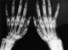
Radiographs revealed bilateral erosive arthritis of hands and wrists (Fig. 1). Magnetic resonance imaging (MRI) of lumbar discs showed posterior bulging, including L4-5 and L5-S1 segments. Anteroposterior and lateral radiographs of the pelvis, right hip and lumbosacral region showed no abnormalities. Pelvic ultrasound was normal. In nerve conduction study (NCS), the right femoral motor amplitude and conduction velocity were found to be decreased. The left femoral motor and bilateral saphenous nerve conductions were normal. Electromyography (EMG) revealed positive sharp waves and fibrillation potentials in the right vastus lateralis muscles with a moderate decrease in recruitment and a slight enlargement of the motor unit potentials. The thigh adductors, tibialis anterior, and gluteus medius were normal.
In light of these clinical findings, a diagnosis of a partial right femoral neuropathy was confirmed. The patient was started on rehabilitation, and 20 mg daily oral methylprednisolone and vitamin B (including B1and B6) were added her present therapy. After two months this therapy resulted in a significant improvement of both walking and muscle strengths of quadriceps and iliopsoas muscles (MRC 5/5). EMG and NCS were unable to be performed after treatment, because the patient refused both tests.
DISCUSSION
Nerve compression is a common cause of neurologic impairment in RA. Peripheral entrapment neuropathies tend to correlate with the degree and severity of the local synovitis, but are not related to the duration, level of activity, or severity of extra-articular manifestations of RA. They generally occur when the nerve is compressed by the inflamed synovium against a fixed structure.12 In patients who had clinical evidence of systemic arteritis superimposed on rheumatoid arthritis, autopsy studies have shown a wide spectrum of arteriel lesions, ranging from fibrinoid necrosis involving the arterial wall with infiltration of polymorphonuclear leukocytes to more chronic lesions of perivascular fibrosis and intimal proliferation. These changes have been found in the heart, muscle, nerve, skin, testes, and viscera. Therefore, in RA, a spectrum of vasculitis occurs that ranges from venulitis and capillaritis in early synovitis to widespread arteritis, the primary clinical manifestation of which may be a neuropathy and which may involve other organs and terminate fatally.13
The femoral nerve is formed by the combination of the posterior divisions of the ventral rami of L2, L3 and L4 spinal roots. It passes through the psoas major and supplies the iliacus and pectineus muscles before passing beneath the inguinal ligament. In the thigh, the anterior branch supplies the sartorius and gives off the intermediate and median cutaneous branches to the thigh. The posterior branch forms the saphenous nerve and also supplies the the quadriceps and knee joint. Because of its short course, the main trunk of the femoral nerve is usually injured at one of two sites: the retroperitoneal pelvic space or the inguinal ligament.3,14 In an anatomic study, Muellner et al. reported that the critical area prone to impingement is about 18 cm above the patella.15
Femoral mononeuropathy has many causes. Most of them are iatrogenic, occuring during intra-abdominal, intrapelvic, inguinal, or hip surgical or diagnostic procedures. Clinical and cadaveric studies have demonstrated that FMN may result from direct compression of the nerve, or more frequently from indirect compression between the psoas muscle and the lateral pelvic wall in this situation.5-11,14 However, in RA, FMN may result from not only compression of the nerve, but also a spectrum of vasculitis ranging from venulitis and capillaritis to widespread arteritis. The histologic findings in rheumatoid neuropathy are similar to those of polyartheritis nodosa, with necrotizing vasculitis mostly involving epineural arterioles between 100 and 250 cm in diameter.13
To our knowledge, this is the first observation of femoral mononeuropathy as an extraarticular finding of RA. Our patient had no previous history of trauma or surgical procedure and no palpable or radiologically detected mass in the pelvic and inguinal area. The probable mechanism of FMN was vasculitis. In general, the course of rheumatoid neuropathy appears to parallel that of the associated vasculitis, and the prospects of meaningful neurologic improvement appear to be fairly good. However, the development of multiple mononeuropathies in patients with rheumatoid vasculitis has been thought to predict an even poorer outcome.13 Thus, mononeuropathy needs careful assessment in patients with RA as an important indicator of vasculitis.
 XML Download