

 PDF
PDF ePub
ePub Citation
 Print
Print


INTRODUCTION
β-hemolytic streptococcal (BHS) isolates from humans can be subdivided into large-colony and small-colony (< 0.5 mm in diameter) formers. Streptococcus pyogenes (Lancefield group A), Streptococcus agalactiae (group B), and Streptococcus dysgalactiae subsp. equisimilis (group C, G) belong to large-colony formers.1 Although large-colony-forming β-hemolytic streptococci (LCF-BHS) are still susceptible to β-lactams, macrolides or lincosamides are recommended as alternative choices when indicated.1-3 However, recent studies have shown that changes in the susceptibility of LCF-BHS to erythromycin and clindamycin have been substantial, although differences in resistance rates to these agents exist according to geographical variation and investigators.4-8 The high transmissibility of LCF-BHS, including resistant clones and the association of increased macrolide usage, may play a significant role in the variable resistance rates that have been reported during the last decade.9-11
In Korea, resistant bacteria are more prevalent than in other industrialized countries, and their presence suggests a high level of antimicrobial selective pressure as well as the nosocomial spread of resistant bacteria.12 In response to this public health problem, the Korean government instituted a new health policy, 'the separation of prescribing and dispensing (SPD) of medications', on July 1, 2000. The purpose of this policy was to provide greater differentiation between the roles of physicians and pharmacists than had historically existed in South Korea. In our previous study,13 however, the resistance rates to erythromycin and clindamycin among Streptococcus pyogenes, Streptococcus agalactiae, and group C streptococci isolates were still high during the period of 2001-2002.
Two major mechanisms account for erythromycin resistance in many gram-positive bacteria: target site modification and active efflux. Target site modification is mediated by erythromycin resistance methylase that is encoded by erm class genes. Methylases cause a conformational change in the prokaryocytic ribosome, leading to reduced binding of macrolide-lincosamide-streptograminB (MLSB) antibiotics to the target site in the 50S ribosomal subunit. The phenotype expression of MLSB resistance in streptococci can be either constitutive or inducible. Macrolide efflux, which is effected by a membrane protein encoded by the mef class genes, has recently emerged among Streptococcus pyogenes and Streptococcus pneumoniae in many countries.14 It has been well documented that the frequency of MLSB resistance phenotypes among streptococci varies considerably between countries.14
The objectives of the present study were to investigate the incidence and trend in susceptibility among the LCF-BHS isolated from clinical specimens in a Korean hospital and to clarify the phenotypes and genotypes of erythromycin-resistant LCF-BHS. We also explored the correlation between serotypes and genotypes of erythromycin-resistant Streptococcus agalactiae.
MATERIALS AND METHODS
A total of 204 strains of LCF-BHS were obtained from various clinical specimens between January 2003 and December 2004 at Wonju Christian Hospital in Korea. Multiple isolates from the same patient were avoided. The isolates were identified by standard criteria on the basis of hemolytic patterns on 5% sheep blood agar, colony morphology, Gram stain, catalase reaction, Streptex latex agglutination assay (Murex Biotech Limited, Dartford, England), and API Rapid ID32 STREP system (bioMérieux, Marcy l'Etoile, France).
The strains were stored in thioglycollate broth with 20% glycerol at -70℃ until analyzed. The frozen isolates of LCF-BHS were thawed, inoculated onto a 5% sheep blood agar plate and incubated at 35℃ overnight. Pure isolates of LCF-BHS obtained from three consecutive subcultures were tested for susceptibility and polymerase chain reaction (PCR).
Susceptibility to penicillin G, erythromycin, clindamycin, tetracycline, ceftriaxone, chloramphenicol (Sigma Chemical Co, St. Louis, MO, USA) and vancomycin (Daewoong Lilly, Seoul, Korea) was tested by the agar dilution method according to the recommendations of the Clinical and Laboratory Standards Institute.15 The Streptococcus pneumoniae ATCC 49619 strain was simultaneously tested to monitor the accuracy of minimal inhibitory concentrations of LCF-BHS. The resistance phenotypes of erythromycin-resistant isolates were determined by the doubledisc test with erythromycin (15 µg) and clindamycin (2 µg) disks.13
The genomic DNA extractions were carried out with the Easy-DNA kit (Invitrogen, Carlsbad, CA, USA) according to the manufacturer's instructions. The presence of erm and mef class genes was determined by PCR amplification using previously described primers specific for erm(A), erm(B), erm(C), erm(TR), and mef(A).13
GBS serotypes Ia, Ib, and II~VIII were determined by use of a coagglutination test (ESSUM Group B Streptococcus Serotyping Test; Bacterum AB, Umeå, Sweden).13
RESULTS
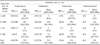
The overall non-susceptible (intermediate and resistance) rates of LCF-BHS were 67.6% to tetracycline, 23.5% to clindamycin, 22.5% to erythromycin and 9.8% to chloramphenicol, whereas all isolates were susceptible to penicillin G, ceftriaxone, and vancomycin. Resistant rates to tetracycline, erythromycin, and clindamycin of Streptococcus agalactiae and Streptococcus pyogenes isolates were 95.4% versus 19.0%, 36.7% versus 4.8%, and 43.1% versus 0%, respectively. Three isolates of group C LCF-BHS were susceptible to all tested antimicrobial agents. Resistant rates to chloramphenicol, erythromycin, and clindamycin of group G LCF-BHS were higher than those of Streptococcus pyogenes (Table 1).
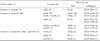
Of the 46 erythromycin-resistant LCF-BHS isolates (Table 2), 37 isolates (80.4%) had the constitutive macrolide-lincosamide-streptograminB (cMLSB)phenotype, six isolates (13.0%) had the M phenotype, and three (6.5%) isolates had the inducible MLSB (iMLSB) phenotype. Of the 40 erythromycin-resistant Streptococcus agalactiae strains, the most prevalent gene was erm(B) (92.5%). All three erythromycin-resistant Streptococcus pyogenes isolates had mef(A) gene. Four isolates of Streptococcus agalactiae had both of erm(B) and erm(TR) genes. Three isolates of Streptococcus dysgalactiae subsp. equisimilis had different resistance genes.
The serotype frequency of 103 Streptococcus agalactiae isolates was V (32.0%), III (22.3%), Ia (15.5%), and Ib (14.6%). The resistance rates to erythromycin by serotypes were 85% (V), 22% (III), 13% (Ib), and 0% (Ia) (Table 3).
Among the LCF-BHS isolates, 72 (35.3%) were from genitourinary specimens, 68 (33.3%) from wounds, 32 (15.7%) from blood, 18 (8.8%) from lower respiratory tract specimens, 11 (5.3%) from throat and 3 (1.5%) from other body fluids. Streptococcus pyogenes were frequently isolated from the throat, blood, and wounds, whereas Streptococcus agalactiae and Streptococcus dysgalactiae subsp. equisimilis were prevalent in genitourinary tract specimens and lower respiratory tract specimens, respectively (Table 4).
DISCUSSION
Until the 1980s, LCF-BHS were generally considered uniformly susceptible to erythromycin and clindamycin, but resistance spread rapidly in the 1990s. The prevalence of erythromycin-resistant LCF-BHS has been reported to be variable and depends on the country, selective pressure, serogroup, serotype, age, and season. Compared with our previous study,13 we observed that resistance among Streptococcus pyogenes isolates decreased from 25.7% to 4.8% in erythromycin, 15.8% to 0% in clindamycin, and 47.1% to 19.0% in tetracycline. In addition, the prevalent phenotypes and genotypes of MLSB resistance in Streptococcus pyogenes isolates have changed from the cMLSB phenotype carrying erm (B) to the M phenotype with the mef(A) gene. The determination of antibiotic prescriptions in outpatient clinics is an important factor to consider when decreasing resistance rates to commonly used antimicrobial agents, especially in skin and upper respiratory infections, are observed.
The isolation rate of Streptococcus pyogenes from throat specimens was 2.0% (2/102) in our hospital during the period of 1997-2000.16 These results suggested that resistance rates to commonly-used antimicrobial agents in outpatient clinics and the distribution of MLSB resistance phenotypes were partly influenced by selective pressure.
In contrast with Streptococcus pyogenes, resistance rates to erythromycin, clindamycin, and tetracycline in Streptococcus agalactiae isolates did not show decreasing trends in this study. The continued high resistance rates to erythromycin, clindamycin, and tetracycline are considered related to the clonal spread of serotype V with a multi-drug resistance phenotype.17 The resistance rates to clindamycin of our serotypes Ib and III isolates were higher than that of erythromycin, while the other serotypes were nearly equal in susceptible rates to erythromycin and clindamycin. Our results show resistance to clindamycin to be more common than resistance to erythromycin, and similar results have been reported in Taiwan and New Zealand.18,19 The distribution of MLSB resistant genes and the isolation frequency of serotypes of GBS may be major factors contributing to the difference between erythromycin and clindamycin resistance in different countries.
Malbruny et al. have reported that a new LSA (lincosamide-streptogramin A) phenotype was noted in erythromycin-susceptible, clindamycinresistant Streptococcus agalactiae isolates from New Zealand, and that III (13/19) and I (5/19) were the main serotypes of GBS with LSA phenotype.19 However, in spite of their extensive molecular studies, the resistance mechanism of LSA in Streptococcus agalactiae was not elucidated.
The overall resistance rates to erythromycin and clindamycin in group C and G BHS seemed to be somewhat lower than those of our previous results.13 Streptococcus dysgalactiae subsp. equisimilis colonizes and causes various infections in humans.20,21 Zaoutis et al. reported that three isolates (group G; 2, group C; 1) of 23 Streptococcus dysgalactiae subsp. equisimilis were resistant to erythromycin.21 Hashikawa et al. documented that all eleven of the Streptococcus dysgalactiae subsp. equisimilis strains were sensitive to β-lactam antibiotics, vancomycin, and chloramphenicol, whereas about half of the strains were tetracycline resistant, and one strain was resistant to erythromycin and clindamycin harbored erm(B).22 Our findings were similar to those of the aforementioned investigators' reports.
Continual monitoring of antimicrobial resistance among LCF-BHS is needed to provide the medical community with current data regarding the resistance mechanisms that are most common to their local or regional environments. Additionally, further epidemiologic studies are needed to confirm whether or not our susceptibility data on LCF-BHS are restricted to our geographic area.


 XML Download