Abstract
Purpose
Postoperative intraabdominal adhesion formation is a major clinical problem. No previous study was found, reporting the relationship between adhesion formation and melatonin administration, but melatonin, a strong antioxidant, is recognized to have certain effects on the progression of adhesion formation mechanism. It was therefore decided to investigate the effects of melatonin on postoperative adhesion formation.
Materials and Methods
A total number of 24 Spraque-Dawley rats were utilized. Three groups, described as: Group A, sham laparatomy (n = 8), Group B, rats that underwent only ischemia-reperfusion (n = 8) and Group C, rats that underwent ischemia- reperfusion and were given 10 mg/kg melatonin solution i.v. (n = 8). For Groups B and C, the ileocolic vessels were clamped. Blood glutathione peroxidase levels of all study groups were assessed, then microscopic and macroscopic adhesion scores were evaluated.
Adhesions subsequent to abdominal surgery occur as a reaction of the peritoneum to trauma resulting from abnormal connection of the tissues and the organs in the abdomen. The rate of intraabdominal adhesions, following various cases of abdominal surgery, has been reported to be as high as 60-95%.1-3 Adhesions are the most frequent cause of medical problems, such as, intestinal obstruction, infertility and pelvic pain, which invariably require surgical intervention or hospitalization.4
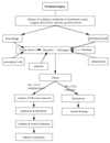
Major factors recognized in the etiology of adhesions, such as, ischemia, infection, presence of a foreign body, mechanical trauma and other factors, are known to trigger an inflammatory reaction in the peritoneum. This leads to loss of mesothelial cells in the peritoneal tissue and then to a discharge of fibrin-rich exudative fluid.4,5 Coagulation of intraabdominal exudate and the reduced fibrinolytic activity of the injured peritoneum occurs within hours after peritoneal injury. This sequence of events quickly leads to a fibrinous connection between adjacent intraabdominal viscera. Light and electron microscopic studies reveal fibroblast infiltration and collagen deposition following the peritoneal injury.5 Fig. 1 shows the adhesion formation steps and the factors affecting the adhesion formation mechanism.6 Melatonin inhibits fibroblast proliferation, platelet aggregation and secretion, and prostaglandin synthesis; which all belong to the adhesion formation mechanism.7-10
Recently oxygen-derivated free radicals, resulting from ischemia-reperfusion, have been reported to trigger the tissue injury. Melatonin, as well as a direct oxygen-derivated free radical scavenger, indirectly prevented oxygen-derivated free radical formation by increasing the activity of antioxidant enzymatic systems (superoxide dismutase and glutathione peroxidase among others).11-13
In the light of the above considerations, melatonin was hypothesized to assist in the prevention of postoperative intraperitoneal adhesion formation. No previous study was found that reporting adhesion formation and melatonin administration relationship. Melatonin, a strong antioxidant, is recognized to have certain effects on the sequence of the adhesion formation mechanism. This study was undertaken to investigate the effects of melatonin administration on intraabdominal adhesion formation resulting from ischemia-reperfusion (I-R).
A total number of 24 Spraque-Dawley rats recruited from the Animal Research and Care Unit of Trakya University Faculty of Medicine, weighing 180-220g, were included in this study. Trakya University Local Ethical Committee approved the animal experiments described in this study. Prior to and throughout the research, the rats were housed in (40 × 60 × 40cm) zinc cages at a room temperature of (24℃) and were fed with standard rat chow and water ad lib. The animals were kept under 12hr light/12hr dark conditions, and after 15 days of acclimatization they were divided into 3 groups. Table 1 presents the three experimental groups.
Feeding was ceased 12 hours before the time of the operation. After premedication with 1mL/kg Xylazine (Rompun-Bayer), the rats were anaesthetized with 10mg/kg Ketamine hydrochloride (Ketalar-Eczacıbaşı). Talcum powder on the gloves was washed off with sterile saline water. The abdominal area of each rat was shaved and prepped with povidon iodine and a 3cm midline incision was made for abdominal surgery.
In research Groups B and C, the omentum and the small intestines were explored. Ileocolic vessels were found, prepared and occluded with a vascular clamp for 45 minutes.14 Five minutes before the release of the clamp and the reperfusion of the intestine, the tail veins of Group C rats were injected with melatonin solution at 10mg/kg. For glutathione peroxidase levels, 1cc blood samples were collected from each rat in Group A following exploration, and from Group B and C rats at the 60th. min of reperfusion.
Melatonin (Sigma Chemical Co., M 5250) was kept at 0℃. The stock melatonin solution was diluted in ethanol and 0.9% NaCl (1/5) mixture.15 The solution was freshly prepared before use. Glutathione peroxidase levels were assessed with RanSOD kit (Randox Comp., SD 125) in a UV-120 spectrophotometer (Shimadzu). Glutathione peroxidase values were calculated and expressed as the U/gr.Hb values.
15 days later, all of the animals underwent a secondary surgery via an incision made on the left side of the first incision. Gross adhesive areas were removed. The examiner evaluated the adhesions, without knowing to which group the animals belonged. Nonparametric ANOVA test (Kruskal Wallis-ANOVA) performed to compare the results from three groups.
Oelsner et al.16 has described a scale for postoperative adhesion formation. Oelsner's scale was graded separately for each uterine horn. Oelsner's scale was used as a guide and a modified macroscopic adhesion scale is given in Table 2. In this scale, the thickness of the adhesion was taken as the major criterion instead of vascularity and starting point.
The samples for histopathological evaluation were taken from the most dense adhesion areas. Microscopic assessment of the adhesions, was done by another investigator who was unaware as to which adhesion came from which animal. The tissue samples were fixed with 10% formaline and paraffin blocks were prepared. The cross-sections were stained with Hematoxylene-Eosin (H & E) and were examined under light microscopy. They were evaluated according to fibroblast proliferation, as slight (+), moderate (++) or intense (+++). The data obtained was statistically evaluated by the Chi-Square test. Evaluation of the entire statistical data was carried out by Trakya University, Faculty of Medicine, Public Health Department of T.U. SPSS for Windows ver. 8.0 (licence no: 10592). All data is expressed as mean ± SD.
The average adhesion scores were found to be 2.1 ± 0.6 in Group A, 3.4 ± 0.7 in Group B and 2.5 ± 0.7 in Group C (Table 3). Upon macroscopic evaluation of all of the groups, the adhesion scale for both Group A and Group C adhesion formation was found to be significantly reduced compared to Group B (X2 = 8.591, p = 0.014). Group A presented no difference in adhesion formation compared to Group C (p > 0.05). This indicated that melatonin administration prevented adhesion formation.
The glutathione peroxidase levels were 49.1 ± 15.5; 28.3 ± 10.7 and 47.3 ± 15.3 for Groups A, B, and C, respectively (see Table 4 for individual values). When all groups were evaluated according to their glutathione peroxidase levels, the glutathione peroxidase level of Group B was found to be significantly lower than those of both Group A and Group C (X2 = 7.293, p = 0.026). No statistical difference has been found between Groups A and C in terms of glutathione peroxidase levels (p > 0.05).
The results obtained from the fibroblast proliferation evaluation of the tissue samples of the gross adhesive areas of peritoneum, viscera and fibrous bands, are presented in Table 5.
For the fibroblast proliferation in Group B, a statistically significant increase was detected when compared to Group A (X2 = 13.9, p = 0.001). A statistically significant increase was also detected when Group B was compared to Group C (X2 = 7.2, p = 0.03). There was no significant difference between Group A and Group C (p > 0.05). Consequently, for fibroblast proliferation, in the ischemia-reperfusion group, a statistically significant increase was detected when compared to the sham laparatomy group. In Group C, there was no significant increase in fibroblast proliferation when the ischemia-reperfusion was established after melatonin administration.
The formation of intraabdominal adhesions following abdominal surgery is inevitable in many cases, despite the improvement of surgical technique and equipment. Intra-abdominal adhesions can result in intestinal obstruction, infertility and chronic pelvic pain. In many cases, surgery is required and that increases the rate of morbidity and mortality.4
Substances such as octreotid, nonsteroidal anti-inflammatory drugs, corticosteroids, calcium channel blockers, L-arginine and pentoxifylline which are also successful in reducing postoperative adhesions, inhibit fibroblastic proliferation and the inflammatory reaction.14,17,18 Çubukçu et al.19 demonstrated the decrease of postoperative intraabdominal adhesion and fibroblast proliferation in their study concerning the use of Mitomycin-C. Carissino et al.,7 in their research on healthy individuals and patients with systemic sclerosis, reported that high doses of melatonin reduced fibroblast proliferation. In this present study, it was observed that in the melatonin group, fibroblast proliferation and intraabdominal adhesion were reduced, compared to the ischemia-reperfusion group (Fig. 2).
In addition, antioxidants such as vitamin E, L-arginin and pentoxifylline have been used successfully. Melatonin is one of the most powerful antioxidants known to date. Both in vivo and in vitro studies have showed that melatonin not only serves particularly as a direct free radical scavenger but also as an indirect antioxidant.11,20-22
Melatonin is known to be effective in reducing oxidative damage in ischemia-reperfusion. Sawerynek et al.21 have reported that exogenous melatonin effectively reduces oxidative damage in ischemia-reperfusion damage of the liver. Cabeza et al.15 reported that in rats subjected to gastric ischemia-reperfusion damage, the melatonin treated group showed a higher antioxidant enzyme activity than the untreated group. De La Lastra et al.23 reported that in rats subjected to gastric ischemia reperfusion, there was no decrease in the glutathione peroxidase levels with melatonin use. In this present study, glutathione peroxidase levels of the melatonin group were not reduced compared to the ischemia-reperfusion group.
In conclusion, the glutathione peroxidase levels of the melatonin-treated group were significantly higher and fibroblast proliferation and macroscopic adhesion scores were significantly lower, than in the melatonin-free group. These findings suggest that melatonin administration has beneficial effects on postoperative intraabdominal adhesions.
Figures and Tables
Fig. 2
An area of the fibrous band and/or peritoneum. H & E stain (original magnification 100 ×). (A) Microscopic view of a subject from the ischemia-reperfusion group. (B) Microscopic view of a subject in which ischemia-reperfusion was established after melatonin administration. Significant fibroblast proliferation was observed in A compared to B.

References
1. DeCherney AH, diZerega GS. Clinical problem of intraabdominal postsurgical adhesion formation following general surgery and use of adhesion prevention barriers. Surg Clin North Am. 1997. 77:671–688.

2. Bigatti G, Boeckx W, Gruft L, Segers N, Brosens I. Experimental model for neoangiogenesis in adhesion formation. Hum Reprod. 1995. 10:2290–2294.
3. Menzies D. Postoperative adhesions: Their treatment and relevance in clinical practice. Ann R Coll Surg Engl. 1993. 75:147–153.
4. Ellis H. The cause and prevention of intestinal adhesions. Br J Surg. 1982. 69:241–243.
5. Wurster SH, Bonet V, Mayberry A, Hoddinott M, Williams T, Chaudry I. Intraperitoneal sodium carboxymethylcellulose administration prevents reformation of peritoneal adhesions following surgical lysis. J Surg Res. 1995. 59:97–102.

6. Holmdahl LE, Al-Jabreen M, Risberg B. Role of fibrinolysis in the formation postoperative adhesions. Wound Rep Regen. 1992. 4:171–176.
7. Carrossino AM, Lombardi A, Matucci-Cerinic M, Pignone A, Cagnoni M. Effect of melatonin on normal and sclerodermic skin fibroblast proliferation. Clin Exp Rheumatol. 1996. 14:493–498.
8. Leach C, Thorburn GA. Comparision of the inhibitory effectsof melatonin and indomethacin on plateletaggregation and thromboxane release. Prostaglandins. 1980. 20:51–56.

9. Gimeno MF, Landa A, Sterin-Speziale NS, Cardinali DP, Gimeno AL. Melatonin bloks in vitrogeration of prostaglandins by the uterus and hypotalamus. Eur J Pharmacol. 1980. 62:309–317.

10. Gimeno MF, Ritta MN, Bonacossa A, Lazzari M, Gimeno AL, Cardinali DP. Birau , editor. Inhibition by melatonin of prostaglandin synthesis in hypotalamus, uterus and platelets. Melatonin: Current Status and Perspective. 1980. Pergamon Press;147–150.

11. Reiter RJ. Functional pleiotropy of the neurohormone melatonin: antioxidant protection and neuroendocrine regulation. Front Neuroendocrinol. 1995. 16:383–415.

12. Reiter RJ, Carneiro RC, Oh CS. Melatonin in relation to cellular antioxidative defense mechanisms. Horm Metab Res. 1997. 29:363–372.

13. Reiter RJ, Guerrero JM, Garcia JJ, Acuna Castroviejo D. Reactive oxygen intermediates, moleculer damage, and aging: relation to melatonin. Ann N Y Acad Sci. 1998. 854:410–424.

14. Baykal A, Özdemir A, Renda N, Korkmaz A, Sayek I. The effect of octreotide on postoperative adhesion formation. Can J Surg. 2000. 43:43–47.
15. Cabeza J, Motilva V, Martin MJ, de la Lastra CA. Mechanisms involved in gastric protection of melatonin againts oxidant strees by ischemia-reperfusion in rats. Life Sci. 2001. 68:1405–1415.

16. Oelsner G, Graebe RA, Pan SB, Haseltine FP, Barnea ER, Fakih H, et al. Chondroitin sulfate: a new intraperitoneal treatment for postoperative adhesion prevention in the rabbit. J Reprod Med. 1987. 32:812–814.
17. Holtz G. Prevention and management of peritoneal adhesions. Fertil Steril. 1984. 41:472–504.
18. Kaleli B, Özden A, Aybek Z, Bostancı B. The effect of L-arginin and pentoxyfilline on postoperative adhesion. Acta Obstet Gynecol Scand. 1998. 77:377–380.

19. Çubukçu A, Alponat A, Gönüllü NN, Özkan S, Erçin C. An experimental study evaluating the effect of mitomycin C on the prevention of postoperative intraabdominal adhesions. J Surg Res. 2001. 96:163–166.

20. Tan DX, Chen LD, Poeggeler B, Manchester LC, Reiter RJ. Melatonin: a potent, endogenous hydroxyl radical scavenger. Endocr J. 1993. 1:57–60.
21. Sewerynek E, Reiter RJ, Melchiorri D, Ortiz GG, Lewinski A. Oxidative damage in the liver induced by ischemia-reperfusion: protection by melatonin. Hepatogastroenterology. 1996. 43:898–905.
22. Beyer CE, Steketee JD, Saphier D. Antioxidant properties of melatonin- an emerging mystery. Biochem Pharmacol. 1998. 56:1265–1272.
23. De La Lastra CA, Cabeza J, Motilva V, Martin MJ. Melatonin protect againts gastric ischemia-reperfusion injury in rats. J Pineal Res. 1997. 23:47–52.


 PDF
PDF ePub
ePub Citation
 Print
Print


 XML Download