

 PDF
PDF ePub
ePub Citation
 Print
Print


INTRODUCTION
Sclerotic fibroma (SF) is a unique benign fibrous tumor that may develop anywhere on the skin. SF was originally recognized as a cutaneous manifestation of Cowden disease.1-3 Although SF was first described in a patient with Cowden's disease, solitary and sporadic cases of SF of the skin in patients without Cowden's disease have been reported.4-6 However, to the best of our knowledge, only seven cases of sporadic SF of the oral cavity have been described.7-9
Solitary fibrous tumor (SFT) usually arises in the pleura and it was first described by Klemperer.10 With the increasing number of reported cases in a variety of extrapleural sites, it was recently suggested that extrapleural SFTs may actually develop more frequently than the pleural tumors.11 SFTs occur rarely within the oral cavity, and only about 37 cases of SFTs in the oral cavity have been reported.12-26
In this report, we present two cases of oral pathology, one involving SF and the other involving SFT, and compare the histopathologic findings.
CASE REPORT
Case 1 (A case of sclerotic fibroma)
In June 2005, a 36-year-old woman visited the Department of Otolaryngology-Head and Neck Surgery with complaints of a small mass in the right buccal mucosa that she had noted one year prior. The lesion was asymptomatic and had not increased in size. Intraoral examination revealed a 1-cm-sized polypoid nodule. She did not have any of the known signs or symptoms associated with Cowden's disease.
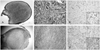
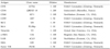
Grossly, the lesion was a relatively well-demarcated, firm, flesh-colored nodule. Histopathologic examination showed a relatively well demarcated, non-capsulated, round hypocellular and eosinophilic nodule, and the overlying mucosa was attenuated (Fig. 1A). The nodule was composed of hyalinized sclerotic collagen bundles arranged in a whorled pattern. Prominent clefts between the collagen bundles were observed. The scanty tumor cells were spindle shaped and entrapped among the thick collagen bundles (Fig. 1C). The antibodies listed in Table I were used for the immunohistochemical staining with the streptavidin-biotin method. The tumor cells stained intensely for CD34 and vimentin, and several CD99-positive cells were seen (Fig. 1E). The immunoreactivity for bcl-2, α-SMA, S-100, CD68, EMA, CD31, factor VIII and bcl-6 was negative (Fig. 1E).
Case 2 (A case of solitary fibrous tumor)
In May 2005, a 28-year-old woman visited the Department of Otolaryngology-Head and Neck Surgery. She had noticed a painless mass in her oral cavity about three months prior to our examination. Intraoral examination revealed a 1.5-cm-sized elevated nodule in the hard palate.
Grossly, the lesion was sharply circumscribed and firm in consistency. Histopathologic examination showed a well demarcated submucosal mass with ulceration of the overlying mucosa (Fig. 1B). The tumor showed a predominantly haphazard or short fascicular arrangement of the spindle cells that had fusiform or oval vesicular nuclei, inconspicious nucleoli, and scant cytoplasm. The cellularity of the tumor varied from area to area, and the cellularity was inversely related to the amount of collagen (Fig. 1D). In the areas with less cellularity, the tumor cells were embedded in a collagenous matrix. The tumor was richly vascularized and occasionally contained areas with dilated vessels. Focal calcification was seen, but neither mitosis nor necrosis was found. On immunohistochemical staining, the tumor cells were positive for vimentin, CD34, bcl-2 and CD99, and were negative for α-SMA, S-100, CD68, EMA, CD31, factor VIII and bcl-6 (Fig. 1F).
DISCUSSION
Although SF and SFT are very rarely found in the oral cavity, there are several fibrous lesions that may be included in the histologic differential diagnosis of tumors in the oral cavity. These lesions are traumatic fibroma (TF), giant cell fibroma (GF), and benign fibrous histiocytoma (BFH). TFs are the most commonly occurring oral soft tissue lesion. The microscopic features of TF include dense collagen, numerous mature fibroblasts, and chronic inflammatory cells. Unlike in SF and SFT, the fibrous tissue of TF blends into the surrounding connective tissue. The collagen bundles of GF are rarely thickened, and clefts are not seen. The sharp circumscriptions of SF and SFT are the most important histologic features that distinguish these lesions from other oral fibrous lesions. Moreover, the fibroblasts and myofibroblasts of TF, GF and BFH do not react with anti-CD34 antibodies.7
Rapini and Golitz4 coined the term "sclerotic fibroma (SF)" when they described a series of 11 solitary skin lesions with histology similar to that of the cutaneous neoplasms in Cowden's disease. Microscopically, these tumors are well circumscribed, paucicellular, dermal nodules that are composed of thick collagen bundles separated from one another by prominent clefts, and the collagen bundles are arranged in a whorled or storiform pattern.2-4 Spindle- and stellate-shaped, occasionally multinucleated CD34-positive dendritic cells are haphazardly scattered throughout the lesions.3-6 Intraorally, the patients with Cowden's disease typically present with numerous pink or mucosa-colored papular lesions that often involve the tongue, gingiva and lips.27,28 In most cases, the oral papules reveal non-specific histologic features that are consistent with epithelial and fibrous hyperplasia.27,28 Alawi et al.7 described the first documented series of solitary SF of the oral soft tissues. The authors reported five cases of isolated SF, of which four arose in the buccal mucosa and one arose in the lower lip. Ide et al.8 and Gonzalez-Vela et al.9 reported one case of SF in the lip and one case of SF in the buccal mucosa. Ours is the seventh case of SF in the absence of Cowden's disease.
Suster et al.12 were the first to describe an SFT in the oral cavity. To date, we have found only 37 reported cases in our literature search.12-26 Microscopically, SFTs are similar at any site in the body and are characterized by the presence of hypocellular and hypercellular areas, a sclerotic collagen matrix and the presence of vessels, mast cells, and nuclei arranged in a palisading fashion. Although the cells are generally distributed haphazardly, they may form fascicles and then present a storiform pattern.
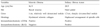
SF and SFT present as sharply circumscribed masses with very similar gross findings. Although SF and SFT exhibit distinct microscopic features, confusion may arise when examining the fragmented biopsy tissue, because the hypocellular, collagen-rich areas of SFT may closely resemble the appearance of a SF. The results of immunohistochemistry on SF and SFT are very similar. Like SF, the neoplastic cells of SFT are positive for CD34 and CD99. However, unlike SF, the neoplastic cells of SFT are uniformly bcl-2 positive. The bcl-2 positivity, as well as the microscopic features, are very important for differentiating SFT from SF. Bcl-2, a 26-kD protein associated with programmed cell survival via the inhibition of apoptosis, has been reported to be positive in most SFTs, regardless of benign or malignant histologic features.29
In summary, we presented the clinical, microscopic, and immunohistochemical aspects of one case each of SF and SFT in the oral cavity involving the buccal mucosa and hard palate, and we discussed their differential diagnosis and immunohistochemical markers (Table 2). Although these are rare tumors, SF and SFT must be considered in the differential diagnosis of oral soft tissue tumors.
 XML Download