Abstract
Purpose
Deterioration of local immunity in the adenoids may make them vulnerable to infection by microorganisms, resulting in otitis media with effusion. To determine the factors associated with this condition, we evaluated adenoid size, mucosal barrier, squamous changes of ciliated epithelium, IgA secretion, and BCL-6 expression in adenoids.
Materials and Methods
Seventeen children diagnosed with otitis media with effusion (OME group) and 20 children without any history of OME (control group) were enrolled. Their adenoids were sized by lateral view X-ray and stained with hematoxylin and eosin to detect squamous metaplasia. The adenoids were also stained with cytokeratin to evaluate mucosal barriers, and with anti-IgA antibody and anti- BCL-6 antibody to determine expression of IgA and BCL-6.
Otitis media with effusion (OME), which accompanies fluid accumulation in the middle ear cavity and hearing loss, may be caused by Eustachian tube dysfunction, bacterial infection, allergic and/or immunological factors, adenoid hypertrophy, nasopharyngeal infection, or sinusitis.1 The adenoids, which are pharyngeal tonsils in the upper posterior wall of the nasopharyngeal cavity, are important in OME caused by bacteria.2,3 The adenoids help induce immune reactions against external pathogens, and thus act as a first line of defense against infection. Primarily involved in humoral immune reactions (including the production of antibodies),4 the adenoids are also involved in the lymphatic system related to the mucosa, such as Peyer's patches, and in cellular immune reactions involving lymphocytes and other immune system cells.5 Adenoids also act as reservoirs for viruses and bacteria, however, and may thus frequently develop inflammation of various kinds, including sinusitis and otitis media. In addition, inflammation of the adenoids may be accompanied by nasal obstruction, causing symptoms such as chronic mouth breathing, snoring, hyponasality, and obstructive sleep apnea syndrome.6 With repeated or continuous inflammatory response to infection, the adenoids may deteriorate, leading to an increase in the incidence of OME. To determine the factors associated with OME in children, we evaluated adenoid size, mucosal barrier, squamous changes in ciliated epithelium, IgA secretion, and expression of B cell leukemia lymphoma-6 (BCL-6) in children with and without OME.
Between March 2005 and January 2006, pediatric patients underwent adenoidectomy and tonsillectomy in the Department of Otolaryngology at the Kyung Hee University Hospital were enrolled in this study. Of these, 17 had OME (10 males, 7 females, mean age 4.5 years, range 3-7 years) and had undergone bilateral or unilateral ventilation tube insertion when they had the adenoidectomy and tonsillectomy (OME group), and 20 (15 males, 5 females, mean age 5.2 years, range 4-7 years) had no history of OME (control group). Tonsillectomy and adenoidectomy were indicated for children in the OME group because they had not responded to treatments during the 3-month period following tube insertion or had experienced otitis media repeatedly. The surgeries were indicated for children in the control group because they experienced mouth breathing, snoring, or sleep apnea. In the OME group, diagnosis and surgery recommendation were made on the basis of disease history, otoscopic examination, illness duration, pure tone audiometry, and impedance audiometry. In the control group, these factors (except illness duration) confirmed the absence of otitis media. Children with congenital anomaly, systemic diseases, or suspected congenital or acquired immunodeficiency were excluded from this study. The purpose of the study was explained to each child's parents or guardians, and written informed consent was obtained from each.
Adenoidectomy was performed carefully, using a curette, so that each adenoid was resected as a single mass if possible. Half of each sample was used for biopsy and the remaining half was used in our experiments.
Collected adenoid tissues were fixed in 10% neutral formalin and embedded in paraffin. Following deparaffinization and rehydration, each sample was cut into 3-µm sections. Hematoxylin and eosin (H & E) staining was performed, and squamous metaplasia was measured under a microscope at 40 × magnification.
Samples were fixed in 10% neutral formalin, embedded in paraffin, and cut into 4-µm sections. The slides were heated in a 60℃ oven, deparaffinized 3 times with xylene for 5 min each, dehydrated gradually with low concentrations of alcohol, and washed with distilled water. Sections were treated with 3% hydrogen peroxide for 10 min and washed with phosphate buffered saline (PBS) to suppress endogenous peroxidases.
Antigen retrieval was performed by heating the samples in a microwave oven (750 W) for 20 min using a pressure container filled with Chem Mate™ target retrieval solution (DAKO, Glostrup, Denmark). The samples were incubated for 20 min at room temperature, digested with pepsin, and soaked in PBS for 5 min. The samples were incubated with DAKO Protein Block Serum- Free (DAKO, Carpinteria, CA, USA) for 30 min at room temperature, and with a 1:500 dilution of murine monoclonal antibody to cytokeratin (DAKO, Glostrup, Denmark) overnight at 4℃.
After antigen retrieval, the slides were cooled for 20 min at room temperature, soaked in PBS for 5 min, and incubated for 20 min at room temperature with DAKO Protein Block Serum-Free. The samples were incubated overnight at 4℃ with a 1:500 dilution of rabbit polyclonal anti-IgA antibody (DAKO, Glostrup, Denmark).
Antigen retrieval was performed by heating in a microwave oven (750W) for 20 min in a container filled with Borgecloaker, pH 9.5 (Biocare Medical, Camio Diablo, Walnut Creek, CA). The slides were cooled for 20 min at room temperature and soaked in PBS for 5 min. Nonspecific activity was suppressed using DAKO Protein Block Serum-Free. Samples were incubated with a 1:20 dilution of mouse monoclonal anti-BCL-6 antibody (DAKO, Glostrup, Denmark) for 24 hr at 25℃ during the day and 4℃ overnight. All antigen-antibody reactions were evaluated using a DAKO ChemMate™ DAKO Envision™ Detection Kit and Peroxidase/DAB K5007 (DAKO, Glostrup, Denmark).
Staining was evaluated by pathologists blinded to histopathological and clinical information. IgA was evaluated by staining the cytoplasm and BCL-6 was evaluated by staining the nucleus. The presence of clear brown pigmentation in the cytoplasm or nucleus indicated a positive result. The percentage of stained cells in one microscopic field was obtained separately for IgA, BCL-6, and squamous epithelial metaplasia, and each was scored as grade 0 (0-10%), grade 1 (10-25%), grade 2 (25-50%), or grade 3 (50%). Squamous metaplasia and mucosal deterioration were quantified by selecting 3 areas randomly and calculating the mean. There were two tissue sections taken from each sample and each section was stained only one, and the average of the two values was used.
Adenoid size was described by the A/N ratio on cervical lateral views of simple X-rays, where N is the distance between the posterior superior edge of the hard palate and the anteroinferior edge of the sphenobasioccipital synchondrosis, and A is the distance between the maximum convexity of the adenoids and a line drawn along the basiocciput. A/N ratios below 25% were scored as 1+, those between 25% and 50% as 2+, those between 50% and 75% as 3+, and those between 75% and 100% as 4+.
Adenoid size was approximately equal in the OME (2.72 ± 0.67) and control groups (2.50 ± 0.67) (p = 0.523) (Fig. 9).
The adenoid is part of Waldeyer's ring, which is located in the upper posterior wall of the nasopharyngeal cavity and at the entrance area of the respiratory and digestive tracts. This organ is one of the first to encounter antigens present in the air or food.7 Adenoids consist primarily of lymphocytes; 50-60% of these are B lymphocytes, 40% are T lymphocytes, and approximately 3% are plasma cells. Adenoids are covered by the respiratory epithelium and are responsible for immune reactions through non-specific anti-bacterial factors, including the secretory IgA precursor system, mucociliary transport, mucin, lysozyme, and lactoferrin.8 Adenoids are also primarily involved in the humoral immune response via the production of antibodies.
Several observations have suggested the involvement of the adenoids in OME. Maximal enlargement of the lymphoid system occurs at the time of highest incidence of OME. In addition, the adenoids can physically obstruct the entrance area of the Eustachian orifice or impair its lymphoid drainage. The adenoids can also be reservoirs of bacteria, inducing inflammation in the Eustachian orifice due to ascending infection.2,3,9
Among the bacteria that cause otitis media with effusion, the most prominent are H. influenzae (8-20%), S. pneumoniae (4-10%), and M. catarrhalis (2-8%).10 H. influenzae has been detected in approximately 30-64% of adenoids in patients with OME,11 suggesting a close association between adenoidal infection and OME.
Adenoidectomy may be correlated with decreased OME. In one study, patients with bilateral OME who received an adenoidectomy showed significant improvement in their otitis media compared with patients who did not receive an adenoidectomy.12 Moreover, in adenoidectomy patients who had a ventilating tube inserted into only one ear, a follow up observation on the other ear 1 year later showed an improvement in otitis media compared with its occurrence in patients who did not receive an adenoidectomy.12-14 In addition to adenoidectomy, factors that may be correlated with healing of OME include patient age, the presence or absence of pain in the ear, and smoking history of parents.15
While most studies have reported that the weight and size of adenoids as assessed radiologically did not have a significant association with the recurrence of OME,16,17 others18,19 have reported that although adenoid size did not correlate with recurrence of OME, the postnasal space between the adenoid and the soft palate correlated with both the incidence of and prognosis for OME. However, there are no standardized criteria for adenoid weight or volume in the diagnosis of adenoid hyperplasia because this condition is primarily a clinical concept. Using simple X-rays, adenoid size was evaluated in all the studies as the A/N ratio. This ratio only provides one-dimensional measurements of depth and height, and cannot provide information on area or volume. Furthermore, adenoid size is not always proportional to the level of adenoid symptoms.8 We did not detect a statistically significant difference in adenoid size, as measured by the cervical simple X-rays, between the OME and control groups, which supports the finding of other studies that adenoid size is not associated with the development of OME.
However, chronic non-specific inflammation has been detected in adenoids in other studies, including an increase in the size of the germinal center and the absence of crypts and vertical furrows. Other chronic inflammation, including fibrosis and retention cysts, has been detected in adenoid hyperplasia and defined as chronic proliferative nasopharyngitis or adenoiditis.8,20 The ciliated columnar epithelium that covers surfaces of normal adenoids is reduced in chronically proliferating adenoids, and diverse squamous epithelial metaplasia appears. Deterioration of the adenoid mucosal defense system ultimately exposes adenoids to bacterial infection.21 Our study also found that the ciliated columnar epithelium in the adenoid was decreased and squamous epithelial metaplasia was increased in the OME group compared with the control group. In addition, the destruction of adenoid mucosa was somewhat higher in the OME group, but the difference was not statistically significant. These results suggest that the elimination of pathogens is not effective in patients with OME because of the squamous epithelial metaplasia in the respiratory epithelium caused by chronic inflammation. In patients with OME, that is, infection could progress or spread, even though destruction of the adenoid mucosa by chronic inflammation might not occur.
Following the invasion of external antigens through the adenoid epithelium, helper T cells are stimulated by antigen presenting cells in association with MHC class II. These stimulated T cells stimulate B lymphocytes in the follicle, leading to the production of specific antibodies and/or the production of memory B cells.22,23 We detected IgA, a class of immunoglobulin associated with mucosal immunity, primarily in the interfollicular areas and/or the germinal centers. IgA was not expressed significantly more in the OME group. However, we assayed total IgA rather than secretory IgA, which is abundant in the epithelial and subepithelial areas. More B-2 cells than B-1 cells are present in the adenoid beginning at 3 years of age, and B-2 cells secrete IgG rather than IgA.24 These findings suggest that IgA secretion did not differ between the two groups, and that IgA expression may not be directly related to adenoid immunity or is ineffective against infection.
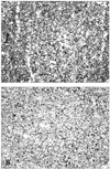
BCL-6, a transcription factor involved in antibody production by B lymphocytes, has been shown to be a major factor regulating the differentiation of B lymphocytes into plasma cells in the germinal centers.25-27 Plasma cells secrete IgA, which is involved in immune reactions that sometimes induce chronic inflammation in the adenoids. BCL-6 was first discovered as a gene translocated in a type of non-Hodgkin's lymphoma, but it has not been found in terminally differentiated plasma cells.28,29 We found that BCL-6 expression was significantly lower in the OME group, and that it was expressed primarily in the germinal centers. One explanation might be that BCL-6 suppresses conversion of B lymphocytes to plasma cells in normal adenoids, but decreased BCL-6 expression during infection allows increased conversion to plasma cells and thus greater production of immunoglobulin. However, IgA secretion was not increased significantly in the OME group, and thus a firm association between IgA secretion and BCL-6 expression could not be made. Since we measured BCL-6 expression only after antigen stimulation, and since BCL-6 staining occurred primarily in the germinal centers, it may be that this transcription factor directly controls IgA production in the germinal centers.
In conclusion, we observed significant decreases in the respiratory epithelium of adenoid mucosa, significant increases of squamous epithelial metaplasia, and significant reductions of BCL-6 expression in OME patients. Therefore, OME may be due to alterations in the mucosal epithelium due to inflammatory reactions in the adenoids, and may influence the expression of BCL-6, which controls antibody production in the germinal centers of adenoids. Since the causes and clinical course of OME are diverse and complex, however, the pathophysiology of this condition should be understood in association with other risk factors, not only adenoid problems.
Figures and Tables
Fig. 1
Squamous metaplasia of adenoid. Upper hinge of box, upper range; midline of box (-☐-), mean. Samples were graded by percent (%) squamous metaplasia, as determine by H-E staining. *p < 0.05.

Fig. 2
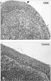
H-E stain for squamous metaplasia. In OME adenoids, thin squamous epithelium (↙) is transposed to thick squamous epithelium (↙) without respiratory ciliated cells. Control adenoids, however, have respiratory ciliated cells without squamous epithelium. Original magnification, × 100.

Fig. 3
Mucosal barrier of adenoid. Upper hinge of box: upper range, midline of box(-☐-): mean. The number of areas of deterioration area were calculated by immunohistochemical staining for cytokeratin.

Fig. 4
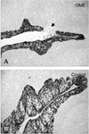
Cytokeratin immunohistochemistry for mucosal barrier. Microscopic deterioration of the mucosal barrier was detected microscopically in OME adenoid (arrow head), but not in control, adenoids. Original magnification, × 40.

Fig. 5
IgA positive cells in adenoids. Upper hinge of box: upper range, midline of box(-☐-): mean. Samples were graded by the percent (%) IgA positive cells.

Fig. 6
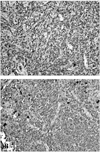
IgA immunohistochemistry. Nearly the same percentage of cells were positive for IgA in control and OME adenoids. Original magnification, × 400.

Fig. 7
BCL-6 positive cells of adenoid. Upper hinge of box, upper range; midline of box (-☐-), mean; lower hinge of box, lower range. Samples were graded by the percent (%) BCL-6 positive cells. *p < 0.05.

References
2. Tomonaga K, Kurono Y, Chaen T, Mogi G. Adenoids and otitis media with effusion: nasopharyngeal flora. Am J Otolaryngol. 1989. 10:204–207.

3. Gates GA, Avery CA, Prihoda TJ. Effect of adenoidectomy upon children with chronic otitis media with effusion. Laryngoscope. 1988. 98:58–63.

4. Chu KC, Chang BC. Diagnosis and treatment of pediatric adenotonsillar disease. J Clin Otolaryngol. 1999. 10:134–146.

6. Brodsky L, Koch RJ. Anatomic correlates of normal and diseased adenoids in children. Laryngoscope. 1992. 102:1268–1274.

7. Ophir D, Gilboa S, Halperin D, Marshak G. Obstructing adenoids in adolescents: changing trends? J Otolaryngol. 1993. 22:91–93.
8. Fujioka M, Young LW, Girdany BR. Radiographic evaluation of adenoidal size in children: Adenoidal-nasopharyngeal ratio. AJR Am J Roentgenol. 1979. 133:401–404.

9. Paparella MM, Jung TT, Goycoolea MV. Paparella MM, Shumrick DA, Gluckman JL, Meyerhoff WL, editors. Secretory otitis media. Otolaryngology. 1991. 3rd ed. Philadelphia: Saunders;1317–1342.
10. Post JC, Prestone RA, Aul JJ, Larkins-Pettigrew M, Rydquist-White J, Anderson KW, et al. Molecular analysis of bacterial pathogens in otitis media with effusion. JAMA. 1995. 273:1598–1604.

11. da Costa JL, Navarro A, Branco Neves J, Martin M. Otitis medias with effusion: association with the Eustachian tube dysfunction and adenoiditis. The case of the Central Hospital of Maputo. Acta Otorrinolaringol Esp. 2005. 56:290–294.

12. Maw AR. Chronic otitis media with effusion (glue ear) and adenotonsillectomy: prospective randomized controlled study. Br Med J (Clin Res Ed). 1983. 287:1586–1158.
13. Gates GA, Avery CA, Prihoda TJ, Cooper JC Jr. Effectiveness of adenoidectomy and tympanostomy tubes in the treatment of chronic otitis media with effusion. N Engl J Med. 1987. 317:1444–1451.

14. Coyte PC, Croxford R, McIsaac W, Feldman W, Friedberg J. The role of adjuvant adenoidectomy and tonsillectomy in the outcome of the insertion of tympanostomy tubes. N Engl J Med. 2001. 344:1188–1195.

15. Maw AR, Bawden R. Factors affecting resolution of otitis media with effusion in children. Clin Otolaryngol Allied Sci. 1994. 19:125–130.

16. Roydhouse N. Adenoidectomy for otitis media with mucoid effusion. Ann Otol Rhinol Laryngol Suppl. 1980. 89:312–315.

17. Gates GA, Avery CA, Prihoda TJ. Effect of adenoidectomy upon children with chronic otitis media with effusion. Laryngoscope. 1988. 98:58–63.

18. Phillips DE, Maw AR, Harvey K. The nasopharynx and adenoid in children with glue ear compared with normal controls. Clin Otolaryngol Allied Sci. 1987. 12:255–260.

19. Maw AR, Parker A. Surgery of the tonsils and adenoids in relation to secretory otitis media in children. Acta Otolaryngol Suppl. 1988. 454:202–207.

20. Kamel RH, Ishak EA. Enlarged adenoid and adenoidectomy in adults: Endoscopic approach and histopathological study. J Laryngol Otol. 1990. 104:965–967.

21. Fujihara K, Fujihara T, Yamanaka N. Secretory IgA and squamous epithelization in adenoids of children with otitis media with effusion. Acta Otolaryngol Suppl. 1996. 523:155–157.
22. Forsgren J, Rynnel-Dagoo B, Christensson B. In situ analysis of the immune microenvironment of the adenoid in children with and without secretory otitis media. Ann Otol Rhinol Laryngol. 1995. 104:189–196.

23. Brandtzaeg P. Immune functions and immunopathology of palatine and nasopharyngeal tonsils. Immunology of the Ear. 1992. New York: Raven;63–66.
24. Arita M, Kodama S, Suzuki M, Mogi G. Single cell analysis of adenoid CD5+B cells and their protective contributions to nasopharyngeal immunity. Laryngoscope. 2003. 113:484–491.

25. Deweindt C, Albagli O, Bernardin F, Dhordain P, Quief S, Lantoine D, et al. The LAZ3/BCL-6 oncogene encodes a sequence-specific transcriptional inhibitor: a novel function for the BTB/ POZ domain as an autonomous repressing domain. Cell Growth Differ. 1995. 6:1495–1503.
26. Chang CC, Ye BH, Chaganti RS, Dalla-Favera R. BCL-6, a POZ/zinc-finger protein, is a sequence-specific transcriptional repressor. Proc Natl Acad Sci U S A. 1996. 93:6947–6952.

27. Seyfert VL, Allman D, He Y, Staudt LM. Transcriptional repression by the proto-oncogene BCL-6. Oncogene. 1996. 12:2331–2342.


 PDF
PDF ePub
ePub Citation
 Print
Print


 XML Download