

 PDF
PDF ePub
ePub Citation
 Print
Print


INTRODUCTION
For many years, spontaneous dissecting aneurysms of the intracranial vertebral artery (VA) were believed to be quite rare; however these aneurysms are now being reported more frequently and are increasingly recognized as a source of appreciable morbidity and mortality.1-6 Since these lesions can be difficult to diagnose on an angiography and clinical manifestations may be delayed or overlooked, their prevalence is not precisely known.7 Furthermore, the indications and methods of treatment remain controversial.
In the present study, we have reviewed cases at our institute and those reported in the literature to provide proper management strategies and to improve the management outcome of patients on the basis of experience over the past 14 years.
MATERIALS AND METHODS
This study represents a retrospective analysis of all patients admitted to and treated at our institute from February 1992 to June 2005 with a diagnosis of spontaneous dissecting aneurysm of the intracranial VA. During this period, a total of 1,990 patients were treated for intracranial aneurysms by surgery or neurointervention. Of these, 194 (9.7%) had aneurysms of the posterior circulation. Fifty-four (27.8%) of the 194 patients had aneurysms of the VA, and 28 (51.8%) of the 54 patients with aneurysms of the VA had dissecting aneurysms. The mean follow up period was 33.6 months.
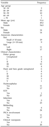
The study therefore involved a total of 28 patients ranging in age from 34 to 71 years (2 in their 30s; 10 in their 40s; 10 in their 50s; 5 in their 60s; and 1 in their 70s), with a median age of 51.6 years. Fourteen (50%) patients were male and 14 (50%) were female (Table 1).
All patients were investigated by computed tomography (CT), cerebral angiography, and magnetic resonance imaging (MRI). Magnetic resonance angiography (MRA) and/or diagnostic lumbar spinal tapping were also used to diagnose some patients.
Nineteen (67.9%) of the 28 patients had small (diameter < 10mm) aneurysms, and the remaining 9 (32.1%) patients had large (diameter ≥ 10mm) aneurysms. The aneurysms were located on the right side in 17 patients and on the left in 11 patients. In 4 (14.3%) of 28 patients, aneurysms involved the posterior inferior cerebellar artery (PICA) (Table 1). Thirteen (46.4%) patients had lesions proximal to PICA, and 11 (39.3%) patients had lesions distal to PICA.
Of the 28 patients, 22 (78.6%) presented with subarachnoid hemorrhage (SAH), and the remaining 6 patients (21.4%) had unruptured lesions. The Fisher group was I in 2 patients, II in 4 patients, III in 12 patients, and IV in 4 patients. The initial Hunt & Hess grade on admission was 0 in 6 patients, I in 4 patients, II in 6 patients, III in 8 patients, IV in 3 patients, and V in 1 patient. Four (18.2%) of the 22 patients presented with recurrent SAH before admission, 1 day, 2 days, and 6 days respectively after the first ictus. Hydrocephalus was identified in 11 (50%) of 22 patients. One patient presented with both SAH and intracerebral hemorrhage (ICH). Cerebral or cerebellar infarction was identified in 4 (18.2%) of 22 patients with ruptured aneurysms, and one (16.7%) of 6 patients with unruptured aneurysms. Fourteen (50%) patients had current medical history of essential hypertension (Table 1).
Neurointervention was performed in 20 patients (71.4%) and involved the use of a detachable coil, a detachable balloon, and self-expandable stent. The methods used were proximal VA occlusion in 16 patients via a detachable coil with/without a detachable balloon, and intra-aneurysmal packing of detachable coil with or without stent in 4 patients (Table 2).
Surgery was performed in 8 patients (28.6%). The methods used were: wrapping in 3 patients, proximal VA occlusion with clipping in 3 patients, clipping of aneurysmal neck in one patient, and trapping in one patient. All surgical approaches were by the lateral suboccipital route (Table 2).
Indications of surgery or neurointervention were analyzed for each case.
The management outcome was assessed as favorable (good, fair) or unfavorable (poor, dead) using the Modified Glasgow Outcome Scale (GOS) at a 6-month follow-up.
RESULTS
In selection of the therapeutic option, the following patients were initially evaluated as possible candidates for neurointervention (occlusion of the affected VA proximal to the PICA): 1) poor clinical grade; 2) advanced age; 3) medical illness; 4) unruptured aneurysm; 5) equal or larger opposite VA; 6) anticipated surgical difficulty due to a deep location of the VA-PICA junction. Surgery (occlusion of the affected VA distal to the PICA or clipping and reinforcement of the dissecting aneurysms) was considered for patients with: 1) high-risk aneurysms (large or irregular shaped); 2) smaller opposite VA; 3) failed neurointervention; 4) dissection involving the PICA.
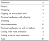
Favorable outcome was obtained in 25 (89.3%) of the 28 patients (23 good, 2 fair). The outcome for the remaining 3 patients was poor in 2 patients due to the initial insult, and death in one patient suffering from acute respiratory distress syndrome within days of neurointervention (Table 3).
Among 20 patients treated by neurointervention, there were no major complications attributable to the procedure. One patient suffered acute respiratory failure that was not attributable to the interventional procedure (Table 3).
Two (25%) of 8 patients undergoing surgery experienced significant complications directly attributable to the procedure: one suffered postoperative cerebrospinal fluid collection with infection, and the other suffered postoperative temporary facial palsy. One patient suffered from post- operative pneumonia that was not attributable to surgery (Table 3).
DISCUSSION
Spontaneous dissecting aneurysm of the intracranial VA observed by angiography, surgery, and autopsy was reported by Yonas et al. in 1977.8 Provenzale JM et al. made the same diagnosis based on characteristic radiologic findings, appropriate clinical presentation, and the demonstration of the absence of atherosclerotic disease elsewhere in the cerebrovascular circulation.9
The annual incidence of spontaneous dissecting aneurysm is 1 to 1.5 per 100,000, and the lesion is known to be an important cause of ischemic stroke in young and middle-aged patients.10 The lesions have been reported to occur in a relatively young age group,5,11 with a mean age of 45.6 ± 8.7 years, and a peak age of onset in the 40s for the 38 reported lesions.5,11,12 The data in this study showed similar results. In contrast to female predominance (70%) in saccular aneurysms of the VA,13 dissecting aneurysms showed a male predominance among both SAH and non-SAH patients.5,11 However, our data did not show any sexual predominance. Our study indicated a right-side dominance similar to that reported by Yamaura A et al.5 Sasaki et al. reported that the right VA was more frequently involved in SAH patients, whereas involvement of the left VA was more common in non-SAH patients.13 However, bilateral involvement of the VA was not rare, and there were 5 instances in the 36 collected cases.5 In our series, one patient had bilateral lesions although the asymptomatic side was not treated. Hypertension was recorded in 29% of one reported series,5 but in a separate series hypertension and atherosclerosis were shown to be rare.12 Our results indicated a comorbid incidence of hypertension in 33.3% of patients.
The first step in clinical management of spontaneous dissecting aneurysm of the intracranial VA is to recognize early symptoms and to make the correct initial diagnosis based on characteristic symptomatology.6 Patients usually present with a variable combination of neck pain or headache, transient loss of consciousness, SAH, and embolic stroke.7 Mizutani T et al. reported that patients presented with pain and embolic neurological deficits if the dissection was confined by the arterial media, but rupture of the thin layer of intradural adventitia usually occurred and resulted in SAH.3 The reported incidence of SAH in the literature is similar to that of our data. Yamaura A et al. reported that the incidence of SAH was 86% (21 of 24 cases).5 In a study by Kitanaka C et al., 16 out of 24 patients treated during the last 12 years were admitted with SAH.14 Fourteen of 16 patients reported by Sano H et al. developed SAH, and 4 of these 14 patients complained of headaches for several days (2-10 days) prior to the onset of SAH.6 Although in one series where the preoperative rebleeding rate was not very high (4 of 16, 25%) probably because many patients underwent early surgery,14 subsequent rebleeding is thought to occur frequently especially within 24 hours in ruptured lesions.12 Further hemorrhage is associated with high mortality (45%) and poor neurological outcome.3 All patients, including those who presented with ischemia, complained of sudden and severe headache,5,14 and the occipital headache or neck pain intensified immediately before clinical deterioration.5 The pain waxed and waned in some cases and in others appeared repeatedly at intervals of several hours to weeks.5 Friedman AH and Drake CG reported that headache might precede neurological deficits by days or weeks, and that occasionally the stroke was not completed at its onset but proceeded in a stepwise fashion.1 Many authors consider that a history of prodromal neck pain is a characteristic finding, and that a neurosurgeon should suspect dissecting aneurysm of the VA in such cases.1,5,11 Differential diagnosis includes brain stem infarctions, Wallenberg's syndrome, cerebellar infarction, central vertigo, and transient ischemic attacks.2
Cerebral angiography is necessary for definitive diagnosis and preoperative assessment, although MRI (plus MRA) offers a non-invasive means of ensuring satisfactory parent vessel occlusion in treated patients.7 Even with angiography the diagnosis is not easy, and CT and MR examinations of the head are frequently normal.9 The most common erroneous diagnosis was that of a ruptured saccular aneurysm of unusual shape with intraluminal thrombus, associated with vasospasm of its parent artery.1,8 The most common angiographic finding is irregular narrowing and fusiform dilatation of the vessel (pearl and string sign) with retention of contrast medium into the late arterial phase.2,3,5 Kitanaka C et al. reported that the angiographic findings of SAH patients were uniform compared with those of non-SAH patients, with a typical pattern of a pearl and string appearance. Most of the string signs in SAH patients showed a short, mild tapering 'constriction' in contrast to the typical string sign commonly seen in non-SAH patients. This constriction sign was helpful in differentiating dissecting aneurysms from fusiform aneurysms. They correlated the angiographic findings with the intraoperative findings in their series using the origin of the PICA as the landmark. As a result, the constriction sign was considered to represent the very end of the dissected portion. In general, because of the presence of constriction signs, they felt that the extent of VA abnormalities in SAH patients was localized and easy to predict preoperatively.14 Angiographically normal portions also appeared to be normal intraoperatively.14,15 Angiographic change due to the progression of minimal dissection not found at the time of the first angiogram was also reported by Ito Y et al.15 A saccular aneurysm at the site of dissection is uncommon and is more typical of dissecting aneurysm of the extracranial VA.16
General methods of surgical treatment are proximal occlusion of the VA, trapping, and direct clipping and/or vertebral arterial reconstruction. The first surgical procedure was performed by Yonas et al. in 1977.8 The principal therapeutic aim was the proximal occlusion of the VA adjacent to the dissection without manipulation of the aneurysm,7 and this procedure has been regarded as the standard surgical technique for treatment of this condition.8 Trapping and wrapping have also been used in a small number of patients.14 In 1989, Tanaka et al. reviewed the literature and summarized 22 surgical cases. The surgical techniques used were proximal clipping in 18 cases, and trapping in 4 cases. Because the incidence of postoperative deterioration was lower after proximal clipping (2 of 18) than after trapping (2 of 4), they concluded that proximal clipping of the parent artery was the treatment of choice.17 However, since bleeding after appropriated proximal clipping is possible, Kitanaka C et al. argued that proximal clipping is not a perfect treatment, and that a combination of trapping and base clipping is more satisfactory with respect to prevention of recurrent SAH or dissection.18
With respect to surgical complications, Kitanaka C et al. reported that lower cranial nerve (glossopharyngeal, vagus, accessory nerve) palsy was seen very frequently (in 13 of 17 cases, 76%), although there was no apparent cerebellar dysfunction.14 They also observed that trapping procedures were associated with a higher incidence and a more severe degree of lower cranial nerve palsy, and that abducens palsy and hypoglossal palsy were also frequent after this procedure.14 The far lateral inferior suboccipital approach, which provides better visualization of the distal VA with less retraction,18 did not prove to be superior to the classic lateral suboccipital approach in reducing postoperative neurological complications.14 They considered that sacrificing functioning perforating arteries, which may be end arteries supplying the brain stem, has a high risk of brain stem ischemia, whereas sacrificing the PICA is not always dangerous because of the rich pial anastomoses.14 However, when it is necessary to sacrifice the PICA in a case with poor collateral network covering the PICA territory, the anastomosing technique must be taken into consideration.19,20
Infarction of the perforating branches may occur by retrograde thrombosis of the VA due to occlusion of the vascular lumen between the VA and the perforating branches. In one series, brain stem infarction occurred in 1 of 16 patients.6 Although VA reconstruction is difficult, blood flow through parent arteries should be preserved as much as possible.6
Direct clipping may be preferable in cases with unilateral aneurysms and VA reconstruction was successful in 8 of 16 patients reported by Sano H et al.6
Rebleeding occurred in 5 patients (24%) of one series within 17 days of the initial ictus, and the outcome of these patients was extremely poor. Surgery is required to prevent such early rebleeding; however, dissecting aneurysms are naturally unclippable and the occlusion of the affected VA would pose a considerable risk in the acute stage.5
Yamaura A et al. reported that the most characteristic operative findings were a fusiform or tubular enlargement of the affected artery and discoloration due to intramural hematoma.5 The ruptured lesion becomes a firm whitish gray mass, probably due to an organized intramural clot, approximately 1 month after the ictus and rebleeding is less likely to occur at this stage.5
The indications for surgical treatment remain controversial. Yamaura et al. insisted that a longer follow-up study and analysis of a larger number of untreated cases are required to identify when surgical treatment is indicated.5 There is no single recommended surgical treatment because each technique has its advantages and disadvantages,14 therefore it is essential to individualize surgical treatment on a case by case basis.14
A convincing argument for early neurointervention also exists.7 Since dissecting aneurysms are extremely fragile and easily ruptured at operation, proximal occlusion of the parent vessel by neurointervention is considered to be the safest method.2,4 However, a case showing refilling of the aneurysm by the contralateral VA has been reported.21 Recently Iihara et al. reported the optimal management strategy of neurointervention for VA dissecting aneurysms including the most serious lesions involving the PICA. They concluded that aneurysms that did not involve the PICA could be safely treated by internal trapping. For lesions that involved the PICA, they described a decision-making algorithm to maximize the efficacy of the treatment as well as minimize the risks of treatment-related morbidity based on balloon test occlusion (proximal endovascular occlusion performed during the acute stage followed by internal trapping of the dissected site).21 We treated some selected lesions (Case No. 24 and 27) by stenting with self-expanding, flexible stent (Neuroform stent®) followed by coiling. The stent can prevent coil herniation into the parent artery.22 But, anticoagulation medication and post treatment follow-up imaging study should be performed for possibility of thromboembolism and in-stent stenosis.22
Prognosis is also controversial. Among the 20 surgically treated cases reported in the 1970's and 1980's and the 19 surgical cases in another series, there was only one death, reported by Yonas et al.5,8 The clinical course of the untreated cases is very important. In 16 untreated cases described in the report by Yamaura A et al., 3 achieved good recovery after SAH, 2 others were moderately disabled after ischemia, and the remaining 11 patients died without surgery: 2 of rebleeding at 5 days and 15 days respectively after initial hemorrhage, and the other 9 from the initial ictus.5 During long-term observation in the report by Nakagawa K et al., good or excellent recovery was achieved in 14 (87.5%) of 16 patients.23 These authors also insisted that a follow-up angiography must be performed during the early stage (within approximately 3 weeks of the onset of symptoms) to confirm the formation or enlargement of an aneurysm because such conditions might be amenable to surgical treatment, and unruptured lesions could otherwise be treated and followed conservatively.23 Kitanaka et al. reported that the long-term outcome of surgically treated lesions was favorable, but there was a very high rate of postoperative neurological complications, and the major causes of disability were attributable to lower cranial nerve palsy and associated events in half of the patients with poor outcome.14 Prolonged intubation or tube feeding is an extreme burden on conscious patients.14 They insisted that the appropriate selection of surgical techniques and intensive postoperative care, including deliberate respiratory management, is necessary to improve the outcome of surgical treatment and shorten hospitalization.14
The overall management outcome in our series was satisfactory. Ruptured aneurysms must be treated as soon as possible to prevent early rebleeding. For unruptured aneurysms, a follow-up angiography is necessary to detect formation or growth of aneurysm. The treatment modality of dissecting aneurysm of the VA should be selected according to the clinical characteristics of each patient and close collaboration between neurosurgeons and neurointerventionists is essential to improve the management outcome.
 XML Download