

 PDF
PDF ePub
ePub Citation
 Print
Print


INTRODUCTION
Syringoma is a benign adnexal tumor derived from intraepidermal eccrine duct, which occurs predominantly in women at puberty or later in life. It typically presents as small, skin-colored or slightly yellow, soft papules, usually only 1mm or 2mm in diameter on the lower eyelids.1 Even though syringoma is a common benign eccrine gland neoplasm, there have been not enough studies on the clinical or histopathological features of this disease. The aim of our study is to describe clinical and histopathological features of sixty one patients with histological diagnosis of syringoma over four year period in our dermatology clinic in Korea. We also performed statistical analysis to find out correlations between clinical and histopathological features of syringoma.
MATERIALS AND METHODS
Sixty one cases with histologically confirmed diagnosis of syringoma observed in the department of dermatology (Severance Hospital, Seoul, Korea) between 2000 and 2003 were selected for this retrospective study. Clinical features of age, sex, age of onset, family history, associated diseases, subjective symptoms, aggravating factors, clinical impressions, clinical characteristics of lesions such as color, location, and distribution, and histopathological characteristics including acanthosis, basal hyperpigmentation, proliferation of fibrous stroma, vacuolization of inner layer of ductal cells, keratin-filled cyst, and tadpole appearance were reviewed and analyzed by hospital medical records and biopsy slides using t-test, Mann-Whitney U test and chi-square test. Clinical course of twenty three patients treated with CO2 laser were also investigated during follow-up visits. The multiple section specimens stained with haematoxylin-eosin were reviewed.
Additional sections were cut from 56 paraffin blocks from 56 patients for immunohistochemistry. Deparaffinization and rehydration of tissue sections were done with xylenes or other clearing agents and graded alcohol series. After microwave pretreatment in citrate buffer (pH 6) for antigen retrieval, slides were immersed in 0.3% H2O2 for 20 minutes to block the endogenous peroxidase activity. After washing, slides were incubated for 1 hour at room temperature with monoclonal antibody against estrogen receptor (ER) and progesterone receptor (PR) (NovoCastra, Newcastle, United Kingdom) in a dilution of 1:100.
RESULTS
Clinical features
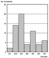
Sixty one Korean patients (53 females and 8 males; ratio 6.6:1) were included in this study. The mean age of the patients was 40 years (range 10-72) with average duration of 8 years and mean age of onset of 32 years. Distribution of age of onset showed 2 patients (3.3%) in the 1st decade, 14 patients (23.0%) in the 2nd decade, 20 patients (32.8%) in the 3rd decade, 5 patients (8.2%) in the 4th decade, 11 patients (18.0%) in the 5th decade, and 4 patients (6.6%) in the 6th decade, and 5 patients (8.2%) in the 7th decade (Fig. 1). Two peaks were observed in the 3rd decade and 5th decade. There were seven patients (11.5%) with family history of syringoma. Associated diseases included hypertension in 3 cases (4.9%), diabetes mellitus in one case (1.6%), B-viral hepatitis in one case (1.6%), and parathyroid adenocarcinoma in one case (1.6%). Nine patients (14.8%) complained of pruritus in the skin lesion. Aggravating factors were reported to be summer season in eight cases (13.1%), stress in two cases (3.3%), and menstruation in one case (1.6%). Clinical impressions were syringoma with concordance rate of 68.9%, verruca plana in nine cases, and various disease entities including bowenoid papulosis, keratosis pilaris, sarcoidosis, xanthelasma, and sebaceous hyperplasia. The most frequently involved site was eyelids (43 cases, 70.5%), followed by forehead (14 cases, 23.0%), cheek (9 cases, 14.8%), genital area (8 cases, 13.1%), abdomen (6 cases, 9.8%), upper extremities (6 cases, 9.8%), neck (5 cases, 8.2%), chest (3 cases, 4.9%), lower extremities (1 case, 1.6%), upper lip, nose, and chin (1 case, 1.6%). 8 patients (13.1%) were categorized in generalized type whereas 53 patients (86.9%) were categorized in localized type. The color of lesions were skin-colored (30 cases, 49.2%), brownish (16 cases, 26.2%), erythematous (13 cases, 21.3%), yellowish (1 case, 1.6%), whitish (1 case, 1.6%). Out of 11 patients (18%) who were treated with CO2 laser and followed up, 7 patients (63.6%) reported clinical improvements whereas 4 patients (36.4%) reported recurrences.
Histopathologic features
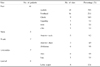
We could observe five cases with acanthosis (8.2%), 16 cases with basal hyperpigmentation (26.2%), 16 cases with proliferation of fibrous stroma (26.2%), 20 cases with vacuolization of lining cells (32.8%), 17 cases with keratin-filled cysts (27.9%), 34 cases with characteristic tad-pole appearances (55.7%) (Table 1). Immunohistochemical stains for the presence of PR and ER were performed in 56 cases with all negative results.
Correlations between clinical features and histopathologic features
We performed statistical comparison analysis between clinical features (age, sex, age of onset, duration, presence of family history, presence of past history, symptom, color, genital involvement and distribution) and histopathologic features including acanthosis, basal hyperpigmentation, fibrosis, vacuolization of duct, keratin cyst and tad-pole appearance. Basal hyperpigmentation was observed more frequently in brown-colored lesion (Chi-Square Tests, p = 0.005; skin-colored, 12.5%; erythematous, 23.1%; brown-colored, 56.3%). Fibrosis was observed more frequently in erythematous lesion (Chi-Square Tests, p = 0.033; skin-colored, 15.6%; brown-colored, 31.3%; erythematous, 53.8%). Keratin cyst was observed less frequently in genital involved group (Chi-Square Tests, p = 0.006; genital involved, 75.0%; genital uninvolved, 22.6%). Other comparisons were statistically insignificant (Table 2).
DISCUSSION
Syringoma is a benign adnexal tumor derived from intraepidermal eccrine duct that may present as single or multiple papules predominantly in women at puberty or later in life, commonly on the eyelids, in 0.6% of the population.1,2 Selection bias should be considered since only patients who sought consultation with dermatologist and, furthermore, who were subjected to biopsy were included in our study.
Several selection biases may be responsible for exaggerated female:male ratio of 6.6:1 in our study compared with 1.2:1 reported by Patrizi et al.3 It is speculated that females with syringoma seek medical care more frequently for cosmetic reasons. More than half of the patients reported the age of onset during 2nd and 3rd decades. Two peaks were observed in the 3rd decade (32.8%) and 5th decade (18.0%) in a similar bipolar pattern of age of onset distribution (2nd, 4th) observed in Patrizi's study.3
Associated diseases in our study were hypertens diabetes mellitus, B-viral hepatitis and parathyroid adenocarcinoma. We could not observe vacuolization in a case associated with diabetes mellitus although the relationship between clear-cell syringoma and diabetes mellitus is well established.4 The patient was diagnosed of diabetes mellitus 5 years after the appearance of syringoma. Hypertension, diabetes mellitus and B-viral hepatitis do not seem to be related to syringoma considering the fact that hypertension, diabetes mellitus and B-viral hepatitis are common in Korean general population. Neoplasms including carcinoid tumor and tubular breast adenoma were reported.3,5 However, parathyroid adenocarcinoma has not reported as an associated disease with syringoma in the literature until now. Since aggravating factor may provide a clue in the pathogenesis of syringoma, it has been closely surveyed in many studies. Hwang et al.6 previously reported that 7 patients out of 18 vulvar syringoma patients experienced aggravation during summer or during menstruation. Summer season or high temperature was also the most common aggravating factor (eight patients, 13.1%) in our study. The report that topical atropine is effective in syringoma also supports the hypothesis that high temperature induced sweat secretion may worsen the symptom.7 Only one patient reported aggravation associated with menstruation in our study. This may explain the negative immunohistochemical stains for ER and PR in our cases.
The differential diagnosis of vulvar syringoma includes Fox-Fordyces disease, epidermal cysts, lichen simplex chronicus, and steatocystoma multiplex.8 Clinical impressions in our study were syringoma in 42 cases with concordance rate of 68.9%, verruca plana in 9 cases and other various diseases including bowenoid papulosis, keratosis pilaris, sarcoidosis, xanthelasma and sebaceous hyperplasia. Even though syringoma is relatively common, we think that skin biopsy is needed especially in nonfacial areas due to around 70% diagnostic concordance rate.
Friedman & Butler9 proposed a classification of syringoma consisting of four principal clinical variants: a localized form, a familial form, a form associated with Down's syndrome and a generalized form that encompasses multiple and eruptive syringoma. Study of 29 syringoma patients by Patrizi et al. revealed 11 cases of localized form and 18 cases of generalized form.3 However, 53 patients (86.9%) were localized form and 8 patients (13.1%) were generalized form and the most frequently involved site was eyelids (43 cases, 70.5%) in our study. This result can be explained by the fact that our study population mostly comprised of the most frequent typical clinical variant involving the eyelid in middle-aged women.
Even though Patrizi et al.3 could not find association between hyperpigmentation of basal layer and a brownish or a yellowish color of the lesions, we could find an association between basal hyperpigmentation and brown-colored lesion (Chi-Square Tests, p = 0.005; skin-colored, 12.5%; erythematous, 23.1%; brown-colored, 56.3%).
Previously reported methods for the treatment of syringoma include surgical methods such as electrodissection, cryotherapy, CO2 laser ablation, and excision or chemical therapy such as topical or systemic retinoids.10,11 A recent report suggests the use of topical atropine to alleviate the pruritus in symptomatic eruptive syringoma.7 There is also a report of vulval syringoma successfully treated with tranilast.12 In our series, 63.6% of patients out of 11 patients who were treated with CO2 laser and followed up showed clinical improvements compared with 100% resolution rate reported by Hwang et al.6 This may be due to the differences in the distribution and severity of conditions.
The influence of gonadal steroid hormones on the physiology and pathology of human skin have been studied extensively. However, the details of gonadal steroidal receptors in human skin are not well known. Immunohistochemical markers for estrogen receptor (ER) and progesterone receptor (PR) are routinely used in diagnostic surgical pathology to determine hormone receptor status for breast carcinoma.13 It has been theorized that syringomas are at least partially under estrogen or progesterone hormonal influences, as they are common in women and aggravate during pregnancy and menstruation.13 Wallace and Smoller13 reported that eight of nine syringoma cases showed strong nuclear and cytoplasmic progesterone receptor positivity, supporting the hypothesis of hormonal influence on syringoma. Yorganci et al.14 reported a vulvar syringoma showing progesterone receptor positivity. Timpanidis et al.15 also reported an eruptive clear cell syringoma associated with diabetes in which immunohistochemical staining showed negativity for ER and strong positivity for PR. However, there have been reports in which increase in ER and PR cannot be demonstrated. Trager et al.8 reported a case of syringoma showing neck and vulvar papules in an 8-year-old girl with negative results for both ER and PR by immunohistochemical stains. Recently, Huang et al.6 reported that all 15 specimens failed to stain with the ER and PR markers on syringoma and peripheral normal vulvar tissue. They also suggested that the lesions might not be under estrogen or progesterone control, or regulated by hormone not through the mechanism of increasing hormonal receptors. Later, Bal et al.16 also reported negativity for ER and PR in a case of vulvar syringoma aggravated by pregnancy.
In our study, we performed immunohistochemical stain on syringoma specimens from various anatomical sites in the largest number of cases reported until now. The results were all negative for ER and PR in fifty six specimens. As mentioned above, there are only two possibilities if ER or PR are not increased in syringoma. The first possibility is that syringoma is not under the influence of estrogen or progesterone. The second possibility is that if syringoma is influenced by estrogen or progesterone, the hormonal control is not through increasing hormonal receptors. Only one patient reported aggravation associated with menstruation in our study. There were no other clinical evidences of hormonal influence in our cases. Statistical analysis also revealed association between fibrosis and erythematous lesion. Since skin lesion with fibrosis may not respond well to ablative treatment, this correlation may be important in clinical aspect.
In conclusion, Review of clinicopathologic features of sixty one cases and immunohistochemical features of fifty six cases with histologically confirmed diagnosis of syringoma observed in the department of dermatology (Severance Hospital, Seoul, Korea) between 2000 and 2003 revealed that syringoma involves abdomen, chest and genitalia in addition to face with female preponderance without hormonal influence or control. The clinicopathological correlation also revealed that basal hyperpigmentation was observed more commonly in brown-colored skin lesion, that fibrosis was observed more commonly in erythematous lesion and that the keratin cyst was observed less frequently in genital lesion.

 XML Download