Journal List > Yonsei Med J > v.48(3) > 1030158

Cho: Current Issues on Molecular and Immunological Diagnosis of Tuberculosis

Abstract

Laboratory diagnosis of tuberculosis (TB) traditionally relies on smear microscopy and culture of Mycobacterium tuberculosis from clinical samples. With recent advances in technology, there have been numerous efforts to develop new diagnostic tests for TB that overcome the low sensitivity and specificity and long turnover time associated with current diagnostic tests. Molecular biological tests based on nucleic acid amplification have brought an unprecedented opportunity for the rapid and specific detection of M. tuberculosis from clinical specimens. With automated sequencing analysis, species identification of mycobacteria is now easier and more accurate than with conventional methods, and rapid detection of mutations in the genes associated with resistance to TB drugs provides early information on the potential drug resistance for each clinical isolate or for clinical samples. In addition, immunological tests for the detection of M. tuberculosis antigens and antibodies to the antigens have been explored to identify individuals at risk of developing TB or with latent TB infection (LTBI). The recent introduction of commercial IFN-γ assay kits for the detection of LTBI provides a new approach for TB control even in areas with a high incidence of TB. However, these molecular and immunological tools still require further evaluation using large scale cohort studies before implementation in TB control programs.

MOLECULAR DIAGNOSIS OF TUBERCULOSIS

Molecular tests have brought unprecedented opportunities for the rapid diagnosis of tuberculosis (TB) and can be incorporated into control programs for the disease. Most research on molecular tests has focused on the following three areas: (a) detection of Mycobacterium tuberculosis in clinical specimens, (b) identification of Mycobacterium species, and (c) detection of mutations in genes associated with resistance to drugs against TB.

Detection of M. tuberculosis in clinical samples

Over the last 20 years, nucleic acid amplification (NAA) tests have been studied for the ability to rapidly detect nucleic acids of pathogens in clinical samples for the diagnosis of infectious diseases including TB. NAA tests were particularly attractive for the diagnosis of TB because of the slow growth of TB, and these tests have advantages such as greater sensitivity and specificity and faster results than conventional laboratory diagnostics tests. Among the NAA tests, the polymerase chain reaction (PCR) has been most widely used for the detection of M. tuberculosis in clinical specimens including sputum, blood, bone marrow, and biopsy samples. Subsequently, several commercial kits have been developed using different targets from the M. tuberculosis genome and amplification formats, and these kits have been evaluated extensively in various clinical settings and samples with periodic reviews of their accuracy.1-5 Despite numerous claims that commercial kits have greater sensitivity than do culture methods, a series of technical problems have been encountered when PCR is employed in clinical mycobacteriology laboratories. A recent meta-analysis on commercially-based NAA tests including Amplicor-MTB (PCR), Cobas Amplicor-MTB (PCR), BDProbeTecET (strand displacement amplification: SDR), E-MTD (transcription mediated amplification; TMD), LCx (ligase chain reaction: LCR) showed marked variation in the diagnostic accuracy of the tests with regard to sensitivity and specificity.1 In the case of AFB smear-positive samples, the mean sensitivity ranged from 0.96 for the lowest kit to 0.98 for the highest, but the mean specificity varied markedly from 0.71 for the lowest kit to 0.96 for the highest. With AFB smear-negative samples, however, the mean sensitivity was only 0.57 for the lowest kit and 0.76 for the highest, and the mean specificity was 0.97 for the lowest and 0.99 for the highest. Overall, the pooled sensitivity and specificity were 0.96 and 0.85 for AFB smear-positive samples and 0.66 and 0.98 for AFB-negative samples, respectively. The high variability in sensitivity and specificity was also noted in a previous meta-analysis of commercially available kits.2-5
There have also been tremendous efforts to develop and evaluate the use of in-house PCR particularly in a research-based laboratory, where commercial kits are not affordable. In most cases, the variability in sensitivity and specificity for in-house PCR is even higher than that for commercial kits.2-4 With in-house PCR, the sensitivity varied from 9.4% to 100%, and the specificity varied from 5.6% to 100%.2 Therefore, careful interpretation of the NAA test results is important due to potentially confounding factors that may influence the results, such as the targets of amplification, the nature and amount of specimens, the storage method and duration before experiment, the specimen processing procedure, DNA extraction procedures, the presence of inhibitors, amplification methods, detection methods of amplified products, the contamination by amplicons, and cross-contamination between samples, etc.6,7
Besides the problems in accuracy and confounding factors associated with NAA tests, the relatively high expense is one of the major drawbacks for implementing these tests in field settings where TB is a major public health problem. Most NAA tests require expensive equipment and trained personnel as well as an expensive supply of materials. In order to overcome these drawbacks, another commercial kit, the loop-mediated isothermal amplification (LAMP) test,8 was recently developed to detect M. tuberculosis.9 The major advantages of the LAMP test include a room temperature reaction mode, obviating the need for thermocyclers, and visual reading of the results, obviating the need for electrophoresis steps or hybridization steps. A recent multi-center study involving countries with a high burden of TB including Tanzania, Peru, and Bangladesh was performed with technicians who had one week of LAMP test training, and the sensitivity of the LAMP test was 97.7% for smear- and culture- positive samples and 48.8% for smear-negative and culture-positive samples, and the specificity was 99%.10 Thus, the LAMP test looks promising for application in effective control programs in field settings with a high burden of TB.

Molecular tests for species identification of mycobacteria

Species identification of Mycobacterium species has become more important recently because of the steady increase in non-tuberculosis mycobacteria infections, particularly among HIV infected and elderly people and because of the wide use of liquid culture systems. Traditionally, species identification of mycobacteria relies on biochemical tests such as growth characteristics, pigmentation, the niacin test, the nitrate reduction test, the Tween 80 hydrolysis test, among others. Because these test procedures are often cumbersome, time consuming and inaccurate, several other methods such as HPLC analysis of mycolic acids,11,12 DNA probes,13,14 and sequence-based species identification systems have been developed and are widely used. HPLC analysis of mycolic acids provides a discrimination power of more than 50 species and can be run semi-automatically with minimum processing of cultures. One drawback of HPLC is the expensive instrumentation and the cost of machine and system maintenance. The AccuProbeR has been most widely used for species identification in laboratories that run a liquid culture system. At lease five species, including M. tuberculosis complex, M. avium, M. intracellulare, M. kansasii, and M. gordonae, constitute the majority of isolates from clinical samples.
With a marked increase in HIV infections, NTM infection has also increased as well and the number of Mycobacterium species has grown substantially. To cope with the increased number of Mycobacterium species, sequence information from the hypervariable region of the 16S rRNA of mycobacteria has been utilized for species identification.15,16 The polymorphic sites of the rpoB gene have been also used to differentiate among Mycobacterium species, and simple restriction enzyme analysis of the PCR products of the polymorphic sites makes it possible to differentiate more than 30 species.17,18 Oligonucleotide probes from the polymorphic site of the rpoB gene can also be used with a dot-blot hybridization assay for species identification.18 In addition, the polymorphic sequences of hsp65 can be successfully used for Mycobacterium species identification.19,20 Therefore, molecular techniques are more often used for Mycobacterium species identification and can be used to develop microarray-based assays in the near future.
One advantage of PCR-based methods over HPLC or AccuProbe is its direct applicability to clinical specimens, such as sputum samples, where the samples have enough bacilli to be AFB smear-positive. In the case of smear-negative and culture-positive specimens, the amplified products may not be enough to perform sequencing or hybridization with Mycobacterium species-specific probes. Since smear-positive specimens are indicative of transmitting agents, the rapid and correct identification of Mycobacterium species is very important.

Detection of mutations in the genes associated with resistance to TB drugs

The recent emergence of multidrug-resistant tuberculosis (MDR-TB)21 and extremely drug-resistant tuberculosis (XDR-TB)22 requires the rapid and accurate determination of drug-resistant M. tuberculosis. The recent emergence of MDR-TB may be due to easy access to the first-line TB drugs in most countries with a high prevalence of TB, including 22 high burden countries. The conventional methods for DST include proportion methods, absolute concentration methods, the drug resistance ratio, and the use of LJ and Middlebrook 7H10 agar. In addition, liquid culture systems, notably BACTEC MGIT960 and MB/BacT systems, have been evaluated extensively and seem to provide comparable results to the solid media-based tests with the advantage of rapid results.23 However, these culture-based methods still require 3-4 weeks including the original culture of organisms from clinical specimens. In order to reduce culture time, therefore, various molecular tests have been explored. Considering that the majority of drug resistance is due to mutations in genes encoding drug targets, one can develop molecular tests to identify gene mutations that are associated with drug resistance.24 The best examples are mutations in the rpoB gene, which are responsible for more than 95% of rifampin (RIF) resistance in M. tuberculosis. Some commercial hybridization assay kits, such as the INNO-LiPA Rif.TB kit25 and the GenoTyper MTBDR assay,26 can provide rapid results on RIF-resistance. A meta-analysis of the commercial probe assays gave a sensitivity of over 95% and specificity of 100% in 12 of 14 studies when cultured M. tuberculosis was used.27 However, there was a lower sensitivity of 82% to 92% and a specificity of 92% to 94% in the other two studies using the same commercial kits, indicating that careful interpretation of the results is required.
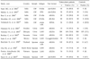
Likewise, resistance to isoniazid (INH) has been attributed to mutations in the katG, inhA, kasA, and/or ahpC genes of M. tuberculosis.24,28,29 However, the molecular mechanisms behind some INH- resistant M. tuberculosis isolates are not known. Other mutations in genes associated with resistance to other first line drugs and some second line drugs are listed in the Table 1. For EMB, while mutations at the embB codon 306 site have been implicated in causing resistance to the drug, only 40 to 70% of EMB-resistant isolates show mutations. Interestingly, mutations at the pncA gene match well with resistance to PZA because over 90% of PZA resistant isolates had mutations. The molecular mechanisms of drug resistance for most second-line drugs are only partially understood and the mutations that are responsible for the drug resistance have not been identified.

IMMUNODIAGNOSTIC TESTS

In general, immunodiagnostic tests can provide indirect evidence of current or past infections in organisms of interest. With the exception of the tuberculin skin test (TST), immunodiagnosis has not been widely used for chronic TB infections because of the low sensitivity and specificity. However, since immunological tests have the advantages of simplicity and rapid turnover over time, there have been tremendous efforts to develop serological tests for identifying people at risk of developing overt TB in the near future. In addition, since latent TB infection has become a hot issue for TB control programs, T cell-based assays such as the QuantiFERON test and T-SPOT.TB assay were developed as commercial kits and have been evaluated in field settings.

Antibody detection

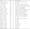
Antibody-based immunodiagnostic tests have been widely explored for detection of TB. With the whole genome sequence of M. tuberculosis available, it is now possible to identify M. tuberculosis-specific antigens that can be used to detect antibodies. In the literature, there are numerous M. tuberculosis antigens, including native, semi-synthetic, and recombinant ones, that have been shown to be useful for serologic diagnosis of pulmonary and/or extrapulmonary TB. Some of the antigens such as lipoarabinomman, cord factor, A60, 38kDa, 16kDa have been used to develop commercial kits, and the kits have been evaluated in areas with a low and high burden of TB (Table 2). As noted in Table 2, the results for all commercial kits varied markedly with sensitivity ranging from 15.7% to 89.2% and specificity ranging from 50% to 100%. In general, higher sensitivity comes at the cost of lower specificity. One of the major problems in the serodiagnosis of TB has been heterologous antibody responses to various M. tuberculosis antigens. It seems, therefore, that more than one antigen is required for increased sensitivity. This approach may result in a lower specificity despite the use of M. tuberculosis specific antigens.
The antibody detection method also evolved into various formats, although enzyme-linked immunosorbent assay (ELISA) is still the predominant one. Lateral flow tests or rapid tests, developed as commercial kits, have the advantages of rapidness and simplicity. However, subjective reading of the flow test results is a major drawback. Recently, a fluorescence polarization assay74 and surface plasmon resonance systems75 have been introduced for the detection of antibodies, which can be objectively read. However, the sensitivity and specificity have not been determined in serologic tests for TB. Likewise, protein chip-based assays are being developed that use a battery of M. tuberculosis antigens.76 Despite all the new technical developments in detecting antibodies for M. tuberculosis antigens, however, research should be conducted about the interpretation of the results for diagnosis or monitoring chemotherapy. A series of cohort studies is necessary to address these issues in the near future.

Antigen detection

Unlike antibody detection, M. tuberculosis-specific antigen detection provides direct evidence of current M. tuberculosis infection, thus warranting immediate chemotherapy against TB. However, since M. tuberculosis antigen-specific antibodies are used for the detection of antigens, there may be some intrinsic problems with non-specific positive results, even if for only a small fraction of them. Another limitation of antigen detection is the low sensitivity due to the scarcity of M. tuberculosis antigens in body fluids such as cerebrospinal fluid (CSF), pleural fluid, blood, and urine. Antigen detection formats include sandwich ELISA (sELISA) and dot-ELISA.
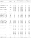
Target antigens vary markedly depending on the nature of the clinical samples and the test format. Lipoarabinomanna (LAM) has been most frequently used as the target antigen, and its monoclonal and polyclonal antibodies have been used to detect antigen in sputum samples (Table 3). One problem with using LAM as the target antigen is the non-specificity due to the presence in other mycobacterial species. Other antigens, such as whole cells, PPD, culture filtrate protein (CFP), 65kDa, 14kDa, among others, have been used as target antigens for antigen detection in CSF and sputum samples. Selection criteria for target antigens may include abundance, secretion, stability during specimen processing, and specificity to M. tuberculosis.
Sensitivity and specificity of antigen detection varies depending on the clinical specimens and the target antigen. In CSF samples, the sensitivity ranged from 50%81 to 90%79 with the specificity ranging from 80%81 to 100%77 (Table 3). Interestingly, LAM antigen was detectable in 74-93% of urine samples from TB patients and in 4-13% of those from healthy controls in the study areas. Since urine samples are easy to obtain and minimal processing is required, the antigen detection assay is promising for the rapid and simple diagnosis of tuberculosis in areas with a high prevalence of TB. However, urine antigen detection methods need to be evaluated further before they can be fully implemented in TB control programs. In sputum samples, the sensitivity of antigen detection varied from 71% to 91% with a specificity ranging from 91% to 100% (Table 3). Due to the complexity of sELISA and sputum processing steps, however, further studies are necessary to develop simple and highly specific antigen detection methods. In order to overcome the limitation of non-specificity, analytical tools, such as liquid chromatography-mass spectrometry, MRI, and NMR, among others, need to be explored to determine if they can directly detect mycobacterial antigens in clinical samples.

IFN-γ assays

The tuberculin skin test (TST) has been widely used to detect latent infection of M. tuberculosis to justify preventive therapy.90 The major drawback of the TST is the positive reaction due to recent BCG immunization or exposure to NTM that are present in the environment. To overcome such non-specific positive reactions with TST, ex vivo T cell-based assays have been explored in many laboratories using M. tuberculosis-specific antigens such as ESAT-6, CFP-10, and TB7.7 antigens whose genes are located in the RD1 or RD11 region, which is deleted in BCG vaccines.91,92 There are two commercial kits available in the market that claim to provide efficient and specific detection of latent M. tuberculosis infection: the QuantiFERON tests (QFT), including QuantiFERON-Gold (QFT-G), and QuantiFERON-Gold In Tube (QFT-GIT), and the T-SPOT.TB (SPOT-TB) assays.93,94 QFT quantitatively measures the amount of IFN-γ released by effector T cells after a 16-24 hour exposure to M. tuberculosis-specific antigens or peptides derived from ESAT-6, CFP-10, and TB7.7 proteins. The SPOT-TB assay was designed to count the number of effector T cells producing IFN-γ after stimulating T cells with the ESAT-6 and CFP-10 antigens overnight.
Both assays were excellent in detecting latent infection of M. tuberculosis.93,95 However, there was a subtle difference in the results due to the nature of testing formats. Since QFT measures the total amount of IFN-γ in the culture supernatant, certain T cells producing a large amount of IFN-γ can influence the total amount, while other T cells may produce little IFN-γ. In contrast, the SPOT-TB assay counts the effector T cells producing any level of IFN-γ without knowing the total amount of IFN-γ. It is not known yet which measurements are most relevant for the diagnosis of active or latent M. tuberculosis infection (LTBI) in terms of sensitivity and specificity, and which measurement is the best indicator of effective chemotherapy in the early phase of treatment.
Both the QFT and the SPOT-TB assay have been evaluated for the diagnosis of active TB and detection of LTBI among TB patients and healthy controls from areas with low, medium, and high prevalence of the disease.93,95-97 In a meta-analysis of numerous studies on IFN-γ assays, the sensitivity was 0.76 (95% confidence interval, CI: 0.7-0.83) by QFT and 0.88 (95% CI: 0.81-0.95) by the SPOT-TB assay when active TB patients were used as the gold standard for LTBI, indicating that the sensitivity of the SPOT assay is greater than that of QFT.95 On the other hand, the specificity of the IFN-γ assays was very high at 0.97 (95% CI: 0.95-0.99) by QFT and 0.92 (95% CI: 0.88-0.95) by the SPOT-TB assay when healthy controls with a low risk of LTBI were used as the negative control, indicating a greater specificity with QFT than with the SPOT-TB assay.95 In comparison, TST showed a sensitivity of 0.71 (95% CI: 0.65-0.74) and a specificity of 0.66 (95% CI: 0.41-0.84). The results indicate that, despite the high specificity, the sensitivity of IFN-γ assays is suboptimal and may be due to immune suppression among TB patients, which was evident in patients with cavity formation.98 The specificity of TST was very low, largely due to BCG vaccination in the study populations.95
When healthy controls (n = 3,216) with a varying risk for LTBI were examined, the overall QFT positive rate was 26.6%, ranging from 5% in the U.S. to 56% in South Africa, while TST gave a positive rate of 45.6%, ranging from 9% in the U.S. to 81% in South Africa.95,99,100 In a Korean population comprised of close contacts of TB patients and healthy controls, the QFT positive rate was 23% compared with a TST positive rate of 64%.96 In comparison, the SPOT-TB assay gave an overall positive rate of 31.8% among healthy controls from various geographical areas (n = 2,916), while the positive rate by TST was 37.4%.95 When the two IFN-γ assays were compared using the same study subjects (n = 633), the overall agreement rate was 83%, ranging from 76% to 93%, with a discordant rate of 6-21%.95,101,102 In a contact tracing of TB patients, the overall agreement rate was 90%, but this could be increased to 94% if different cut-off values are used.103
Test failure and indeterminate results have been noted with IFN-γ assays. In a prospective study on 393 subjects who were exposed to TB patients or who were suspected of having TB, five samples failed with QFT and another five failed with the SPOT-TB assay, indicating a failure rate of 1.3% in each assay.102 Of 383 subjects with satisfactory results, 144 (38%) tested positive on the SPOT-TB assay and 100 (26%) by QFT. The lower positive rate with QFT may be due to a higher rate of indeterminate results, which occurred more frequently with QFT (11%) than with the SPOT-TB assay (3%).102 The indeterminate results were due to immunosuppression from underlying diseases such as cancer, diabetes, HIV infection, among others, and were found more frequently when subjects were TST negative.104
The IFN-γ assays have been employed for diagnosis of active TB and for monitoring patients undergoing chemotherapy. When both IFN-γ assays were employed for the detection of TB among people suspected of having the disease, the results lead to other medical examinations such as CT, which in turn verified the diagnosis of TB.105-107 In addition, IFN-γ assays have been analyzed for use in monitoring TB patient progress with chemotherapy treatment. While QFT failed to show a difference in values before and after chemotherapy in TB patients,108,109 the SPOT-TB assay showed a significant decrease in counts after the completion of chemotherapy in TB patients, indicating that the SPOT-TB assay can be used as a biomarker for treatment efficacy.110 In addition, QFT values decreased when individuals with LTBI were given INH for six months for chemoprophylaxis, indicating that QFT can be also be used to monitor the efficacy of prophylactic treatment of LTBI.111 Likewise, IFN-γ assays were employed to monitor subjects at high risk of M. tuberculosis infection in order to identify newly infected persons between test intervals. In a cohort study on 216 health care workers, Pai et al.112 showed that 12% of the cohort subjects were converted to positive by QFT in 18 months. In comparison, only 4.1% had TST positive conversion, indicating that QFT is more sensitive than TST in detecting new M. tuberculosis infection.
More research on IFN-γ assays is necessary before they can be fully applied to detect LTBI. Future research topics should include: (a) reproducibility or validation of the assays, including variation in test results due to the delay between blood collection and assays, intra-laboratory reproducibility, inter-laboratory reproducibility, endotoxin contamination during assays, antigen types (protein vs. peptides), among others; (b) influence of repeated skin testing113 and exposure to non-tuberculosis mycobacteria such as M. kansasii, M. marinum, M. szulgai,114 which share the antigens and peptides used in the IFN-γ assays; (c) effects of immunosuppression by HIV infection115,116 and other underlying diseases such as cancer, diabetes and other concurrent infections, like pneumonia;117 and (d) the positive predictive values of IFN-γ assay results for the progression of IFN-γ positive subjects to overt TB, which will require a large scale cohort study. In the end, the interpretation of results from IFN-γ assays will vary depending on several confounding factors associated with the individuals or populations of interest.

Figures and Tables

Table 1
Mutations in the Genes Associated with Resistance to TB Drugs
ymj-48-347-i001
Table 2
Evaluation of Serological Test Kits for Diagnosis of Tuberculosis
ymj-48-347-i002

ICT, immunochromatography test; TBGL, tuberculous glycolipid, cord factor.

Table 3
Detection of M. tuberculosis Antigens in Clinical Samples for Diagnosis of Tuberculosis
ymj-48-347-i003

CSF, cerebrospinal fluid; LAM, lipoarabinomannan; sELISA, sandwich ELISA; dELISA, direct ELISA; RPHA, reverse passive haemagglutination; RIA, radioimmunoassay; PPD, purified protein derivatives; CFP, culture filtrate protein.

References

1. Greco S, Girardi E, Navarra A, Saltini C. Current evidence on diagnostic accuracy of commercially based nucleic acid amplification tests for the diagnosis of pulmonary tuberculosis. Thorax. 2006. 61:783–790.
crossref
2. Flores LL, Pai M, Colford JM, Riley LW. In-house nucleic acid amplification tests for the detection of Mycobacterium tuberculosis in sputum specimens: meta-analysis and meta-regression. BMC Microbiol. 2005. 5:55.
3. Pai M, Flores LL, Pai N, Hubbard A, Riley LW, Colford JM. Diagnostic accuracy of nucleic acid amplification tests for tuberculous meningitis: a systematic review and meta-analysis. Lancet Infect Dis. 2003. 3:633–643.
crossref
4. Pai M, Flores LL, Hubbard A, Riley LW, Colford JM. Nucleic acid amplification tests in the diagnosis of tuberculous pleuritis: a systematic review and meta-analysis. BMC Infect Dis. 2004. 4:6.
crossref
5. Piersimoni C, Scarparo C. Relevance of commercial amplification methods for direct detection of Mycobacterium tuberculosis complex in clinical samples. J Clin Microbiol. 2003. 41:5355–5365.
crossref
6. Pai M, Kalantri S, Dheda K. New tools and emerging technologies for the diagnosis of tuberculosis: part I. Latent tuberculosis. Expert Rev Mol Diagn. 2006. 6:413–422.
crossref
7. Tiwari RP, Hattikudur NS, Bharmal RN, Kartikeyan S, Deshmukh NM, Bisen PS. Modern approaches to a rapid diagnosis of tuberculosis: promises and challenges ahead. Tuberculosis (Edinb). 2007. 87:193–201.
crossref
8. Notomi T, Okayama H, Masubuchi H, Yonekawa T, Watanabe K, Amino N, et al. Loop-mediated isothermal amplification of DNA. Nucleic Acids Res. 2000. 28:e63.
crossref
9. Iwamoto T, Sonobe T, Hayashi K. Loop-mediated isothermal amplification for direct detection of Mycobacterium tuberculosis complex, M avium, and M. intracellulare in sputum samples. J Clin Microbiol. 2003. 41:2616–2622.
crossref
10. Boehme CC, Nabeta P, Henostroza G, Raqib R, Rahim Z, Gerhardt M, et al. Operational feasibility of using loop-mediated isothermal amplification for diagnosis of pulmonary tuberculosis in microscopy centers of developing countries. J Clin Microbiol. 2007. 45:1936–1940.
crossref
11. Butler WR, Jost KC, Kilburn JO. Identification of mycobacteria by high-performance liquid chromatography. J Clin Microbial. 1994. 29:2468–2472.
crossref
12. Thibert L, Lapierre S. Routine application of high-performance liquid chromatography for identification of mycobacteria. J Clin Microbiol. 1993. 31:1759–1763.
crossref
13. Goto M, Oka S, Okuzumi K, Kimura S, Shimada K. Evaluation of acridinium-ester labeled DNA probes for identification of Mycobacterium tuberculosis and Mycobacterium avium-Mycobacterium intracellulare complex in culture. J Clin Microbiol. 1991. 29:2473–2476.
crossref
14. Lebrun L, Espinasse F, Poveda JD, Vincent-Levy-Frebault V. Evaluation of nonradioactive DNA probes for identification of mycobacteria. J Clin Microbiol. 1992. 30:2476–2478.
crossref
15. Rogall T, Flohr T, Bottger EC. Differentiation of Mycobacterium species by direct sequencing of amplified DNA. J Gen Microbiol. 1990. 136:1915–1920.
crossref
16. Rogall T, Wolters J, Flohr T, Bottger EC. Towards a phylogeny and identification of species at the molecular level within the geneus Mycobacterium. Int J Syst Bacteriol. 1990. 40:323–330.
crossref
17. Lee H, Park HJ, Cho SN, Bai GH, Kim SJ. Species identification of mycobacteria by PCR restriction fragment length polymorphism of the rpoB gene. J Clin Microbiol. 2000. 38:2966–2971.
crossref
18. Lee H, Bang HE, Bai GH, Cho SN. Novel polymorphic region of the rpoB gene containing Mycobacterium species-specific sequences and its use in identification of Mycobacteria. J Clin Microbiol. 2003. 41:2213–2218.
crossref
19. Plikaytis BB, Plikaytis BD, Yakrus MA, Butler WR, Woodley CL, Silcox VA, et al. Differentiation of slowly growing Mycobacterium species, including Mycobacterium tuberculosis, by gene amplification and restriction fragment length polymorphism analysis. J Clin Microbiol. 1992. 30:1815–1822.
crossref
20. Kim HJ, Mun HS, Kim H, Oh EJ, Ha Y, Bai GH, et al. Differentiation of mycobacterial species by hsp65 duplex PCR-based restriction analysis and direct sequencing. J Clin Microbiol. 2006. 44:3855–3862.
crossref
21. Dye C, Espinal MA, Watt CJ, Mbiaga C, Williams BG. Worldwide incidence of multidrug-resistant tuberculosis. J Infect Dis. 2002. 185:1197–1202.
crossref
22. Gandhi NR, Moll A, Sturm AW, Pawinski R, Govender T, Lalloo U, et al. Extensively drug-resistant tuberculosis as a cause of death in patients co-infected with tuberculosis and HIV in a rural area of South Africa. Lancet. 2006. 368:1575–1580.
crossref
23. Garrigo M, Aragon LM, Alcaide F, Borrell S, Cardenosa E, Galan JJ, et al. Multicenter laboratory evaluation of the MB/BacT Mycobacterium detection system and the BACTEC MGIT 960 system in comparison with the BACTEC 460TB system for susceptibility testing of Mycobacterium tuberculosis. J Clin Microbiol. 2007. 45:1766–1770.
crossref
24. Zhang Y, Telenti A. Hatfull GR, Jacobs WR, editors. Genetics of drug resistance in Mycobacterium tuberculosis. Molecular Genetics of Mycobacteria. 2000. Washington, DC: ASM Press;235–254.
25. Cooksey RC, Morlock GP, Glickman S, Crawford JT. Evaluation of a line probe assay kit for characterization of rpoB mutations in rifampin-resistant Mycobacterium tuberculosis isolates from New York City. J Clin Microbiol. 1997. 35:1281–1283.
crossref
26. Cavusoglu C, Turhan A, Akinci P, Soyler I. Evaluation of the Genotype MTBDR assay for rapid detection of rifampin and isoniazid resistance in Mycobacterium tuberculosis isolates. J Clin Microbiol. 2006. 44:2338–2342.
crossref
27. Morgan M, Kalantri S, Flores L, Pai M. A commercial line probe assay for the rapid detection of rifampicin resistance in Mycobacterium tuberculosis: a systematic review and meta-analysis. BMC Infect Dis. 2005. 5:62.
28. Barry CE, Slayden RA, Mdluli K. Mechanisms of isoniazid resistance in Mycobacterium tuberculosis. Drug Resist Updat. 1998. 1:128–134.
29. Timmins GS, Deretic V. Mechanisms of action of isoniazid. Mol Microbiol. 2006. 62:1220–1227.
crossref
30. Kiepiela P, Bishop KS, Smith AN, Roux L, York DF. Genomic mutaitons in the katG, inhA, and ahpC genes are useful for the prediction of isoniazid resistance in Mycobacterium tuberculosis isolates from Kwazulu Natal, South Africa. Tuber Lung Dis. 2000. 80:47–56.
crossref
31. van Soolingen D, de Haas PE, van Doorn HR, Kuijper E, Rinder H, Borgdorff MW. Mutations at amino acid position 315 of the katG gene are associated with high-level resistance to isoniazid, other drug resistance, and successful transmission of Mycobacterium tuberculosis in the Netherlands. J Infect Dis. 2000. 182:1788–1790.
crossref
32. Lee AS, Lim IH, Tang LL, Telenti A, Wong SY. Contribution of kasA analysis to detection of isoniazid-resistant Mycobacterium tuberculosis in Singapore. Antimicrob Agents Chemother. 1999. 43:2087–2089.
crossref
33. Lee H, Cho SN, Bang HE, Lee JH, Bai GH, Kim SJ, et al. Exclusive mutations related to isoniazid and ethionamide resistance among Mycobacterium tuberculosis isolates from Korea. Int J Tuberc Lung Dis. 2000. 4:441–447.
34. Guo H, Seet Q, Denkin S, Parsons L, Zhang Y. Molecular characterization of isoniazid-resistant clinical isolates of Mycobacterium tuberculosis from the USA. J Med Microbiol. 2006. 55:1527–1531.
crossref
35. Miotto P, Piana F, Penati V, Canducci F, Migliori GB, Cirillo DM. Use of genotype MTBDR assay for molecular detection of rifampin and isoniazid resistance in Mycobacterium tuberculosis clinical strains isolated in Italy. J Clin Microbiol. 2006. 44:2485–2491.
crossref
36. Mokrousov I, Otten T, Vyshnevskiy B, Narvskaya O. Detection of embB306 mutations in ethambutol- susceptible clinical isolates of Mycobacterium tuberculosis from Northwestern Russia: implications for genotypic resistance testing. J Clin Microbiol. 2002. 40:3810–3813.
crossref
37. Lee HY, Myoung HJ, Bang HE, Bai GH, Kim SJ, Kim JD, Cho SN. Mutations in the embB locus among Korean clinical isolates of Mycobacterium tuberculosis resistant to ethambutol. Yonsei Med J. 2002. 43:59–64.
crossref
38. Sreevatsan S, Stockbauer KE, Pan X, Kreiswirth BN, Moghazeh SL, Jacobs WR, et al. Ethambutol resistance in Mycobacterium tuberculosis: critical role of embB mutations. Antimicrob Agents Chemother. 1997. 41:1677–1681.
crossref
39. Ramaswamy SV, Amin AG, Goksel S, Stager CE, Dou SJ, El Sahly H, et al. Molecular genetic analysis of nucleotide polymorphisms associated with ethambutol resistance in human isolates of Mycobacterium tuberculosis. Antimicrob Agents Chemother. 2000. 44:326–336.
crossref
40. Plinke C, Rusch-Gerdes S, Niemann S. Significance of mutations in embB codon 306 for prediction of ethambutol resistance in clinical Mycobacterium tuberculosis isolates. Antimicrob Agents Chemother. 2006. 50:1900–1902.
crossref
41. Louw GE, Warren RM, Donald PR, Murray MB, Bosman M, Van Helden PD, et al. Frequency and implications of pyrazinamide resistance in managing previously treated tuberculosis patients. Int J Tuberc Lung Dis. 2006. 10:802–807.
42. Martin A, Takiff H, Vandamme P, Swings J, Palomino JC, Portaels F. A new rapid and simple colorimetric method to detect pyrazinamide resistance in Mycobacterium tuberculosis using nicotinamide. J Antimicrob Chemother. 2006. 58:327–331.
crossref
43. Suzuki Y, Suzuki A, Tamaru A, Katsukawa C, Oda H. Rapid detection of pyrazinamide-resistant Mycobacterium tuberculosis by a PCR-based in vitro system. J Clin Microbiol. 2002. 40:501–507.
crossref
44. Morlock GP, Crawford JT, Butler WR, Brim SE, Sikes D, Mazurek GH, et al. Phenotypic characterization of pncA mutants of Mycobacterium tuberculosis. Antimicrob Agents Chemother. 2000. 44:2291–2295.
crossref
45. Hirano K, Takahashi M, Kazumi Y, Fukasawa Y, Abe C. Mutation in pncA is a major mechanism of pyrazinamide resistance in Mycobacterium tuberculosis. Tuber Lung Dis. 1997. 78:117–122.
crossref
46. Scorpio A, Lindholm-Levy P, Heifets L, Gilman R, Siddiqi S, Cynamon M, et al. Characterization of pncA mutations in pyrazinamide-resistant Mycobacterium tuberculosis. Antimicrob Agents Chemother. 1997. 41:540–543.
crossref
47. Sreevatsan S, Pan X, Zhang Y, Kreiswirth BN, Musser JM. Mutations associated with pyrazinamide resistance in pncA of Mycobacterium tuberculosis complex organisms. Antimicrob Agents Chemother. 1997. 41:636–640.
crossref
48. Sreevatsan S, Pan X, Stockbauer KE, Williams DL, Kreiswirth BN, Musser JM. Characterizaton of rpsL and rrs mutations in streptomycin-resistant Mycobacterium tuberculosis isolates from diverse geographic localities. Antimicrob Agents Chemother. 1996. 40:1024–1026.
crossref
49. Katsukawa C, Tamaru A, Miyata Y, Abe C, Makino M, Suzuki Y. Characterization of the rpsL and rrs genes of streptomycin-resistant clinical isolates of Mycobacterium tuberculosis in Japan. J Appl Microbiol. 1997. 83:634–640.
crossref
50. Suzuki Y, Katsukawa C, Tamaru a, Abe C, Makino M, Mizuguchi Y, et al. Detection of kanamycin-resistant Mycobacterium tuberculosis by identifying mutations in the 16S rRNA gene. J Clin Microbiol. 1998. 36:1220–1225.
crossref
51. Pitaksajjakul P, Wongwit W, Punprasit W, Eampokalap B, Peacock S, Ramasoota P. Mutations in the gyrA and gyrB genes of fluoroquinolone-resistant Mycobacterium tuberculosis from TB patients in Thailand. Southeast Asian J Trop Med Public Health. 2005. 36:Suppl 4. 228–237.
52. Giannoni F, Iona E, Sementilli F, Brunori L, Pardini M, Migliori GB, et al. Evaluation of a new line probe assay for rapid identification of gyrA mutations in Mycobacterium tuberculosis. Antimicrob Agents Chemother. 2005. 49:2928–2933.
crossref
53. Singh P, Baveja CP, Talukdar B, Kumar S, Mathur MD. Diagnostic utility of ELISA test using antigen A60 in suspected cases of tuberculous meningitis in paediatric age group. Indian J Pathol Microbiol. 1999. 42:11–14.
54. Swaminathan S, Umadevi P, Shantha S, Radhakrishnan A, Datta M. Sero diagnosis of tuberculosis in children using two ELISA kits. Indian J Pediatr. 1999. 66:837–842.
crossref
55. Kunter E, Cerrahoglu K, Ilvan A, Isitmangil T, Turken O, Okutan O, et al. The value of pleural fluid anti-A60 IgM in BCG-vaccinated tuberculous pleurisy patients. Clin Microbiol Infect. 2003. 9:212–220.
crossref
56. Kalantri Y, Hemvani N, Bhatia GC, Chitnis DS. Elisa kit evaluation for IGG and IGM antibodies to A-60 tubercular protein antigen. Indian J Med Sci. 2005. 59:337–346.
crossref
57. Wu HP, Hua CC, Yu CC, Wu SY. Comparison of plasma interferon-gamma and antigen 60 immunoglobulin G in diagnosing pulmonary Mycobacterium tuberculosis infection. Chang Gung Med J. 2005. 28:779–785.
58. Chang CL, Lee EY, Son HC, Park SK. Evaluating the usefulness of the ICT tuberculosis test kit for the diagnosis of tuberculosis. J Clin Pathol. 2000. 53:715–717.
crossref
59. Mathur ML, LoBue PA, Catanzaro A. Evaluation of a serologic test for the diagnosis of tuberculosis. Int J Tuberc Lung Dis. 1999. 3:732–735.
60. Bartoloni A, Strohmeyer M, Bartalesi F, Messeri D, Tortoli E, Farese A, et al. Evaluation of a rapid immunochromatographic test for the serologic diagnosis of tuberculosis in Italy. Clin Microbiol Infect. 2003. 9:632–639.
crossref
61. Perkins MD, Conde MB, Martins M, Kritski AL. Serologic diagnosis of tuberculosis using a simple commercial multiantigen assay. Chest. 2003. 123:107–112.
crossref
62. Ongut G, Ogunc D, Gunseren F, Ogus C, Donmez L, Colak D, et al. Evaluation of the ICT Tuberculosis test for the routine diagnosis of tuberculosis. BMC Infect Dis. 2006. 6:37.
crossref
63. Ratanasuwan W, Kreiss JK, Nolan CM, Schaeffler BA, Suwanagool S, Tunsupasawasdikul S, et al. Evaluation of the MycoDot test for the diagnosis of tuberculosis in HIV seropositive and seronegative patients. Int J Tuberc Lung Dis. 1997. 1:259–264.
64. Tsubura E, Yamanaka M, Sakatani M, Takashima T, Maekura R, Nakatani K. A cooperative clinical study on the evaluation of an antibody detection test kit (MycoDot Test) for mycobacterial infections. Comparative Study Group for MycoDot Test. Kekkaku. 1997. 72:611–615.
65. Del Prete R, Picca V, Mosca A, D'Alagni M, Miragliotta G. Detection of anti-lipoarabinomannan antibodies for the diagnosis of active tuberculosis. Int J Tuberc Lung Dis. 1998. 2:160–163.
66. Lawn SD, Frimpong EH, Nyarko E. Evaluation of a commercial immunodiagnostic kit incorporating lipoarabinomannan in the serodiagnosis of pulmonary tuberculosis in Ghana. Trop Med Int Health. 1997. 2:978–981.
crossref
67. Somi GR, O'Brien RJ, Mfinanga GS, Ipuge YA. Evaluation of the MycoDot test in patients with suspected tuberculosis in a field setting in Tanzania. Int J Tuberc Lung Dis. 1999. 3:231–238.
68. Imaz MS, Comini MA, Zerbini E, Sequeira MD, Latini O, Claus JD, et al. Evaluation of commercial enzyme- linked immunosorbent assay kits for detection of tuberculosis in Argentinean population. J Clin Microbiol. 2004. 42:884–887.
crossref
69. Butt T, Malik HS, Abbassi SA, Ahmad RN, Mahmood A, Karamat KA, et al. Genus and species-specific IgG and IgM antibodies for pulmonary tuberculosis. J Coll Physicians Surg Pak. 2004. 14:105–107.
70. Demkow U, Ziólkowski J, Filewska M, Bialas-Chromiec B, Zielonka T, Michalowska-Mitczuk O, et al. Diagnostic value of different serological tests for tuberculosis in Poland. J Physiol Pharmacol. 2004. 55:Suppl 3. 57–66.
71. Senol G, Erer OF, Yalcin YA, Coskun M, Gunduz AT, Bicmen C, et al. Humoral immune response against 38-kDa and 16-kDa mycobacterial antigens in tuberculosis. Eur Respir J. 2007. 29:143–148.
crossref
72. Maekura R, Okuda Y, Nakagawa M, Hiraga T, Yokota S, Ito M, et al. Clinical evaluation of anti-tuberculous glycolipid immunoglobulin G antibody assay for rapid serodiagnosis of pulmonary tuberculosis. J Clin Microbiol. 2001. 39:3603–3608.
crossref
73. Fujita Y, Ogata H, Yano I. Clinical evaluation of serodiagnosis of active tuberculosis by multiple-antigen ELISA using lipids from Mycobacterium bovis BCG Tokyo 172. Clin Chem Lab Med. 2005. 43:1253–1262.
74. Jolley ME, Nasir MS, Surujballi OP, Romanowska A, Renteria TB, De la Mora A, et al. Fluorescence polarization assay for the detection of antibodies to Mycobacterium bovis in bovine sera. Vet Microbiol. 2007. 120:113–121.
crossref
75. Kim TJ, Cho HS, Park NY, Lee JI. Serodiagnostic comparison between two methods, ELISA and surface plasmon resonance for the detection of antibody titres of Mycoplasma hyopneumoniae. J Vet Med B Infect Dis Vet Public Health. 2006. 53:87–90.
crossref
76. Sartain MJ, Slayden RA, Singh KK, Laal S, Belisle JT. Disease state differentiation and identification of tuberculosis biomarkers via native antigen array profiling. Mol Cell Proteomics. 2006. 5:2102–2113.
crossref
77. Sumi MG, Mathai A, Reuben S, Sarada C, Radhakrishnan VV, Indulakshmi R, et al. A comparative evaluation of dot immunobinding assay (Dot-Iba) and polymerase chain reaction (PCR) for the laboratory diagnosis of tuberculous meningitis. Diagn Microbiol Infect Dis. 2002. 42:35–38.
crossref
78. Mathai A, Radhakrishnan VV, Sarada C, George SM. Detection of heat stable mycobacterial antigen in cerebrospinal fluid by Dot-immunobinding assay. Neurol India. 2003. 51:52–54.
79. Bera S, Shende N, Kumar S, Harinath BC. Detection of antigen and antibody in childhood tuberculous meningitis. Indian J Pediatr. 2006. 73:675–679.
crossref
80. Mudaliar AV, Kashyap RS, Purohit HJ, Taori GM, Daginawala HF. Detection of 65 kD heat shock protein in cerebrospinal fluid of tuberculous meningitis patients. BMC Neurol. 2006. 6:34.
crossref
81. Venkatesh K, Parija SC, Mahadevan S, Negi VS. Reverse passive haemagglutination (RPHA) test for detection of mycobacterial antigen in the cerebrospinal fluid for diagnosis of tubercular meningitis. Indian J Tuberc. 2007. 54:41–48.
82. Hamasur B, Bruchfeld J, Haile M, Pawlowski A, Bjorvatn B, Kallenius G, et al. Rapid diagnosis of tuberculosis by detection of mycobacterial lipoarabinomannan in urine. J Microbiol Methods. 2001. 45:41–52.
crossref
83. Tessema TA, Hamasur B, Bjun G, Svenson S, Bjorvatn B. Diagnostic evaluation of urinary lipoarabinomannan at an Ethiopian tuberculosis centre. Scand J Infect Dis. 2001. 33:279–284.
crossref
84. Boehme C, Molokova E, Minja F, Geis S, Loscher T, Maboko L, et al. Detection of mycobacterial lipoarabinomannan with an antigen-capture ELISA in unprocessed urine of Tanzanian patients with suspected tuberculosis. Trans R Soc Trop Med Hyg. 2005. 99:893–900.
crossref
85. Sada E, Aguilar D, Torres M, Herrera T. Detection of lipoarabinomannan as a diagnostic test for tuberculosis. J Clin Microbiol. 1992. 30:2415–2418.
crossref
86. Banchuin N, Wongwajana S, Pumprueg U, Jearanaisilavong J. Value of an ELISA for mycobacterial antigen detection as a routine diagnostic test of pulmonary tuberculosis. Asian Pac J Allergy Immunol. 1990. 8:5–11.
87. Cho SN, Shin JS, Kim JD, Chong Y. Production of monoclonal antibodies to lipoarabinomannan-B and use in the detection of mycobacterial antigens in sputum. Yonsei Med J. 1990. 31:333–338.
crossref
88. Pereira Arias-Bouda LM, Nguyen LN, Ho LM, Kuijper S, Jansen HM, Kolk AH. Development of antigen detection assay for diagnosis of tuberculosis using sputum samples. J Clin Microbiol. 2000. 38:2278–2283.
crossref
89. Stavri H, Moldovan O, Mihaltan F, Banica D, Doyle RJ. Rapid dot sputum and serum assay in pulmonary tuberculosis. J Microbiol Methods. 2003. 52:285–296.
crossref
90. Lardizabal AA, Reichman LB. Schlossberg D, editor. Diagnosis of latent tuberculosis infection. Tuberculosis & Nontuberculous Mycobacterial Infections. 2006. 5th ed. New York: McGraw-Hill;61–70.
crossref
91. Arend SM, Engelhard AC, Groot G, de Boer K, Andersen P, Ottenhoff TH, et al. Tuberculin skin testing compared with T-cell reponses to Mycobacterium tuberculosis-specific and nonspecific antigens for detection of latent infection in persons with recent tuberculosis contact. Clin Diagn Lab Immunol. 2001. 8:1089–1096.
crossref
92. Leyten EM, Mulder B, Prins C, Weldingh K, Andersen P, Ottenhoff TH, et al. Use of enzyme-linked immunospot assay with Mycobacterium tuberculosis-specific peptides for diagnosis of recent infection with M. tuberculosis after accidental laboratory exposure. J Clin Microbiol. 2006. 44:1197–1201.
crossref
93. Connell TG, Rangaka MX, Curtis N, Wilkinson RJ. QuantiFERON-TB Gold: state of the art for the diagnosis of tuberculosis infection? Expert Rev Mol Diagn. 2006. 6:663–677.
crossref
94. Pai M, Kalantri S, Dheda K. New tools and emerging technologies for the diagnosis of tuberculosis: part I. Latent tuberculosis. Expert Rev Mol Diagn. 2006. 6:413–422.
crossref
95. Menzies D, Pai M, Comstock G. Meta-analysis: new tests for the diagnosis of latent tuberculosis infection: areas of uncertainty and recommendations for research. Ann Intern Med. 2007. 146:340–354.
crossref
96. Kang YA, Lee HW, Yoon HI, Cho B, Han SK, Shim YS, et al. Discrepancy between the tuberculin skin test and the whole-blood interferon gamma assay for the diagnosis of latent tuberculosis infection in an intermediate tuberculosis-burden country. JAMA. 2005. 293:2756–2761.
crossref
97. Pai M, Gokhale K, Joshi R, Dogra S, Kalantri S, Mendiratta DK, et al. Mycobacterium tuberculosis infection in health care workers in rural India: comparison of a whole-blood interferon gamma assay with tuberculin skin testing. JAMA. 2005. 293:2746–2755.
crossref
98. Wu HP, Hua CC, Chuang DY. Decreased in vitro interferon gamma production in patients with cavitary tuberculosis on chest radiography. Respir Med. 2007. 101:48–52.
crossref
99. Porsa E, Cheng L, Seale MM, Delclos GL, Ma X, Reich R, et al. Comparison of a new ESAT-6/CFP-10 peptide-based gamma interferon assay and a tuberculin skin test for tuberculosis screening in a moderate-risk population. Clin Vaccine Immunol. 2006. 13:53–58.
crossref
100. Mahomed H, Hughes EJ, Hawkridge T, Minnies D, Simon E, Little F, et al. Comparison of mantous skin test with three generations of a whole blood IFN-gamma assay for tuberculosis infection. Int J Tuberc Lung Dis. 2006. 10:310–316.
101. Lee JY, Choi HJ, Park IN, Hong SB, Oh YM, Lim CM, et al. Comparison of two commercial interferon- gamma assays for diagnosing Mycobacterium tuberculosis infection. Eur Respir J. 2006. 28:24–30.
crossref
102. Ferrara G, Losi M, D'Amico R, Roversi P, Piro R, Meacci M, et al. Use in routine clinical practice of two commercial blood tests for diagnosis of infection with Mycobacterium tuberculosis: a prospective study. Lancet. 2006. 367:1328–1334.
crossref
103. Arend SM, Thijsen SF, Leyten EM, Bouwman JJ, Franken WP, Koster BFP, et al. Comparison of two interferon-γ assays and tuberculin skin test for tracing tuberculosis contacts. Am J Respir Crit Care Med. 2007. 175:618–627.
crossref
104. Ferrara G, Losi M, Meacci M, Meccugni B, Piro R, Roversi P, Bergamini BM, D'Amico R, Marchengiano P, Rumpianesi F, Fabbri LM, Richeldi L. Routine hospital use of a new commercial whole blood interferon-gamma assay for the diagnosis of tuberculosis infection. Am J Respir Crit Care Med. 2005. 172:631–635.
crossref
105. Arend SM, Ottenhoff TH, Andersen P, van Dissel JT. Uncommon presentations of tuberculosis: the potential value of a novel diagnostic assay based on the Mycobacterium tuberculosis-specific antigens ESAT-6 and CFP-10. Int J Tuberc Lung Dis. 2001. 5:680–686.
106. Richeldi L, Ewer K, Losi M, Hansell DM, Roversi P, Fabbri LM, et al. Early diagnosis of subclinical multidrug-resistant tuberculosis. Ann Intern Med. 2004. 140:709–713.
crossref
107. Gooding S, Chowdhury O, Hinks T, Richeldi L, Losi M, Ewer K, et al. Impact of a T cell-based blood test for tuberculosis infection on clinical decision-making in routine practice. J Infect. 2007. 54:e169–e174.
crossref
108. Pai M, Joshi R, Dogra S, Mendiratta DK, Narang P, Dheda K, Kalantri S. Persistently elevated T cell interferon-gamma responses after treatment for latent tuberculosis infection among health care workers in India: a preliminary report. J Occup Med Toxicol. 2006. 1:7.
109. Pai M, Joshi R, Bandyopadhyay M, Narang P, Dogra S, Taksande B, et al. Sensitivity of a whole-blood interferon-gamma assay among patients with pulmonary tuberculosis and variations in T-cell responses during anti-tuberculosis treatment. Infect. 2007. 35:98–103.
crossref
110. Aiken AM, Hill PC, Fox A, McAdam KP, Jackson-Sillah D, Lugos MD, et al. Reversion of the ELISPOT test after treatment in Gambian tuberculosis cases. BMC Infect Dis. 2006. 6:66.
crossref
111. Goletti D, Parracino MP, Butera O, Bizzoni F, Casetti R, Dainotto D, et al. Isoniazid prophylaxis differently modulates T-cell responses to RD1-epitopes in contacts recently exposed to Mycobacterium tuberculosis: a pilot study. Respir Res. 2007. 8:5.
112. Pai M, Joshi R, Dogra S, Mendiratta DK, Narang P, Kalantri S, et al. Serial testing of health care workers for tuberculosis using interferon-gamma assay. Am J Respir Crit Care Med. 2006. 174:349–355.
crossref
113. Richeldi L, Ewer K, Losi M, Roversi P, Fabbri LM, Lalvani A. Repeated tuberculin testing does not induce false positive ELISPOT results. Thorax. 2006. 61:180.
crossref
114. Andersen P, Munk ME, Pollock JM, Doherty TM. Specific immune-based diagnosis of tuberculosis. Lancet. 2000. 356:1099–1104.
crossref
115. Brock I, Ruhwald M, Lundgren B, Westh H, Mathiesen LR, Ravn P. Latent tuberculosis in HIV positive, diagnosed by the M. tuberculosis specific interferon-gamma test. Respir Res. 2006. 7:56.
116. Luetkemeyer AF, Charlebois ED, Flores LL, Bangsberg DR, Deeks SG, Martin JN, et al. Comparison of an interferon-gamma release assay with tuberculin skin testing in HIV-infected individuals. Am J Respir Crit Care Med. 2007. 175:737–742.
crossref
117. Wu HP, Hua CC, Liu HY, Chuang DY. Comparison of interferon-gamma response between tuberculosis and non-tubercular pneumonia. Inflamm Res. 2007. 56:11–16.
crossref
TOOLS
Similar articles