

 PDF
PDF ePub
ePub Citation
 Print
Print


INTRODUCTION
Photodynamic therapy (PDT) has proven to be effective in the treatment of predominant classic choroidal neovascularization (CNV) of age-related macular degeneration and myopic CNV and is now used in the treatment of a wide range of retinochoroidal disorders. PDT was also shown to be effective in the obliteration of polypoid vascular lesions of polypoidal choroidal vasculopathy (PCV)1 and in a case of subretinal neovascularization associated with radiation retinopathy.2 We report 2 cases of an unusual pattern of subretinal, new vessel growth with excellent outcomes after PDT with verteporfin.
CASE REPORT
Case 1
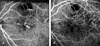
A 55-year-old male presented to our clinic complaining of decreased visual acuity in his left eye. The visual acuity in the left eye at presentation was 0.2. The anterior segments of both eyes were unremarkable. Fundus examination revealed submacular hemorrhage of about 4 disc areas in diameter in the left eye. No abnormal findings were observed in the right eye other than tiny pigment epithelial atrophy in the macula. Intravitreal injection of 25 µg of tPA and 0.3cc of SF6 was performed to remove the subretinal hemorrhage. The hemorrhage was significantly absorbed 4 days after the injection and fluorescein angiography (FAG) and indocyanine green angiography (ICGA) were performed. FAG & ICG showed that an aberrant new subretinal vessel with large vessel diameter traversed the macula in a triangular shape. Superior to the macula, an abnormal vessel with terminal polypoid dilation that is considered to be of choroidal origin was observed above the retinal vessel. Multiple hyperfluorescent bleb-like lesions were observed inferior to the fovea and showed leakage during the late phase (Fig. 1A). Optical coherence tomography (OCT) confirmed that the hyper-reflective lesion is located above retinal pigment epithelium (RPE). The patient received PDT with verteporfin. The polypoid lesions and aberrant subretinal new vessels observed on FAG and ICGA disappeared following PDT (Fig. 1B). The visual acuity had improved to 0.8 five months after PDT and the patient is currently being followed up.
Case 2
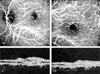
A 50-year-old female presented at our department with decreased visual acuity in her left eye. Visual acuity was 0.2 at the time of initial visit. The findings of the anterior segment of both eyes were not remarkable. Fundus examination revealed no significant findings in the right eye and a grayish lesion with serous elevation in the macula of the left eye. FAG revealed atypical vascular branches with a distinct margin. Leakage from these lesions was observed in the late phase. ICGA also showed new vessels in the subretina (Fig. 2A). A hyper-reflective lesion from the subretinal vascular complex was also confirmed on OCT (Fig. 2C). The patient was treated with PDT with verteporfin and the visual acuity had improved to 0.4 three months after PDT. Fundus examination showed fibrotic changes to the subretinal neovascularization with shallow serous elevation. Three months after PDT, FAG showed the disappearance of abnormal vascular branches and no leakage from the previously observed lesions. On ICGA, abnormal vascular branches were not seen in the early phase, and mild hyper-fluorescent lesions typical of stained fibrotic tissue were observed (Fig. 2B). On OCT, the previous hyper-reflective lesion disappeared and newly developed hyper-reflective lesions due to fibrosis were observed (Fig. 2D). The patient has been followed up for 9 months with no further change.
DISCUSSION
Subretinal new vessels may evolve from angiomatous proliferation within the retina or from choroidal neovasclarization penetrating the retinal pigment epithelium. The subretinal location of the new vessels observed in our cases was confirmed by OCT and these vessels are believed to originate from the choroids. In Case 1, the presence of leaking dilated lesions superior and nasal to the thick subretinal vessel may be interpreted as another form of polypoidal choroidal vasculopathy. However, there is no evidence from FAG and ICGA to prove that the polypoid lesions are terminal dilations of the branching vessels. Furthermore, the fact that the polypoid lesion is located above the retinal vessel and atypical vessels are located at the subretinal level, distinguishes this case from normal PCV. In case 2, new vessels originating from choroids run at the subretinal level as confirmed by the observation of hyper-reflective lesions in OCT.
A possible explanation for the good outcome in these cases is that the effect of PDT was maximized in the treatment of immature subretinal new vessels and new vessels close to the retinal pigment epithelium. Classic CNV responds to PDT more favorably than occult CNV and the fact that polypoidal lesions in PCV were obliterated readily with PDT supports the excellent outcomes from the above cases.
It should be noted that although PDT resulted in excellent outcomes in the current two cases, PDT may not be effective in all cases of retinal new vessels. As reported previously, PDT was not effective in chorioretinal anastomosis and may cause photochemical damage due to intraretinal leakage of the photosensitizing dye. This initial encouraging outcome should be confirmed through a series of studies with longer follow-up periods.
 XML Download