Journal List > Yonsei Med J > v.48(1) > 1030148

Kim, Jung, Lee, Huh, and Lee: A Clinical Analysis of Twelve Cases of Ruptured Cerebral De Novo Aneurysms

Abstract

Formation of cerebral de novo aneurysms (CDNA) is rare, and the pathogenesis remains obscure. In this study, we investigated the factors that contribute to the formation of CDNA and suggest guidelines for following patients treated for cerebral aneurysms. We retrospectively reviewed 2,887 patients treated for intracranial aneurysm at our institute from January of 1976 to December of 2005. Of those patients, 12 were readmitted due to recurrent rupture of CDNA, which was demonstrated by cerebral angiography. We assessed clinical characteristics, such as gender, size and site of rupture, past history, and the time to CDNA rupture. Of the 12 patients, 11 were female and 1 was male, with a mean age at rupture of the first aneurysm of 44.7 years (range: 30-69 years). The mean time between the first episode of subarachnoid hemorrhage (SAH) and the second was 8.9 years (range: 1.0-16.7 years). The most common site of ruptured CDNA was the internal carotid artery (5 patients, 41.7%), followed by basilar artery bifurcation (3 patients, 25.0%). In the remaining 4 patients, rupture occurred in the anterior communicating, middle cerebral, anterior cerebral (A1), or posterior cerebral (P1) arteries. In 5 cases (41.7%), the CDNA occurred contralateral to the initial aneurysm. Eleven patients (91.7%) had a past history of arterial hypertension. There was no history of habitual smoking or alcohol abuse in any of the patients. Eight patients underwent clipping for CDNA and three patients were treated with coiling. One patient who had multiple aneurysms was treated with clipping following intra-aneurysmal coiling. Assessment according to the Glasgow Outcome Scale (GOS) of the patients after the treatment was good in 10 cases (83.3%) and fair in 2 cases (16.7%). Although formation of CDNA after successful treatment of initial aneurysm is rare, several factors may contribute to recurrence. In our study, female patients with a history of arterial hypertension were at higher risk for ruptured CDNA. We recommend follow-up imaging studies every five years after treatment of the initial aneurysm, especially in women and those with a history of arterial hypertension.

INTRODUCTION

Cerebral de novo aneurysms (CDNA) are either new ruptures at sites separate from an initial aneurysm1-4 or aneurysms that were not evident upon preoperative digital subtraction angiography and/or computed tomography angiogram.5 Because CDNA formation is rare, the pathogenesis remains obscure. In general, intracranial aneurysms arise from congenital or acquired defects at the internal elastic lamina of the arterial wall. Diseases, such as Ehlers-Danlos syndrome, coarctation of the aorta, and polycystic kidney disease are known risk factors for aneurysm formation.6-8 However, the most common cause of delayed recurrent subarachnoid hemorrhage (SAH) after aneurysm clipping is known to be de novo lesions.9 Aneurysm clipping causes changes in intracranial hemodynamics that may provoke aneurysm formation.10,11 Although the prevalence of unruptured intracranial aneurysm increases with age,12-15 the peak incidence of SAH is around the sixth decade. The reported risk of recurrent SAH is also cumulative over time: approximately 2% in the first 10 years after treatment and 9% after 20 years.1,2 Although the risk of recurrent SAH increases with age, there have been no reports evaluating the risk factors that may contribute to CDNA formation or rupture.
In this study, we investigated factors that may contribute to the formation of CDNA and suggest guidelines for following patients previously treated for cerebral aneurysm.

MATERIALS AND METHODS

2,887 patients were treated for unruptured or ruptured intracranial aneurysm from January 1976 through December 2005, at Yonsei University College of Medicine, Seoul, South Korea. Of those patients, 12 were readmitted due to recurrent SAH. In these cases, cerebral angiography demonstrated CDNA formation. We assessed the clinical characteristics and outcome of these 12 patients. All data were based on medical records, radiological findings, and follow-up observations in the outpatient department.
We determined the location and number of aneurysms at the first SAH incidence and at recurrence. The size of the recurred aneurysms was measured with computed tomography angiography or cerebral angiogram. We excluded regrowing aneurysms from the previous treatment site. Regrowth or de novo formation of aneurysms was examined by comparing follow-up angiography with earlier angiography obtained before and after clipping. We also reviewed surgery reports for complications caused by treatment of the CDNA. Outcome was assessed by the Glasgow Outcome Scale (GOS) on the basis of information from discharge letters.

RESULTS

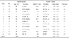
Among the 2,887 patients, there were 1,147 males and 1,740 females. The mean age of all patients was 51.2 years old (Male-47.2 years old, Female-53.9 years old). Sixty percent of the patients had a history of hypertension. Of the 2,887 patients, we identified 12 patients with recurrent SAH. Of the 12 patients, eleven (91.7%) were female and one (8.3%) was male. The mean age at initial SAH was 44.7 years (range: 30-69 years). The mean interval between the first SAH and the recurrent SAH was 8.9 years (range: 1.0-16.7 years). The most common site of ruptured CDNA was the internal carotid artery (5 cases, 41.7%), followed by basilar artery bifurcation (3 cases, 25.0%). In the remaining 4 cases, rupture occurred in the anterior communicating, middle cerebral, anterior cerebral (A1), or posterior cerebral (P1) arteries (Table 1). In 5 cases (41.7%), the CDNA occurred contralateral to the initial aneurysm. The mean size of recurrent ruptured aneurysms was 6.4 × 4.2mm. Eleven patients (91.7%) had a past history of arterial hypertension. There was no history of habitual smoking, connective tissue disease, family history of polycystic kidney disease, or alcohol abuse in any of the patients. Eight patients underwent surgery for CDNA and three patients were treated with the endovascular method. One patient who had multiple aneurysms was treated surgically following intra-aneurysmal coiling with Guglielmi detachable coil (GDC). Overall outcome based on the GOS was good in 10 cases (83.3%) and fair in 2 cases (16.7%).

DISCUSSION

Formation of new aneurysms after successful treatment of the initial aneurysm has rarely been documented in the literature. It is difficult to diagnose CDNA since they are almost always detected after a rupture. The estimated annual rate of development of recurrent CDNA in a previous report ranged from 0.28 to 1.8%.16,17 Only 0.004% of the 2,887 SAH patients in our cohort had a de novo aneurysm, which is a much lower incidence than previously reported. Known or suspected risk factors for multiple intracranial aneurysms include female gender, smoking, hypertension, and a possible family history of intracranial aneurysms or cerebrovascular disease.
Of the 12 patients, recurrence occurred predominantly in women. This finding is in accordance with a recent article that reports an increased risk for aneurysm formation in women after SAH.4 Some authors suggest that, after menopause, women are at even higher risk for SAH,4 and hormonal factors have been implicated.18 Estrogen is known to have an inhibitory effect on aneurysm formation, and cigarette smoking is known for its anti-estrogenic properties. In addition, collagen content of cerebral arteries may also diminish after menopause, further favoring aneurysm formation.19
Risk factors known to induce aneurysm rupture include gender, hypertension, family history of aneurysm, high growth rate of aneurysm, and cigarette smoking.4 Smoking is known to be one of the highest risk factors for aneurysmal SAH; smokers have about 3.7 ± 5.7 times the rate of rupture found in nonsmokers.20-22 In both SAH patients and in the general population, cigarette smoking declines with age and is more frequent among men than women.23-26 In our study, however, smoking was not a risk factor because none of the patients had a history of smoking. This is probably due to the fact that most of the patients diagnosed with ruptured CDNA in our study were female. Our study did confirm female gender and hypertension as risk factors.
Although the pathogenesis of CDNA remains unclear, several hypotheses have been proposed. First, hemodynamic alterations of vessels after treatment may cause CDNA formation.10,11 Second, defects in the tunica muscularis or lamina elastica interna have been reported to cause aneurysm formation.6,8,27 Some also suggest that incomplete treatment or slippage of the clip after the initial operation may cause aneurysm regrowth.28 CDNA is known to be the most common cause of recurrent episodes of SAH, indicating the persistence of an underlying vascular condition. The risk of recurrent SAH appears to increase over time from approximately 2% in 10 years to 9% after 20 years.1,2 In the general population, the risk of SAH in 10 years is approximately 0.072%,29 suggesting that patients who have had SAH are 30 times more likely to experience a new episode. Additional aneurysms that arise in patients with a history of cerebral aneurysms may also carry a higher risk of rupture. David et al.17 reported a 0.52% annual regrowth rate for completely clipped aneurysms and a 1.8% annual rate of CDNA formation. In our study, rupture of CDNA occurred in patients 8.9 years after the initial treatment. Because of the high fatality rate of SAH, follow-up investigation should be performed 8.9 years after surgery even in patients with completely clipped aneurysms, and especially in those with an accumulated risk of aneurysm.
We recommend following patients, especially women or those with a history of arterial hypertension, using magnetic resonance angiography, computed tomography angiography, or angiogram every 5 years after initial treatment.

Figures and Tables

Table 1
Overview of 12 Cases with Ruptured De Novo Intracranial Aneurysms
ymj-48-30-i001

F, female; M, male; ACOM, anterior communicating artery; ICPCOM, posterior communicating artery; MCBIF, middle cerebral artery bifurcation; ICBIF, internal carotid artery bifurcation; PCOM, posterior communication artery; BABIF, basilar artery bifurcation; A1, anterior cerebral artery; P1, posterior cerebral artery; M1, middle cerebral artery; Lt, left; Rt, right.

References

1. David CA, Vishteh AG, Spetzler RF, Lemole M, Lawton MT, Partovi S. Late angiographic follow-up review of surgically treated aneurysms. J Neurosurg. 1999. 91:396–401.
2. Tsutsumi K, Ueki K, Usui M, Kwak S, Kirino T. Risk of recurrent subarachnoid hemorrhage after complete obliteration of cerebral aneurysms. Stroke. 1998. 29:2511–2513.
3. Tsutsumi K, Ueki K, Usui M, Kirino T. Risk of aneurysm recurrence in patients with clipped cerebral aneurysms: results of long-term follow-up angiography. Stroke. 2001. 32:1191–1194.
4. Juvela S, Poussa K, Porras M. Factors affecting formation and growth of intracranial aneurysms: a long-term follow-up study. Stroke. 2001. 32:485–491.
5. Van der Schaaf IC, Velthuis BK, Wermer MJ, Majoie C, Witkamp T, de Kort G, et al. New detected aneurysms on follow-up screening in patients with previously clipped intracranial aneurysms: comparison with DSA or CTA at the time of SAH. Stroke. 2005. 36:1753–1758.
6. Sekhar LN, Heros RC. Origin, growth, and rupture of saccular aneurysms: a review. Neurosurgery. 1981. 8:248–260.
7. Stehbens WE. Etiology of intracranial berry aneurysms. J Neurosurg. 1989. 70:823–831.
8. Suzuki J, Ohara H. Clinicopathological study of cerebral aneurysms. Origin, rupture, repair, and growth. J Neurosurg. 1978. 48:505–514.
9. Yamakawa H, Sakai N, Takenaka K, Yoshimura S, Andoh T, Yamada H, et al. Clinical analysis of recurrent subarachnoid hemorrhage after neck clipping surgery. Neurol Med Chir (Tokyo). 1997. 37:380–386.
10. Rinne JK, Hernesniemi JA. De novo aneurysms: special multiple intracranial aneurysms. Neurosurgery. 1993. 33:981–985.
11. Sakaki T, Tominaga M, Miyamoto K, Tsunoda S, Hiasa Y. Clinical studies of de novo aneurysms. Neurosurgery. 1993. 32:512–517.
12. Epidemiology of aneurysmal subarachnoid hemorrhage in Australia and New Zealand: incidence and case fatality from the Australasian Cooperative Research on Subarachnoid Hemorrhage Study (ACROSS). Stoke. 2000. 31:1843–1850.
13. Horikoshi T, Akiyama I, Yamagata Z, Nukui H. Retrospective analysis of the prevalence of asymptomatic cerebral aneurysm in 4518 patients undergoing magnetic resonance angiography-when does cerebral aneurysm develop? Neurol Med Chir (Tokyo). 2002. 42:105–113.
14. Ohkuma H, Fujita S, Suzuki S. Incidence of aneurysmal subarachnoid hemorrhage in Shimokita, Japan, from 1989 to 1998. Stroke. 2002. 33:195–199.
15. Pobereskin LH. Incidence and outcome of subarachnoid haemorrhage: a retrospective population based study. J Neurol Neurosurg Psychiatry. 2001. 70:340–343.
16. Cheong JJ, Ghinea N, van Gelder JM. Estimating the annual rate of de novo multiple aneurysms: three statistical approaches. Neurosurg Focus. 2004. 17:E8.
17. David CA, Vishten AG, Spetzler RF, Lemole M, Lawton MT, Partovi S. Late angiographic follow-up review of surgically treated aneurysms. J Neurosurg. 1991. 91:396–401.
18. Tonn J, Hoffmann O, Hofmann E, Schlake HP, Sorensen N, Roosen K. De novo formation of intracranial aneurysms: who is at risk? Neuroradiology. 1999. 41:674–679.
19. Kongable GL, Lanzino G, Germanson TP, Truskowski LL, Alves WM, Torner JC, et al. Gender-related differences in aneurysmal subarachnoid hemorrhage. J Neurosurg. 1996. 84:43–48.
20. Sacco RL, Wolf PA, Bharucha NE, Meeks SL, Kannel WB, Charette LJ, et al. Subarachnoid and intracerebral hemorrhage: natural history, prognosis, and precursive factors in the Framingham Study. Neurology. 1984. 34:847–854.
21. Bonita R. Cigarette smoking, hypertension and the risk of subarachnoid hemorrhage: a population-based case-control study. Stroke. 1986. 17:831–835.
22. Morris KM, Shaw MD, Foy PM. Smoking and subarachnoid haemorrhage: a case control study. Br J Neurosurg. 1992. 6:429–432.
23. Juvela S, Hillbom M, Numminen H, Koskinen P. Cigarette smoking and alcohol consumption as risk factors for aneurysmal subarachnoid hemorrhage. Stroke. 1993. 24:639–646.
24. Juvela S. Prevalence of risk factors in spontaneous intracerebral hemorrhage and aneurysmal subarachnoid hemorrhage. Arch Neurol. 1996. 53:734–740.
25. Weir BK, Kongable GL, Kassell NF, Schultz JR, Truskowski LL, Sigrest A. Cigarette smoking as a cause of aneurysmal subarachnoid hemorrhage and risk for vasospasm: a report of the Cooperative Aneurysm Study. J Neurosurg. 1998. 89:405–411.
26. Juvela S, Porras M, Poussa K. Natural history of unruptured intracranial aneurysms: probability of and risk factors for aneurysm rupture. J Neurosurg. 2000. 93:379–387.
27. Jellinger K. Pia HW, Langmaid C, Zierski J, editors. Pathology and aetiology of intracranial aneurysms. Cerebral aneurysms: Advances in Diagnosis and Therapy. 1979. Berlin, Heidelberg, New York: Springer;5–19.
28. Wermer MJ, Rinkel GJ, Greebe P, Albrecht KW, Dirven CM, Tulleken CA. Late recurrence of subarachnoid hemorrhage after treatment for ruptured aneurysms: patient characteristics and outcomes. Neurosurgery. 2005. 56:197–204.
29. Linn FH, Rinkel GJ, Algra A, van Gijn J. Incidence of subarachnoid hemorrhage: role of region, year, and rate of computed tomography: a meta-analysis. Stroke. 1996. 27:625–629.
TOOLS
Similar articles