

 PDF
PDF ePub
ePub Citation
 Print
Print


INTRODUCTION
To detect a hepatocellular carcinoma (HCC) in patients with chronic liver disease, periodic routine examinations, including imaging studies, are becoming more common. As a result, the detection rate of small nodular lesions in patients with chronic liver disease is increasing. Although most of these nodules are small HCCs, a small number of nodules are regenerative or of uncertain malignancy. Hypervascular hyperplastic nodules in patients with chronic liver disease are very rare and clinically resemble HCC.1-3 Thus, the differential diagnosis of hypervascular hyperplastic nodules in a cirrhotic liver and HCC is a difficult and important clinical concern. We report a case of multiple hypervascular liver nodules in a 41-year-old male patient with alcoholic cirrhosis.
CASE REPORT
A 41-year-old man with a history of alcohol intake (90 g ethanol/day for three years) was admitted to Jeju Hospital in June 2004 due to a traffic accident in which he sustained a right distal radius fracture. During the hospital stay, he developed jaundice and was diagnosed with an aneurysm of the distal descending thoracic aorta. He was then transferred to Yonsei Medical Center, where he received further treatment.
The physical examination upon admission revealed the following vital signs: blood pressure 160/110 mmHg, pulse rate 85 beats/minute, respiration rate 18 breaths/minute, and body temperature 36.7℃. There was a slight hepatosplenomegaly without evidence of other abdominal masses. Laboratory tests revealed the following: hemoglobin 13.2 g/dL, hematocrit 38.7%, white blood cell count 5,720/µL with 59.4% polymorphonuclear cell, platelet count 97,000/µL, sodium 136.5 mEq/L, potassium 3.97 mEq/L, chloride 96.7 mEq/L, bicarbonate 23.9 mEq/L, blood urea nitrogen 6.5 mg/dL, creatinine 0.6 mg/dL, total protein 8.8 g/dL, albumin 3.7 g/dL, total bilirubin 2.6 mg/dL, direct bilirubin 1.9 mg/dL, alkaline phosphatase 159 IU/L, AST 82 IU/L, ALT 27 IU/L, gamma-glutamyltranspeptidase 605 IU/L, prothrombin time 70%, c-reactive protein 1.61 mg/dL, HBsAg negative, anti-HBs negative, anti-HBc negative, anti-HCV negative, alpha-fetoprotein (AFP) 7.52 ng/mL, and protein induced by vitamin K absence II 158 mAU/mL.
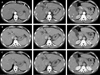
On the first day of hospitalization, a computerized tomography (CT) of the aorta and a dynamic CT of the liver were taken. The CT of the aorta showed a fusiform shaped aneurysm at the distal descending thoracic aorta measuring 4 cm at the maximal diameter, with circumferential calcification of the aneurysm wall. The CT also revealed surface nodularity of the liver, suggesting liver cirrhosis, and numerous small nodules throughout the entire liver. The dynamic liver CT showed multiple nodular lesions with arterial enhancement and delayed washout in both lobes of the liver (Fig. 1). On the fifth day, a US-guided liver gun biopsy was performed on a 2.5 cm-sized hyperechoic nodule with a peripheral halo in the right lobe of the liver (Fig. 2). The liver biopsy showed a hepatocellular nodule with slightly increased cellularity, fatty change, and Mallory bodies (Fig. 3). It also showed unpaired arteries and focal sinusoidal expression of CD34, indicating increased angiogenesis in the nodule; however, both the structural and cytological atypia were unremarkable and there was focal iron deposition. The background revealed alcoholic micronodular cirrhosis. From these observations, the lesion was tentatively diagnosed as a hypervascular hyperplastic nodule in alcoholic cirrhosis. On the twelfth day, a percutaneous transarterial angiography with stent insertion at the aortic aneurysm was performed. Two days later, an aortic CT demonstrated the complete exclusion of the aneurysm. Magnetic resonance imaging (MRI) of the liver was performed on the fifteenth day revealed underlying liver cirrhosis and numerous nodules that showed high signals on T1 weighted images, but there were some with a central low signal portion (Fig. 4). Among those, arterial phase enhancing nodules were present, suggesting overt HCCs. Other nodules also showed central enhancement patterns, suggesting dysplastic nodules with subfocus HCCs (Fig. 5). On the nineteenth day, a second liver gun biopsy was performed on the same lesion as the first biopsy and demonstrated the same pathological features. From these findings, the possibility of mistargeting the biopsy was ruled out and the diagnosis of hypervascular hyperplastic nodules in alcoholic cirrhosis was confirmed. Hepatic angiography performed on the twenty-fourth day showed faint multiple nodular staining of both liver lobes in the early arterial phase (Fig. 6). The patient was discharged three days after the hepatic angiography and scheduled for an outpatient follow-up.
DISCUSSION
Although the terminology of nodular hepatocellular lesions is not yet established, international efforts are underway to develop a uniform terminology. The International Working Party of 19951 classified nodular hepatocellular lesions into two main classes: regenerative lesions and dysplastic or neoplastic lesions, with more detailed subclassifications. Due to the lack of uniform terminology, the most important clinical concern is to distinguish between a regenerative lesion (hyperplastic nodule) and a tumorous lesion (dysplastic nodule or neoplastic lesion).
In 1984, Nagasue et al.2 reported three cases of regenerative hyperplastic nodules in cirrhotic livers and proposed the name 'hepatocellular pseudotumor in the cirrhotic liver'. The patients were all male, heavy drinkers, had chronic liver disease for six to ten years, had slightly elevated AFP levels, were positive for anti-HBs and anti-HBc, and were preoperatively diagnosed with either HCC or highly suspicious HCC. In 1996, Nakashima et al.3 reported five cases of hyperplastic nodules in patients with chronic alcoholic liver disease. In all of the patients, AFP was within the normal range, HBsAg was negative, and the patients were preoperatively diagnosed with HCCs by imaging study and/or needle biopsy. In 2003, Lepreux et al.4 reported focal nodular hyperplasia (FNH)-like nodules in eight patients who underwent hepatic resections for the removal of a mass lesion. In two of the patients, HCC was suspicious with imaging techniques. The various types of small FNH-like nodules were characterized by an association with various degrees of numerous and/or enlarged arteries in the portal tracts or septa, with hyperplastic foci, slight ductular reaction, and regions of sinusoidal dilatation, accompanied by a thin fibrous band. Vascular abnormalities included consisted of unpaired arteries, portal tracts with arteries larger than the associated bile duct, and regions of sinusoidal dilatation. They suggested that these FNH-like nodules could be precursors of the large mass lesions. However, in 2004, Kim et al.5 reported three cases of regenerative hypervascular liver nodules, according to the classification of the International Working Party,1 in patients without hepatitis B or C virus infection, but with a history of alcohol abuse. In 2004, Nakashima et al.6 reported six partial hepatectomy and six biopsy cases of small hypervascular nodules found in alcoholic cirrhosis, which were considered FNH-like nodules. Those nodules showed hypervascularity upon angiography and histologically revealed a moderate increase of cell-density, with an irregular trabecular pattern, scar-like fibrosis with anomalous blood vessels, and unpaired arteries. The nodules also showed a diffuse capillarization of the sinusoids and marked or mild iron deposits in the hepatocytes and/or Kupffer cells.
In our case, the patient had been a heavy drinker for three years. Serum was negative for HBsAg and anti-HCV. AFP was within the normal limit. Imaging studies, including CT and angiography, revealed hypervascular tumors in the liver. The dynamic CT showed multiple nodular lesions with arterial enhancement and delayed washout on both lobes of the liver, while the MRI revealed arterial phase enhancing nodules. These radiologic findings were highly suggestive of a HCC.7-9 However, the liver biopsy revealed a hepatocellular nodule with a slightly increased cellularity and the hepatocytes, which composed the nodule, showed similar features to those of alcoholic cirrhotic nodules in the background. This nodule showed increased angiogenesis of unpaired arteries and sinusoidal capillarization with expression of CD34, which might explain the hypervascularity on radiologic studies. Prussian blue stain showed focal iron deposits in the nodule, but was absent in the cirrhotic nodules. These features were very similar to previous cases of hypervascular hyperplastic nodules;2-6,10 however, scar-like fibrosis was not found in this case.
The pathogenesis of hypervascular hyperplastic nodules in the liver has not yet been explained. Benign nodular hepatocellular lesions, such as FNH, nodular regenerative hyperplasia, and partial nodular transformation, may be caused by abnormal hepatic circulation, including a change in the portal venous or arterial system. One explanation for the pathogenesis of hypervascularity is the proliferation of unpaired arteries, which are found in microregenerative cirrhotic nodules. Generally, hyperplastic changes are usually associated with hypervascularity.11 However, further studies are needed to verify the pathogenesis of hypervascularity and nodule formation in heavy alcohol drinkers.
Regarding a strategy for the management of hyperplastic nodules in heavy drinkers, Suzuki et al.10 suggested the following considerations: (1) hyperplastic nodules are often present as multiple lesions in relatively young patients with portal hypertension, negative hepatitis virus markers, and normal tumor marker values, (2) dynamic CT and MRI studies of hypervascular lesions often show no evidence of decreased portal blood flow; however, there are patients in whom imaging studies reveal findings similar to those in classical HCC, (3) differential diagnosis of hyperplastic versus dysplastic nodules from fine-needle biopsy results can be difficult in some patients, and (4) in patients positive for the hepatitis virus, hyperplastic nodules should be more cautiously diagnosed. In the present case, it was difficult to rule out the possibility of neoplastic lesions, such as HCC or dysplastic nodules, based on the radiological features even though the histological features of the first liver biopsy did not favor a neoplastic lesion. A second biopsy was performed to confirm whether the first biopsy was correctly obtained from the target nodule and the results showed features similar to the first biopsy. Based on this, the diagnosis of hypervascular hyperplastic nodule was made.
We reported a case of hypervascular hyperplastic nodules in alcoholic cirrhosis. Differential diagnosis of hypervascular nodules and HCC, particularly in patients with cirrhosis, is difficult with imaging studies. Thus, histological confirmation is necessary, although fine-needle biopsy results might be difficult in some patients.


 XML Download