Abstract
Idiopathic granulomatous lobular mastitis (IGLM), also known as idiopathic granulomatous mastitis, is a rare chronic inflammatory lesion of the breast that can clinically and radiographically mimic breast carcinoma. The aim of this study was to describe the radiological imaging and clinical features of IGLM in order to better differentiate this disorder from breast cancer. We performed a retrospective analysis of the clinical and radiographic features of 11 women with a total of 12 IGLM lesions. The ages of these women ranged between 29 and 42 years, with a mean age of 34.8 years. Ten patients were examined by both mammography and sonography and one by sonography alone. The sites that were the most frequently involved were the peripheral (6/12), diffuse, (3/12), and subareolar (3/12) regions of the breast. The patient mammograms showed irregular ill-defined masses (7/11), diffuse increased densities (3/11), and one oval obscured mass. In addition, patient sonograms showed irregular tubular lesions (7/12) or lobulated masses with minimal parenchymal distortion (2/12), parenchymal distortion without definite mass lesions (2/12), and one oval mass. Subcutaneous fat obliteration (12/12) and skin thickening (11/12) were also observed in these patients. Contrary to previous reports, skin changes and subareolar involvement were not rare occurrences in IGLM. In conclusion, the sonographic features of IGLM show irregular or tubular hypoechoic masses with minimal parenchymal distortion. Both clinical information and the description of radiographic features of IGLM may aid in the differentiation between IGLM and breast cancer, however histological confirmation is still required for the proper diagnosis and treatment of the disorder.
Idiopathic granulomatous lobular mastitis (IGLM), or idiopathic granulomatous mastitis, is a rare chronic inflammatory lesion of the breast that can clinically and radiographically mimic breast carcinoma.1-4 IGLM was first described by Kessler and Wolloch in 1972.5 This disorder is characterized by the chronic granulomatous inflammation of lobules without caseous necrosis.1,3,6,7 IGLM is of unknown origin, and its diagnosis rests on both the demonstration of a characteristic histological pattern and the exclusion of other possible causes of granulomatous breast lesions. Clinically and radiographically, IGLM is difficult to differentiate from early onset breast cancer. Here we report the clinical and radiographic features of 11 women with a total of 12 IGLM lesions. The aim of our study was to find differential diagnostic patterns that would allow a clear differentiation between IGLM and breast carcinoma.
We performed a retrospective study of records from our institution between January 1993 and December 2000. During this period, 11 women with 12 lesions (7 lesions in the left breast, 3 lesions in the right breast, and one case in both breasts) of IGLM were diagnosed and treated. The mean age of the 11 patients was 34.8 years (ranging from 29 to 42 years old). Ten patients were diagnosed within 15 years of their last pregnancy. One patient was unmarried. A positive diagnosis of IGLM required a characteristic histological pattern of granulomatous inflammation. This inflammation consisted of epithelioid cells, Langhans-type giant cells, and lobular-confined neutrophils. Other possible causes of granulomatous inflammatory lesions of the breast were excluded by serologic tests, histologic analysis by staining with either modified Ziehl-Neelsen, periodic acid-Schiff with diastase predigestion, or Gomori's methenamine silver stains, and by testing the effected tissue for aerobic and anaerobic bacteria, mycobacteria, and fungi.
Ten patients were examined by both mammography and sonography, while one patient was only examined by sonography. We used a CGR 500T or GE DMR mammography unit (GE Medical Systems, Milwaukee, WI, USA), Acuson 128, Acuson 128/xp 3, Acuson 128/xp 10 (Acuson, Mountain View, CA, USA), and a Ultramark 9, HDI 3000 ultrasonography unit (Advanced Technology Laboratory, Bothell, WA, USA). All lesions were examined by routine mammography using both the craniocaudal and the mediolateral oblique views. We also examined the 11 patients (12 lesions) using real-time sonography.
One patient was examined with a Tomoscan SR-7000 spiral CT unit (Phillips Medical Systems, Best, Netherlands). After a non-enhanced study of the entire breast was performed, 100 mL of contrast material (iopromide, [Ultravist]; Schering, Berlin, Germany) was injected at 2.5 mL/sec into an antecubital vein using a mechanical power injector (Medrad, Pittsburgh, PA, USA). Five-millimeter-collimation helical scans were obtained from the apex of the lung to the diaphragm. The scans were obtained by using 120 kVp, 225 mAs, and 1 : 1 pitch. The images were viewed at a window width of 400 HU and a level of +40 HU.
our institution. This Two patients underwent MR breast imaging at analysis was performed with a 1.5-T MR imaging system (Vision, Siemens, Erlangen, Germany). In order to prevent false positive signaling artifacts from the area of contact with the breast coil, we attached a pad to the inner surface of the coil. After an initial localization, a fat-suppressed T2 turbo spin-echo sequence was performed with the following scan parameters: field of view, 320 × 320 mm, contiguous axial slice, 3-mm thickness, matrix size, 256 × 256, TR/TE, 4200/90 msec, scan time, 2 min, acquisition, 35 sec, flip angle, 180°. This was followed by T1-weighted imaging with a conventional spin echo pulse sequence. The scan parameters for this imaging were as follows: field of view, 320 × 320 mm, axial slice, 3-mm thickness, matrix size, 256 × 256, TR/TE, 590/12 msec, scan time, 5 min, acquisition, 5 sec, flip angle, 90°. We then performed a Gd-enhanced dynamic MRI study. For this technique we applied a 2D FLASH with TR/ TE/flip angle of 32 ms/9 ms/30°, a matrix size of 198 × 256/256 × 256, a field of view of 320 × 320 mm, and a slice thickness of 3 mm. In addition, we performed 90 switch phase-encoding swap and presaturation techniques to avoid cardiac motion artifacts. We also performed pre- and post-contrast MRI scans with an intravenous injection of 0.16 mmol gadopentetate dimeglumine (Magnevist; Schering, Berlin, Germany) per kilogram of body weight. After the injection, dynamic images were made at 1 min intervals for 5 min. Time-intensity curves were constructed from signal intensity values obtained from free drawn ROIs, in the most enhanced area of the tumor mass.
All mammograms, US images, CT and MR mammogram images were retrospectively analyzed and assessed by two consenting radiologists (JHL, KKO). The location and imaging features of the lesions were analyzed by mammography The ultrasonographic features of IGLM were analyzed by assessing the internal echoes of the masses, as well as the shape of masses, skin changes, subcutaneous fat obliteration, and parenchymal distortion. Parenchymal distortion was considered to be present when the sonogram depicted the loss or disruption of linear, echogenic fibrous bands and normal Cooper's suspensory ligaments that outlined fat planes. The shape and enhancement of the breast lesions, subcutaneous fat obliteration, and skin changes were all analyzed using the CT and MR images.
Among the 11 patients, two were treated with mastectomy, six with excision, one with incision and drainage, and two with corticosteroid therapy that was diagnosed by core needle biopsy.
Symptoms at the time of admission were a palpable mass (9 of 11 patients) or painful swelling (2 of 11 patients).
Idiopathic granulomatous lobular mastitis involved the following areas of the breast: the upper outer quadrant (2 of 12 lesions), upper medial quadrant (2 of 12 lesions), outer central quadrant (1 of 12 lesions), lower medial quadrant (1 of 12 lesions), diffuse involvement (3 of 12 lesions), and subareolar (3 of 12 lesions) (Table 1). One patient exhibited bilateral lesions.
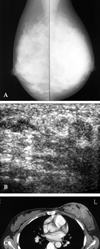
Mammography of the 11 lesions from 10 patients revealed the following: an irregular ill-defined mass (7 of 11 lesions, 63.7%) (Fig. 1A), diffusely increased density (3 of 11 lesions, 27.3%) (Fig. 2A), and one oval, obscured mass (9%). We also observed associated parenchymal distortion (7 of 11 lesions, 63.7%), skin thickening (7 of 11 lesions, 63.7%), and axillary lymph node enlargement of benign appearance (6 of 11 lesions, 54.5%). None of the lesions showed calcification. All except one of the lesions showed heterogeneously dense or extremely dense parenchymal patterns (Table 2).
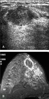
Sonography of 12 lesions from 11 patients showed irregular tubular hypoechoic lesions (7 of 12 lesions, 58.3%) (Fig. 1B) or lobulated hypoechoic masses with minimal parenchymal distortion (2 of 12 lesions, 16.7%) (Fig. 3A), parenchymal distortion without definite mass lesions (2 of 12 lesions, 16.7%) (Fig. 2B), and one oval circumscribed hypoechoic mass (8.3%). We also observed subcutaneous fat obliteration (12 of 12 lesions, 100%) and skin thickening (11 of 12 lesions, 91.7%). The lesions ranged from 1 to 10 cm in diameter. In all cases containing mass lesions (83.3%, 10 of 12 lesions), the axis parallel to the chest wall was greater than the perpendicular axis, thus giving an oval or tubular configuration. Half of the lesions (50%, 6 of 12 lesions) were located at the periphery of the breast, but diffuse involvement (25%, 3 of 12 lesions) and subareolar location (25%, 3 of 12 lesions) were also evident (Table 3).
In the CT scan images, huge heterogeneous enhancing mass lesions were seen in the left breast with prominent skin thickening (Fig. 2C). Subcutaneous fat obliteration was minimal.
In the non-enhanced MR images, the lesions were present as masses with low signal intensity at T1WI and as high signal intensities at T2WI. Gd-DTPA enhanced MRI showed a heterogeneous enhancing mass lesion with spiculated borders of a benign-type time-intensity curve (slow progressive enhancement over time) in one patient and an abscess like lesion with a rim enhancement and an intermediate type time-intensity curve (early enhancement with less signal loss over time) in another patient (Fig. 3B).
IGLM is a rare and benign chronic inflammatory lesion of the breast.1,8,9 These lesions present clinically and radiographically with palpable breast masses in young reproductive women and it is difficult to differentiate IGLM from early onset breast carcinoma.1,3,9 Frequently IGLM is associated with recent childbirth (ranging from 2 months to 15 years since the last delivery). However, no consistent history of breast feeding or oral contraceptives has been associated with IGLM incidence.2 Clinically, the patients have a breast mass that can vary in size from 0.5 to 9 cm and are usually unilateral (left > right > bilateral).1,2 Often there is an inflammation of the overlying skin.1 In addition, regional lymphadenopathy may also be present.2
IGLM is characterized by chronic lobulitis with granulomatous inflammation. The diagnosis of IGLM rests on the documentation of a characteristic histologic pattern, combined with the exclusion of other possible causes of granulomatous lesions of the breast.1 IGLM should be differentiated from other chronic inflammatory breast diseases such as mammary duct ectasia (plasma cell mastitis, subareolar granuloma, and periductal mastitis), Wegener's granulomatosis, sarcoidosis, tuberculosis, and histoplasmosis.3 We think that the term "idiopathic granulomatous lobular mastitis (IGLM)" has the advantage of emphasizing the single most important histologic feature of this condition, and avoids the vagueness of "granulomatous mastitis".
The etiology of IGLM is currently unknown,1-3 but some reports have speculated on several potential causes including a localized autoimmune phenomenon, a reactivation to childbirth, or the prior use of oral contraceptives.2
The reported mammographic features of granulomatous mastitis were a focal asymmetric density or a mass with definable margin and a dense parenchymal pattern without any abnormalities.1,4,10 The most common observation from mammographic examinations was an asymmetric density with no distinct margin or mass effect.10 Asymmetric density is a non-specific mammographic finding that could be seen in clinically normal breasts or in other various disease types. Patients with dense breast parenchyma may also have negative mammograms. In our study, the results of the mammograms were both irregular ill-defined masses, diffuse increased density and oval obscured masses in young women with heterogeneously dense or extremely dense breasts. None of the lesions showed calcification. In our study, benign-looking axillary lymph node enlargements were visible in 54.5% (6 of 11 lesions). Han et al.4 reported no visible changes involving the skin or the nipple in granulomatous mastitis. However, in our study, skin thickening was demonstrated in 63.6% (7 of 11 lesions) of the mammograms. The reported locations of the lesions were usually unilateral and occurred in every quadrant of the breast except for the subareolar region.3 Only one of 15 lesions showed a subareolar location in the report by Memis et al.10 In contrast, three of 12 lesions in our study showed subareolar involvement. Therefore, we believed that the IGLM may involve the subareolar region of the breast.
US features of IGLM have been described relatively frequently.1,4,10,11 Han et al.4 reported multiple clustered, often contiguous, tubular hypoechoic lesions that can be associated with a large hypoechoic mass, and which might suggest the possibility of granulomatous mastitis. Memis et al.10 reported that irregular hypoechoic mass lesions or tubular hypoechoic areas connecting to the mass were frequently sonographic features of IGLM. In our study, the sonogram results showed irregular tubular lesions or lobulated masses, parenchymal distortions without definite mass lesions, and oval masses. In all cases exhibiting mass lesions (10 of 12 lesions), the axis parallel to the chest wall was greater than the perpendicular axis, thus suggesting that an oval or tubular configuration were indicative of benign lesions rather than malignancies. Memis et al.10 reported an unusual US finding of a focally decreased parenchymal echogenicity and acoustic shadowing with no definable masses at the palpable site. Histopathologically, the lesion showed that the granulomas were not confluent and varying degrees of fibrosis accompanied the granulomas. In our study, another sonographic feature of parenchymal distortion without a definite mass lesion was also noted in two out of 12 lesions. Histopathologic findings showed evidence of IGLM with marked granulomatous inflammation and abscess formation with little intervening stroma. In our study, skin thickening and subcutaneous fat obliteration were also frequently noted as associated with IGLM. Sonograms of three cases revealed skin sinus tracts with long, tubular hypoechoic lesions extending from the lesion to the skin. Sinus-forming lesions, such as those associated with tuberculosis or fungal infection, should be included in the differential diagnosis of the patient.4
To our knowledge, radiographic features of mastitis on CT scans have not been reported. In our study, one patient underwent a CT scan. In CT images of this patient, a huge heterogeneously enhancing mass lesion was seen in the left breast as well as prominent skin thickening. Subcutaneous fat infiltration was minimal indicating that the findings were not acute infection.
MRI effectively delineated tissue contrast differences and allowed the use of intravenous contrast agents that further improve mass conspicuity. Rieber et al.12 reported that MRI of the female breast does not provide additional critical information for the differentiation of mastitis from inflammatory carcinoma. In our study, Gd-DTPA enhancing MRI showed a heterogeneous enhancing of a mass lesion with spiculated borders with a benign-type time-intensity curve (slow progressive enhancement over time) in one patient. Another patient had an abscess-like lesion with rim enhancement and intermediate-type time-intensity curve (early enhancement with less signal loss over time). Kocaoglu et al.11 reported that all but one patient exhibited a benign time-signal intensity pattern, and that MR imaging with the measurement of time-signal intensity curves may support the US and mammogram results in their ability to distinguish between benign and malignant inflammatory breast disorders.
The optimal treatment for IGLM has yet to be established. Surgical resection of the affected tissue, with or without corticosteroid therapy, has often been the current method of treatment.3 However, with these treatments, a tendency for local recurrences and delayed wound healing exists.1
In conclusion, clinical information and radiographic features of IGLM may aid in the differentiation between IGLM and early onset breast cancer. However, a histological confirmation is still required for a positive diagnosis and the determination of an appropriate treatment regimen.
Figures and Tables
Fig. 1
Mammogram and ultrasound of a 34-year-old woman 2 months after mass detection. A. The MLO mammogram shows an irregular, indistinct high density mass in the upper left breast. B. The ultrasonography shows an ill-defined, irregular tubular, heterogeneous hypoechoic lesion.

Fig. 2
The mammogram and ultrasound of a 33-year-old woman in 2 months after the detection of a palpable mass in the left breast. A. The mammogram shows a diffuse, increased density in the left breast, but without calcification. B. The ultrasound shows parenchymal distortion without a definite mass lesion. CT scan of a 33-year-old woman in 2 months after the detection of a palpable mass in the left breast. C. The axial CT scan shows heterogeneous enhancing mass lesion with prominent skin thickening.

Fig. 3
The ultrasound and mammogram of a 34 year-old woman 2 months after the detection of a mass in the right breast. A. Ultrasonography shows a well-defined, oval, heterogeneous hypoechoic mass. B. The MR mammogram shows an abscess-like lesion with a rim enhancement in the right breast.

References
1. Van Ongeval C, Schraepen T, Van Steen A, Baert AL, Moerman P. Idiopathic granulomatous mastitis. Eur Radiol. 1997. 7:1010–1012.
2. Jorgensen MB, Nielsen DM. Diagnosis and treatment of granulomatous mastitis. Am J Med. 1992. 93:97–101.
3. Imoto S, Kitaya T, Kodama T, Hasebe T, Mukai K. Idiopathic granulomatous mastitis: case report and review of the literature. Jpn J Clin Oncol. 1997. 27:274–277.
4. Han BK, Choe YH, Park JM, Moon WK, Ko YH, Yang JH, et al. Granulomatous mastitis: mammographic and sonographic appearances. AJR Am J Roentgenol. 1999. 173:317–320.
5. Kessler E, Wolloch Y. Granulomatous mastitis: a lesion clinically simulating carcinoma. Am J Clin Pathol. 1972. 58:642–646.
6. Going JJ, Anderson TJ, Wilkinson S, Chetty U. Granulomatous lobular mastitis. J Clin Pathol. 1987. 40:535–540.
7. Donn W, Rebbeck P, Wilson C, Gilks CB. Idiopathic granulomatous mastitis. A report of three cases and review of the literature. Arch Pathol Lab Med. 1994. 118:822–825.
8. Salam IM, Alhomsi MF, Daniel MF, Sim AJ. Diagnosis and treatment of granulomatous mastitis. Br J Surg. 1995. 82:214.
9. Osborne BM. Granulomatous mastitis caused by histoplasma and mimicking inflammatory breast carcinoma. Hum Pathol. 1989. 20:47–52.
10. Memis A, Bilgen I, Ustun EE, Ozdemir N, Erhan Y, Kapkac M. Granulomatous mastitis: imaging findings with histopathologic correlation. Clin Radiol. 2002. 57:1001–1006.
11. Kocaoglu M, Somuncu I, Ors F, Bulakbasi N, Tayfun C, Ilkbahar S. Imaging findings in idiopathic granulomatous mastitis: a review with emphasis on magnetic resonance imaging. J Comput Assist Tomogr. 2004. 28:635–641.
12. Rieber A, Tomczak RJ, Mergo PJ, Wenzel V, Zeitler H, Brambs HJ. MRI of the breast in the differential diagnosis of mastitis versus inflammatory carcinoma and follow-up. J Comput Assist Tomogr. 1997. 21:128–132.


 PDF
PDF ePub
ePub Citation
 Print
Print


 XML Download