

 PDF
PDF ePub
ePub Citation
 Print
Print


INTRODUCTION
There has been considerable interest in suture adjustment strabismus surgery since it was first described by Jampolsky in 1979.1 It decreases the incidence of reoperation, and increases the accuracy of strabismus correction especially in unpredictable cases.2-4 Generally, adjustable surgery with a two-stage procedure is performed under general or retrobulbar anesthesia; adjustments can be made after the anesthetic effect has faded out.3 This entails an extra visit or a prolonged stay in hospital for the second phase of adjustment.4 One-stage intraoperative adjustment under topical anesthesia has the advantages over a two-stage proedure. There are no complications of general or retrobulbar anesthesia, and less chance of infection, accurate extraocular muscle tests and rapid rehabilitations are possible.
We performed one-stage intraoperative adjustment strabismus surgery under topical anesthesia and analyzed the results.
MATERIALS AND METHODS
The medical records of the patients, who had undergone intraoperative adjustment surgery under topical anesthesia for horizontal or vertical strabismus in our hospital from March 1997 to March 2003 with follow-up of 6 months, were reviewed retrospectively. This study was approved by the ethics committee of Yonsei University Medical Center, Seoul, Korea. Seventy-one patients (29 men and 42 women) underwent an intraoperative adjustment of strabismus surgery under topical anesthesia. The mean age of the patients was 31.4 ± 13.6 years. Forty eight patients had exotropia, 16 had esotropia, and 7 had hyper- or hypotropia (Table 1).
All the procedures were performed by one of authors. Premedication was not given and the local anesthetic consisted of 0.5% proparacaine (Alcaine®, Alcon-Couvreur, Puurs, Belgium) eye drops that were administered prior to surgery. Additional drops were instilled during the incision of conjunctiva and Tenon's capsule, or if the patient complained of discomfort during surgery. The patients were continuously monitored using electrocardiography and pulse oxymetry.
The surgical technique used in all patients consisted of a unilateral or bilateral rectus muscle recession with a fornix-based limbal incision. The muscle was isolated and secured using a double-ended 6/0 Vicryl suture technique and an adjustable suture was tied with a double throw and half bowknot at a point on the sclera according to the estimated amount of recession. If the procedure was performed on two muscles, the first muscle was recessed according to a preoperative estimation using a conventional technique, and an adjustable suture on the second muscle was performed as described above.
A brief period of time was allowed to elapse in order to allow the patient to adjust to the ambient light binocularly, and cover and alternating cover tests with prism were performed by the surgeon. All strabismus measurements were taken in the primary position with the patient fixating at a 20/200 Snellen "E", approximately 5 m away. For patients who required the use of glasses, a pair of disposable contact lenses or their glasses, after being gas sterilized, was used during adjustment. The muscle position was adjusted until there was no deviation or diplopia in the primary position.
In patients with exotropia, surgical success was defined as an angle deviation within 10 Δ of exotropia/phoria, or within 8 Δ of esotropia/phoria without diplopia and asthenopia. In patients with esotropia, surgical success was defined as an angle deviation within 10 Δ of either a heterotropia or phoria. In patients with vertical strabismus, surgical success was defined as an angle deviation within 6 Δ of either a heterotropia or phoria without diplopia. The angle deviation was measured at 1 week, 1 month, 3 months, and 6 months postoperatively.
RESULTS
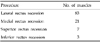
During the study, we recessed 83 lateral rectus muscles, 21 medial rectus muscles, 7 superior rectus muscles, and 3 inferior rectus muscles (Table 2). Of the muscles operated, we had adjusted 41 lateral rectus muscles, 13 medial rectus muscles, and 3 SR muscles.
In the patients with exotropia, 39 out of 48 patients (81.3%) achieved successful results at 1 week after surgery. Thirty four out of 48 patients (70.8%) obtained successful results at 6 months. Table 3 summarizes the postoperative results in exotropia. In the patients with esotropia, fifteen out of 16 patients (93.8%) achieved successful results at 1 week after surgery. Fourteen of 16 patients (87.5%) achieved a successful outcome at 6 months after surgery. Table 4 summarizes the postoperative results in esotropia. Overall, 48 out of 64 patients (75%) with horizontal strabismus achieved successful results at 6 months after surgery. In the patients with vertical strabismus, all of the seven patients (100%) achieved successful results at 1 week and at 1 month after surgery. Six out of 7 patients with a follow-up at 6 months (85.7%) achieved successful results after surgery. Table 5 summarizes the postoperative results in vertical strabismus. The overall success rate for both horizontal and vertical strabismus surgery at 6 months reached up to 76.1%. No operation was stopped in any of the patients, and no serious intraoperative or postoperative complications were noted. All patients who achieved unsuccessful results had undercorrection.
In all the procedures, the patients recorded no pain when an incision was made through the conjunctiva or Tenon's capsule. The most patients complained of discomfort when we hooked the muscle to isolate it and ended by muscle disinsertion. There was no pain on muscle reattachment or suture adjustment. Intraoperative cardiac monitoring did not record positive oculocardiac reflex in any of our patients.
DISCUSSION
The advantage of adjustable surgery, such as decreased frequency of additional surgery and increased accuracy of strabismus correction, has caused a steady increase in its use.2,5 The postoperative adjustment of the rectus muscle is well described and widely used, while an intraoperative adjustment under local anesthesia is less commonly practiced.6 Intraoperative one-stage adjustment has been limited to selected patients because it is a time-consuming procedure, which requires a skilled and experienced surgeon, as well as a cooperative patient.7 However, in a two-stage adjustment procedure, there is usually time lapse between the end of surgery and commencing adjustment usually performed some hours later. Some surgeons have performed adjustable surgery using retrobulbar anesthesia rather than general anesthesia, but the patient and surgeon must wait until the anesthetic effect has faded out, which prolongs the hospital stay.2 Moreover, complications related to retrobulbar anesthesia such as retrobulbar hemorrhage, eyeball perforation, or optic nerve damage can be encountered.8 The disadvantage associated with retrobulbar anesthesia also makes the use of adjustable surgery under topical anesthesia more attractive.
This study showed that one-stage adjustable suture surgery under topical anesthesia was well tolerated with satisfactory results. The overall success rates of adjustable surgery in this study were 85.9% at 1 week, 83% at 1 month, 78.9% at 3 months, and 76.1% at 6 months after surgery. These results are comparable to previous studies on two-stage adjustment under general or retrobulbar anesthesia.9,10 Wygnanski-Jaffe et al.10 reported the surgical success rate of 80% in patients with horizontal strabismus using two-stage adjustment. Our result in vertical strabismus is also comparable to a previous study by Rauz and Govan.6 They performed one stage vertical rectus muscle recession using adjustable sutures under local anesthesia (topical anesthesia supplemented with a subconjunctival injection of local anesthetic over the muscle insertions) on eight patients with vertical diplopia, and six patients became asymptomatic after surgery.
In this study, the success rate appeared to decrease slightly as the follow-up period was extended. This can be explained as postoperative drift, which was reported by Weston et al.2 that this phenomenon can occur within 8 weeks after surgery. This phenomenon was also noted in our study as the survival curve. Postoperative drift occurred more significantly in patients with exotropia compared with other patients, and that was the reason why the surgical success rate of exotropia at 6 months was lower than other types of strabismus. Therefore, the postoperative drift should not be neglected, and be observed more precisely especially for the patients with exotropia. For a more precise alignment, a slight over-correction can be one of the answers to the postoperative drift. However, because most of our patients were adolescents or adults, we tried to adjust not to have unbearable diplopia.
In this study, the use of topical anesthesia can avoid the complications associated with general or retrobulbar anesthesia. In addition, no intraoperative or postoperative complications were encountered during topical anesthesia such as oculocardiac reflex, nausea, or vomiting, which make the use of topical anesthesia more favorable and convenient. Some patients complained of pain during the procedure. This was relieved by the additional instillation of topical anesthesia, and the patients were well tolerated the pain during and after surgery.
In conclusion, intraoperative adjustment strabismus surgery is a simple, tolerable and effective procedure, and is recommended for all cooperative patients, especially when postoperative results would otherwise be unpredictable.


 XML Download