

 PDF
PDF ePub
ePub Citation
 Print
Print


INTRODUCTION
Soybeans are a commonly consumed food product in Korea, where they are also one of the most prevalent food allergens. The reported sensitization rate to soybeans ranges from 5.6-21%1 in pediatric allergy patients with atopic dermatitis and acute urticaria, compared with 1.8%2 in the general Korean population. There is concern about whether genetic manipulation of crops might augment the potential allergenicity of these foods in humans. The FAO/WHO recommends a stepwise assessment of genetically modified (GM) foods using both in vivo and in vitro methods before launching these products in commercial markets.3 Soybean has been the most widely cultivated GM crop since 1997, and the major gene that has been introduced into soybeans is an herbicide resistance gene called EPSPS (5-enolpyruvylshikimate-3-phosphate synthase). This modified type of soybean has been in use for more than 10 years in Korea, but it remains unknown whether GM soybeans are more allergenic than their non-modified counterparts. A recent study4 demonstrated that GM corn did not increase the risk of allergic reactions in patients. Another study, using an in vitro test, reported that transgenic maize and soya seemed to be safe in terms of allergenic potential.5
In this study, we used skin prick testing and IgE ELISA assays to compare the allergenicity of GM and wild-type soybean extracts in a larger cohort of adult allergy patients. We also analyzed the antibody reactive components of the two extracts using 2D gel electrophoresis and IgE immunoblotting.
MATERIALS AND METHODS
Subjects
To evaluate the sensitization to GM and wild-type soybean extracts, 1,716 allergy patients (≥ 15 years old) and 40 healthy non-atopic subjects were enrolled by the Department of Allergy and Rheumatology, Ajou University School of Medicine, Suwon, Korea. The GM soybean extract was Roundup Ready® (Monsanto, St. Louis, MO, USA), into which the 5-enolpyruvylshikimate-3-phosphate synthase (EPSPS) gene had been incorporated. The wild-type soybean extract, called Daewon, was provided by Daewon Co., Suwon, South Korea. Between August 2002 and July 2003, 1,716 patients who had been admitted to Ajou University Hospital for treatment of various allergic problems were skin tested for reactions to GM and wild-type soybean extracts and to common inhaled allergens, including the common dust mite Dermatophagoides pteronyssinus. Skin prick tests were performed on the backs of patients, and responses to 50 common inhalant allergens and 30 food allergens were tested simultaneously. The results were read 15 minutes later. The size of the wheal produced by each allergen and by the histamine control was expressed in terms of maximum diameter and vertical height at the midpoint of the wheal. Skin reactivity was expressed as the ratio of the allergen-induced wheal size to the histamine-induced wheal size (A/H). A/H ratios between 0.1 and 1, with an erythema diameter < 21 mm were scored as 1+. A/H ratios between 0.1 and 1, with an erythema diameter > 21 mm, were scored as 2+. A/H ratios ranging from 1 to 2 were scored as 3+, and A/H ratios ranging from 2 to 3 were scored as 4+. All A/H ratios > 3 were scored as 5+. A positive response was defined as a score greater than 2+ on the skin prick test. This study was reviewed by the institutional review board at Ajou University Medical Center, Suwon, Korea.
Preparation of wild-type and GM soybean extracts
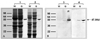
GM soybeans were extracted with phosphate-buffered saline (PBS; pH 7.5, 1:10 w/v), and the extract was incubated at 4℃ overnight and then centrifuged at 12,000-15,000 rpm for 20 minutes. The supernatant was then dialyzed (6,000 Da molecular weight cut-off; Spectrum Medical Industries, Houston, TX, USA) against 4 L of PBS for 72 hours at 4℃ , and the resulting fluid was stored at -20℃ until tested. The protein content, as measured using the Bradford method, was 2.53 mg/mL. For skin prick tests, the protein was mixed with sterile glycerin in a ratio of 1:1. The protein contents of the wild type and GM soybean extracts were compared by sodium dodecyl polyacrylamide gel electrophoresis (SDS-PAGE) under two conditions: with or without β-mercaptoethanol treatment which showed comparable results. An antibody specific for the EPSPS antigen was detected only in the GM soybean extract (Fig. 1). EPSPS antigen and IgG anti-EPSPS antibody used in this study were derived from Monsanto Co., St. Louis, MO, USA.
IgE ELISA assays
The presence of serum IgE specific for the soybean extracts and the EPSPS protein was determined using ELISA, as described previously.6 Briefly, microtiter plates (Corning, NY, USA) were coated with 100 µL/well of soybean extract (GM or wild-type, 10 µg/well) and incubated overnight at 4℃ . Each well was washed three times with 0.05% phosphate-buffered saline-Tween (PBST), and the remaining binding sites were blocked by incubation with 10% fetal bovine serum (FBS)-PBS for 1 h at room temperature. Then 50 µL of patient or control sera were added to wells at 1:2 dilution and incubated for 2 hours at room temperature. The wells were then washed three times with PBST, and biotin-labeled goat anti-human IgE antibody (1:1000 v/v dilution; Vector Labs, Burlin game, CA, USA) was added to each well and incubated for 1 hour. The wells were then washed and incubated with 100 µL of streptavidin-peroxidase (1:1000 v/v; Sigma, St. Louis, MO, USA) for 30 minutes. The colorimetric reaction was developed with TMB (3, 3',5, 5'-tetraethylbenzidine) substrate solution for 15 minutes at room temperature. The reaction was stopped by adding 100 µL of 2 N sulfuric acid and the absorbance was read at 450 nm using an automated microplate reader (Benchmark, Bio-Rad, Hercules, CA, USA). All assays were performed in duplicate. Positive values were set at three standard deviation units above the absorbance values of the controls.
ELISA inhibition test
Competitive ELISAs were performed to determine the specificity of IgE binding to the soybean extracts and to compare the allergenicity of the wild-type extracts, GM extracts, and EPSPS. The sera from four patients with high levels of soybean-specific IgE were pooled and then pre-incubated overnight (4℃) with increasing protein concentrations (1 to 100 µg) of EPSPS, house dust mite, GM soybean extract, or wild-type soybean extract. The sera were then incubated for 12 hours in microtiter plates pre-coated with wild-type or GM soybean extracts. ELISAs were then developed as described above. As a control, samples were pre-incubated with PBS (pH 7.5) instead of EPSPS or soybean extracts. The percentage of inhibition was expressed as (100-[absorbance of the samples pre-incubated with allergens/absorbance of the samples pre-incubated with PBS] × 100).
SDS-PAGE and immunoblot analysis
Four to twenty percent gradient SDS-PAGE and immunoblot analysis were performed under reducing conditions according to methods described previously.6 Briefly, the soybean extracts (24 µg each) were mixed with sample buffer (31 mmol/L Tris-HCl, 10% glycerol, 1% SDS, 0.0025% bromophenol blue, 2.5% β-mercaptoethanol, pH 6.8) and heated in boiling water for 5 minutes. The extracts were then loaded alongside a standard marker (4 to 250 kDa; Novex, San Diego, CA, USA) onto a 4-20% Tris-glycine gel (Novex, San Diego, CA, USA) and run at 125 V for 90 minutes. The gel was fixed and stained with Coomassie brilliant blue. For immunoblotting, the proteins were transferred onto a polyvinylidene fluoride membrane (PVDF; pore size: 0.2 µm; Millipore, Bedford, MA, USA) in transfer buffer (25 mmol/L Tris base, 193 mmol/L glycine, 20% methanol) at 200 mA for 90 minutes. The blotted PVDF membrane was cut into 4-mm widths and the pieces of the membrane were incubated in 5% skim milk in Tris-buffered saline (TBS)-Tween (TBST) for 1 hour to block nonspecific binding. The membranes were then incubated overnight with either patient or control sera diluted 1:2 v/v with 3% skim milk TBST. After a washing step, the membranes were incubated with goat anti-human IgE conjugated with alkaline phosphatase (Sigma, St. Louis, MO, USA, diluted 1:1000 in 3% skim milk TBST). The membranes were developed using BCIP/NBT alkaline phosphatase substrate (Sigma, St. Louis, MO, USA).
2D gel electrophoresis
Soybean extracts (40 µg) were mixed with rehydration buffer (7 mol/L urea, 2 mol/L thiourea, 2% CHAPS, 20 mmol/L DTT, 0.5% IPG buffer, pI 4-7) and applied to an Immobiline(tm) DryStrip (pI 4-7, Amersham). The gel was incubated at room temperature overnight and then run at 50 V for 30 minutes, 500 V for 30 minutes, 1,000 V for 30 minutes, and 5000 V for 90 minutes. The gel was then incubated in equilibration buffer (1.5 mol/L Tris, 2% SDS, 6 mol/L urea, 30% glycerol) for 20 minutes at room temperature, after which the samples were run on a 12% SDS-PAGE with a 4% stacking gel using the aforementioned method. 2D gel electrophoresis and transfer to PVDF membrane were performed in transfer buffer at 200 mA for 90 min. The membrane was then blocked by incubation in 5% skim milk-TBST for 1 hour and then overnight at 4℃ in pooled patient sera that had been diluted 1:2 with TBST. After a wash step, the blots were incubated with goat anti-human IgE antibody conjugated with alkaline phosphatase (diluted 1:1000 in 3% skim milk-TBST). The membrane was then developed using BCIP/NBT alkaline phosphatase substrate.
N-Terminal amino acid sequencing analysis
To confirm the major allergenic components via N-terminal sequencing, the 2D gel electrophoresed proteins were blotted onto a PVDF membrane. The membrane was stained with 0.1% Coomassie blue in 50% methanol, de-stained in 50% methanol and air-dried. The protein spots were excised and micro-sequencing was performed using a Procise 476 A protein sequencer (Applied Biosystems, Foster, CA, USA).
RESULTS
Allergy skin prick test
The skin prick testing conducted throughout the full year of the study revealed that 65 (3.8%) of the 1,716 allergy patients had an A/H score of 2+ in response to the wild-type soybean protein. Similarly, 66 patients (3.8%) had an A/H score of 2+ in response to the GM soybean protein. Only one of the allergy patients failed to respond to both the wild type and GM soybean extracts (Table 1) and none of the subjects had a positive response to EPSPS protein.
Specific IgE to soybean and EPSPS
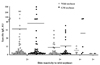
In the subjects who had a positive skin test with an A/H score of 2+, the prevalence of serum IgE specific for wild-type and GM soybean extracts was 80 and 81%, respectively. Of those with an A/H score of 3+, 4+, or 5+, the prevalence of serum IgE specific for wild type and GM soybean proteins was 74 and 66%, 71 and 83%, and 50 and 100%, respectively. The levels of IgE specific for the GM soybean extracts tended to be slightly higher than those specific for the wild-type extracts, and the GM extracts elicited a larger wheal reaction in the skin prick tests (Fig. 2). No subjects had detectable serum IgE specific for EPSPS protein.
ELISA inhibition test
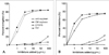
ELISA inhibition assays using pooled IgE-positive sera showed comparable dose-dependent inhibition by both the wild type and GM soybean extracts (Fig. 3). The GM soybean extract showed somewhat greater inhibition than the wild-type extracts. Minimal inhibition was observed when the sera were pre-incubated with an extract from the dust mite allergen D. pteronyssinus, as shown in Fig. 3A.
SDS-PAGE and IgE- immunoblot analysis
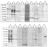
Fig. 4A compares IgE specific for wild type versus GM soybean extracts in the sera of 14 individuals. The control serum was pooled from ten patients who had negative responses in the skin prick test. In the wild-type extracts, 22 protein bands, which ranged in size from 8 to 119 kDa, were detected using the serum IgE. A 32~34-kDa band was the most frequently detected, being observed in ≥ 50% of the sera tested. In the GM soybean extracts, 20 bands, which also ranged in size from 8 to 119 kDa, were detected using the serum antibodies. Similar to the wild-type extracts, a 33-kDa band was the most prevalent, detected in ≥ 50% of the sera tested (Fig. 4B). Therefore, the 33-kDa protein was considered to be the major allergenic protein present in both wild type and GM soybean extracts. No bands were detected when sera were tested against EPSPS protein.
2D gel electrophoresis, IgE immunoblotting, and N-terminal amino acid sequencing
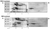
2D gel electrophoresis and IgE immunoblot analysis of pooled sera from patients with high levels of soybean-specific IgE showed that the major 33-kDa allergenic protein had a pI of 4.83, and verified that serum IgE recognized this protein (Fig. 5).
N-terminal amino acid sequencing of the 33-kDa protein revealed the following sequence: Lys-Lys-Met-Lys-Lys-Glu-Gln-Tyr-Ser-Cys-Asp-His-Pro-Pro-Ala.
DISCUSSION
This study demonstrates that among allergic adults in the Korean population, the prevalence of positive responses to wild type and GM soybean extracts is equivalent. With the exception of one individual, all the subjects tested had a positive response to both extracts. Importantly, no subjects responded to GM soybean extracts but not to wild-type extracts, and no subjects responded to the EPSPS protein alone, suggesting that the protein product of the genetically introduced gene was not itself allergenic. The prevalence of serum IgE specific for soybean proteins, as determined by ELISA, increased in parallel with increasing skin reactivity. When the results from ELISA assays and skin tests were compared, soybean-specific IgE antibodies were found in 50-100% of the patients with A/H scores of 2+, with comparable antibody titers specific for wild type and GM extracts. Furthermore, no subjects had detectable EPSPS-specific IgE responses as tested by ELISA.
According to the Codex Committee on Food Labeling, the most common allergenic foods worldwide that are associated with IgE-mediated reactions include peanuts, soybeans, milk, eggs, fish, crustaceans, wheat, and tree nuts.7 As a result, soybeans are considered one of the causative agents of food allergies in Korean adults. However, the results of our study suggest that the genetic manipulation of soybeans does not increase the allergenicity of soybeans, as allergic individuals were found to be equally reactive to wild type and GM soybean extracts.
When the allergenicity of the wild type and GM soybeans was compared using IgE ELISA inhibition tests, the GM extract tended to give higher inhibition percentages than the wild-type extract. IgE immunoblot analysis using sera from five patients (2, 8, 9, 10, and 13) showed strong reactivity to a 33-kDa band from the GM soybean extract. Similarly, a 32~34-kDa band from the wild type extract was detected in the same five patients. 2D gel electrophoresis and IgE blotting demonstrated that these two bands were identical. There were no significant differences in soybean-specific IgE titers between patients that had antibody responses specific for the 33-kDa major allergen and those that did not. No significant changes in IgE binding patterns to another protein (47.5 kDa) were noted, suggesting that the introduction of the gene encoding the EPSPS protein did not affect the allergenicity of the soybean extract, either qualitatively or quantitatively. Consistent with this conclusion, none of the patients had detectable EPSPS-specific IgE antibodies in their serum. These findings suggest that the allergenic risk of GM soybeans is identical to that of wild-type soybeans.
The gapped BLAST and PSI-BLAST programs were used to search for proteins with an amino acid sequence similar to the 33-kDa GM soybean antigen.8 The search revealed that the 33-kDa protein is identical to the P34 protein, which was previously identified as one of the major soybean allergens and is a member of the papain family of cysteine proteases.9 Food allergens commonly exhibit sufficient gastric stability to reach the intestinal mucosa, where absorption and sensitization can occur. Therefore, important food allergens may resist digestion in simulated gastric fluid. For example, soybean β-conglycinin is stable in digestive enzyme for 60 min.8 In conclusion, we evaluated the allergenicity of GM and wild-type soybeans using in vivo and in vitro methods and demonstrated that the introduction of the herbicide-resistance gene EPSPS does not increase allergenic risk in adult allergy patients. Additional studies will be needed to evaluate the allergenic risk of other GM foods in allergy patients before those foods are released to the consumer market.

 XML Download