

 PDF
PDF ePub
ePub Citation
 Print
Print


INTRODUCTION
Intraoperative decisions are often based on the interpretation of results from intraoperative transesophageal echocardiography (TEE). Residual mitral valve insufficiency at the completion of a valve repair is a well-known risk factor for late reoperation, and the use of TEE has decreased the incidence of immediate repair failure.1,2 However, the development of a new mitral valve insufficiency after the removal of the left ventricular vent is rare. This case emphasizes the importance of TEE as a continuous monitor during cardiac surgery. Usually, TEE is used to assess the valve function at the end of the surgery in the operating room, but an unexpected event can occur at any time after the inspection, as in this case, in which the mitral valve was damaged during the left ventricular vent removal.
CASE REPORT
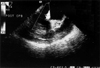
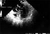
A 53-year-old female patient with mitral valve insufficiency underwent surgery for mitral valve repair. She had no history of medical illness until the day she was diagnosed with mitral valve insufficiency after an incidence of the syncope seven months previously. The dyspnea upon exertion grew worse, and by the time of the operation her symptoms of congestive heart failure were classified as New York Heart Association functional class III. Her transthoracic echocardiography (TTE) revealed a flail posterior mitral leaflet (PML) with an eccentric mitral valve insufficiency of grade IV/IV. The left ventricular ejection fraction was 89%. A preoperative TEE demonstrated the prolapse of the medial scallop (P3) of the PML with grade IV mitral valve insufficiency. In the operating field, a rupture of the posterior chordae was seen, and it was repaired with a prolene suture. The mitral valve annulus was repaired with a Duran ring 610R (#29 mm Medtronic, Minneapolis, MN, USA). Following the release of the aortic cross clamp, pump flow was reduced uneventfully to about 500 ml min-1, and hemodynamics were virtually unchanged due to adjustments of arterial pressure and intravascular volume. A TEE examination was performed to evaluate the result of the surgery. No residual mitral valve insufficiency was found, and the vent in the left atrium was advanced into the left ventricle to remove the air. After removal of the air, the left ventricular vent was removed, and a new mitral valve insufficiency was seen on the TEE (Fig. 1). The cardiopulmonary bypass (CPB) was reinstituted, and the surgeon found tearing of the lateral third part of the anterior mitral leaflet (A1 scallop), which had been normal during the first mitral valve repair. At the end of the second CPB, no residual mitral valve insufficiency was evident (Fig. 2). The patient was discharged without any further events.
DISCUSSION
This case is interesting because the development of a new mitral valve insufficiency after removal of the left ventricular vent is rare. An early reoperation was prevented with the use of TEE, which detected an unpredictable surgical complication. Usually, immediate failure of mitral valve repair is due to residual prolapse, residual annular dilatation, or suture dehiscence, which are procedure-related.1,3 TEE is a mainstay of cardiothoracic anesthesia for the surgical correction of cardiac valve defects.4,5 TEE allows identification of the different mitral valve segments and the precise localization of pathology, and helps to diagnose the mechanism of the mitral valve insufficiency. 6,7 The functional anatomy of the mitral valve insufficiency defined by TEE is strongly and independently predictive of valve reparability and postoperative outcome.8 In this case, the valve function was evaluated before CPB termination and the repair was successful, with no residual mitral valve insufficiency. Since the new mitral valve insufficiency developed after the removal of the left ventricular vent through the mitral valve, it was considered that the left ventricular vent had injured a part of the mitral valve during its removal. During the second CPB, a tearing of A1 was found and repaired with a prolene suture. There was no residual mitral valve insufficiency at the end of the second CPB. The patient was weaned and separated from the second CPB without difficulty. Any procedure related to cardiac manipulation needs TEE as an intraoperative monitor of cardiac function, which must be continued until the end of the surgery.9 This case emphasizes the importance of TEE in the operating room, not only to evaluate the result of the cardiac surgery, but also to detect any unpredictable events during the surgery.
 XML Download