

 PDF
PDF ePub
ePub Citation
 Print
Print


INTRODUCTION
Transepidermal elimination is a phenomenon which occurs spontaneously in certain skin disorders. The affected connective tissue or foreign material is expelled to the exterior via the epidermis. These findings are divided into elastosis perforans serpiginosa, reactive perforating collagenosis, Kyrle's disease, perforating folliculitis, and acquired perforating dermatosis. The perforation can appear as an incidental histologic finding, and can be associated with other cutaneous and systemic disorders.
Membranous lipodystrophy, an uncommon disorder, was described by Nasu et al.1 in 1973 as a form of cyst-like lesions of fat occurring in the long bones together with sudanophilic leukoencephalopathy. Histopathologically, at the border of the lobules with the septa, there were fat cysts lined by a thin homogenous eosinophilic layer of protein that had fine feathery projections into the fat cavity. These peculiar changes in fat tissue have been associated with many local and systemic diseases, including lupus erythematosus, diabetes mellitus, erythema nodosum, morphea, trauma, atypical mycobacterial infection, Behçet's disease, sclerosing panniculitis, Weber-Christian disease, multiple myeloma, vascular disorders, and insulin lipoatrophy. However, in some cases no underlying disease is found.2
CASE REPORT
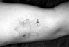
A 43-year-old woman presented with a red to purple indurated plaque approximately seven cm in diameter and located on the left arm, without associated symptoms. This plaque had multiple umbilicated papules with central keratin-filled plugs arranged in arcuate groups (Fig. 1, arrow). She was a chronic hepatitis B antigen carrier and had hypertension for four years. She had been taking antihypertensive drugs intermittently and had no history of diabetes mellitus, chronic renal failure, or trauma.
Liver function tests disclosed the following values: aspartate aminotransferase (AST), 74U/L (normal value: less than 38U/L) and alanine aminotransferase (ALT), 79U/L (normal value: less than 41U/L). However, CBC, BUN, creatinine, serum electrolytes, serum glucose level, antinuclear antibody, and urinalysis were negative or within normal ranges.
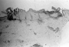
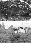
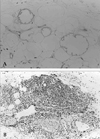
Biopsy specimens from the lesions were stained with hematoxylin-eosin, Masson trichrome, Verhoeff-van Gieson and Periodic Acid-Schiff. These specimens showed a central cup-shaped depression area of the epidermis that contained a solid mass of hyperkeratosis and dense parakeratotic keratinous material (Fig. 2). The epidermis at the base of the crater was ulcerated. Masson trichrome and Verhoeff-van Gieson stained sections showed an invagination that contained altered collagen and elastic fibers, as well as the fibers' transepidermal elimination (Fig. 3A, 3B). The borders of the fat lobules were lined by a thin eosinophilic layer of protein that had fine feathery projections in the fat cavity. The membranes of the microcysts stained positively with Periodic Acid-Schiff stain (Fig. 4A). There was a moderate infiltration of lymphohistiocytes in the dermis and panniculus. In addition to these histopathologic findings, the blood vessels in the dermis and subcutaneous fat layer showed infiltration of the vascular walls by lymphohistiocytes, swelling of the endothelial cells, an increase in vascular wall thickness, and narrowing of the lumen (Fig. 4B).
DISCUSSION
Transepidermal elimination is the mechanism of extrusion of altered dermal substances through epidermal channels.3 This process seems to be the final common pathway for most perforating dermatosis. Disorders chiefly characterized by perforation are divided into elastosis perforans serpiginosa, reactive perforating collagenosis, Kyrle's disease, perforating folliculitis, and acquired perforating dermatosis. The perforation can appear as an incidental histologic finding and can be associated with other cutaneous and systemic disorders, e.g., granuloma annulare, pseudoxanthoma elasticum and chondrodermatitis nodularis chronica helices.
The pathogenesis of acquired perforating dermatosis is thought to be related to pruritus, leading to traumatization by scratching or rubbing, and dermal microdeposit of uric acid or hydroxyl apatite, resulting in an inflammatory reaction, connective tissue degradation, and the release of mediators in patients with chronic renal failure. In our case, connective tissue degeneration found on the biopsied specimen might be a cause of perforating dermatosis.
Histopathologic findings from perforating dermatosis are a combination of features similar to that seen in perforating folliculitis, reactive perforating collagenosis, elastosis perforans serpiginosa, or Kyrle's disease. The combined perforation of both collagen and elastic tissue seen in our case is unusual, and not considered to be characteristic of any of the classic four perforating diseases. Rapini et al.4 reported four patients with renal disease and/or diabetes whose skin biopsy specimens demonstrated combined transepidermal elimination of both collagen and elastic fibers, as in the case of our patient.
Membranous lipodystrophy was initially described as a specific form of fat necrosis involving the bone marrow.1 Later, similar membranous lipodystrophy and lipodystrophy-like changes were observed in the subcutaneous tissue of patients with chronic arterial disturbances, suggesting that these changes, although not specific to a particular disease, were related to vascular impairment of the fatty tissue.5-8 They can be associated with vascular disease, e.g., arterial vascular insufficiency to the lower legs, varicose veins, thrombophlebitis, deep venous thrombosis and stasis dermatitis.
The underlying mechanism of membranous lipodystrophy is unknown, and numerous hypotheses have been proposed. Some of the hypotheses are as follows2: (1) idiopathic processes; (2) enormous proliferation of the fat cell membranes; (3) physiochemical interaction between the ground substance in connective tissue and fat droplets; (4) free fatty acids released from degenerated fat cells processed by macrophages to produce membranous lipodystrophy; (5) metabolic disorders of lipids in mesenchymal cells; (6) loops and fold in the basal laminae of fat cells in varying stages of lipid depletion at the time of necrosis; and (7) ischemic injury of adipose tissue resulting from venous insufficiency.
Hypertension is one of the diseases that induces vascular changes. Histopathologically, hypertension can decrease the diameter of the vascular lumen and increase the ratio of media thickness to luminal diameter in the arteries.9 Distention of the local capillary bed and reduction in the density of capillaries and arterioles can be induced by hypertension because of impaired angiogenesis or apoptosis of capillaries. These changes can impair the perfusion of capillaries.10 Ischemic injury of adipose tissue from venous insufficiency is thought to be the most important factor of membranous lipodystrophy. Machinami6 observed membranous lipodystrophy in 53 patients with chronic circulating disturbances. Alegrae et al.11 observed lipomembranous changes in 13 patients and found that, histopathologically, there was evidence of endarteritis obliterans, hemorrhage, dilated veins, and capillary proliferation in more than half of them. One of the 13 patients had hypertension without any other disease, as did our patient. In summary, we consider that in our case, hypertension gave rise to vascular changes which resulted in ischemic injury, inducing membranous lipodystrophy and connective tissues degeneration, and was extruded by transepidermal elimination.
 XML Download