Abstract
In this paper, we evaluate the radiological features of pregnancy-associated breast lesions and discuss the difficulties in diagnosis by imaging. We selected patients who were diagnosed with pregnancy-associated breast lesions during the previous 5 years. All patients complained of palpable lesions in the breast and underwent ultrasonographic (US) examination, the first choice for examination of pregnancy-related breast lesions. Any suspicious lesions found by the US were recommended for a US-guided core biopsy, US-guided fine needle aspiration (FNA), or surgery. Various breast lesions were detected during pregnancy and lactation, including breast cancer, mastitis and abscesses, lactating adenoma, galactoceles, lobular hyperplasia, and fibroadenomas. The imaging features of pregnancy-associated breast lesions did not differ from the features of non-pregnancy-associated breast lesions; however, some pregnancy-associated benign lesions had suspicious sonographic features. A US-guided core biopsy was necessary for differentiating benign from malignant. In patients with breast cancer, the cancer was often advanced at the time of diagnosis. In conclusion, various pregnancy-related breast lesions were detected and the imaging of these lesions had variable findings. Breast ultrasound could be an excellent imaging modality for diagnosis and differentiation between benign and malignant lesions. However, when the imaging results are suspicious, a biopsy should be performed to obtain a pathologic diagnosis.
Breast disease is pregnancy-associated if the diagnosis is made either during pregnancy or within 1 year following pregnancy.1 During pregnancy and lactation, striking changes take place in the mammary glands. The placenta causes hormone elevation by secreting estrogen and progesterone, the major hormones responsible for full breast development.2 The ductal-lobular-alveolar system undergoes considerable hypertrophy, and prominent lobules are formed.
The safety of mammography during pregnancy or lactation is controversial. One argument is that mammography is of limited benefit due to its reduced sensitivity in the hormonally altered breasts of pregnant and lactating women. Patient sonography could be more valuable during this evaluation period for breast masses because of its safety and ability to detect most of all masses. Radiologists should be aware of the typical imaging results for breasts during normal pregnancy and lactation, including pregnancy-associated breast lesions such as galactoceles,3-6 lactating adenoma7,8 and breast cancer.9-12 The ultimate clinical goal is to differentiate between benign lesions and breast carcinoma during pregnancy. Therefore, the purpose of this study is to evaluate the radiological features of pregnancy-associated breast lesions and analyze the difficulties in diagnosing them.
From 1998 to 2002, we retrospectively reviewed the imaging findings of patients who presented with palpable breast abnormalities during pregnancy and a post-partum period of 1 year. Forty-nine patients were evaluated with breast ultrasound (US) examination. The age of patients ranged from 23 to 37 years (mean 31.4 years). All ultrasound examinations used high resolution sonography units with 10-12 MHz linear transducers (HDI 5000, Advanced Technology Laboratories, Bothell, WA). The sonographic images were assessed for the presence of solid masses, and if masses were present their shapes, margins, orientations, echo patterns, posterior acoustic features, and surrounding tissue effects were recorded according to the American College of Radiology (ACR) Breast Imaging Reporting and Data System (BI-RADS) ultrasound lexicon.13 Eight patients underwent mammography (Senograph DMR, GE, Milwaukee, Wisconsin, USA). The shape, mass, and morphology margins, as well as the distribution of calcifications, were evaluated according to the BI-RADS criteria. Histopathological results were obtained in 35 patients: 22 underwent US-guided automated core biopsies, 10 received US-guided FNA, and 3 patients were treated with incision and drainage. Biopsies or FNAs were recommended either by sonographic findings or the patient's desire. US-guided automated core biopsies were performed using a 14-gauge needle with an automated gun (Bard-Magnum Biopsy Instruments, Covington, GA, USA). Three to five needle passes were given. US-guided FNAs were performed using ultrasound guidance with two needle passes of a 21- or 23-gauge needle. US-guided FNAs were also used for pus drainage. An informed consent was obtained from every patient. The radiologist explained to lactating patients about risk of milk fistula. Six of the patients underwent surgery for breast cancer. The histological, nuclear grading, and pathological staging were then analyzed after surgery. The imaging-histologic correlations were evaluated and we excluded the fourteen patients who were followed by ultrasound for over a 1-year period.
Various breast lesions were detected during pregnancy and lactation. The pathological results of these lesions included ductal epithelium and lobule hyperplasia (n = 4), galactoceles (n = 11), mastitis and abscesses (n = 9), fibroadenomas (n = 2), lactating adenoma (n = 3), and breast cancer (n = 6). Sonographic findings according to ACR BI-RADS as well as the final pathology are listed in Table 1. No malignant lesions were found in BI-RADS category 2 or 3 lesions. Six cases from the BI-RADS category 4 lesions (n = 12) had confirmed malignancy. Breast ultrasonograms taken during pregnancy or lactation were characterized by diffuse, inhomogeneous hypoechogenicity due to lobular hyperplasia and duct dilatation. However, focal, low echoic areas should be differentiated from real mass lesions (Fig. 1).
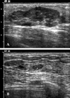
The galactoceles had additional and varying radiologic presentations, i.e. homogeneously anechoic, oval-shaped masses with posterior acoustic enhancements that suggest simple cysts (Fig. 2A), lobulated, fat-fluid level masses (Fig. 2B), or masses with an internally heterogeneous echo (Fig. 3 A, B). Any of these variations suggested a suspicious lesion that could be confused with other malignancies (Fig. 4).
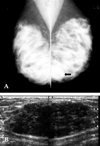
In mammography, lactating adenomas presented as relatively well-circumscribed and partly obscured masses (Fig. 5A) or as asymmetrically increased densities.
Most of these detected tumors were benign and had well-circumscribed margins (Fig. 5A, B). Some lesions, however, had malignant characteristics such as: irregular, angulated, or ill-defined margins. These characteristics gave reason to suspect malignancy (Fig. 6). One of our cases showed regression after the termination of breast-feeding (Fig. 7).
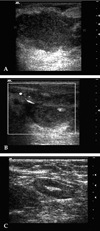
Various sonographic findings were also observed in the mastitis or breast abscesses. Two out of nine cases had suspicious findings and were pathologically confirmed by US-guided core biopsy. US examinations are of great assessment value for clinically suspected mastitis or breast abscesses in acutely inflamed breasts, and therefore prevent unnecessary surgical intervention (Fig. 8A, B, C).
In breast cancer patients, the sonographic findings showed masses with: irregular shape (n = 6), irregular margins (n = 6), non-parallel orientation (n = 5), complex echo patterns (n = 5), or relatively ill-defined margins with lack of spiculation (n = 5). Associated findings were noted in 3 patients, including Cooper's ligament thickening (n = 2), edema (n = 2), skin thickening (n = 1), and axillary lymphadenopathy (n = 3). The mammographic findings included masses (n = 4), masses with microcalcifications (n = 2), masses with axillary lymphadenopathy (n = 3), asymmetric density only (n = 1), and extremely dense breasts with negative findings (n = 2). The advanced stage of PABC typically seen at the time of diagnosis reflects the difficulty in evaluating breasts during pregnancy (Fig. 9). Table 2 lists the imaging findings and histopathological results from the breast cancer patients. In the patients with cancer, the cancer was often advanced at the time of diagnosis.
The imaging features of pregnancy-associated breast lesions did not differ from those of non-pregnancy-associated breast lesions; however, some pregnancy-associated benign lesions had suspicious sonographic features (6/29, 20.68%) and US-guided core biopsies were necessary for differentiating benign from malignant.
During pregnancy, the breast lobules multiply and enlarge in preparation for lactation. Fat droplets begin to accumulate in the epithelial cytoplasm. The intralobular stromal elements diminish, are crowded out, and are replaced by the enlarging lobules. The breast density can also dramatically increase with lactation. This increase in heterogeneous densities likely reduces the sensitivity of mammography for detecting breast cancers.2 The lactating breast presents special problems for breast imaging due to hypertrophic changes. Screening should be avoided while a woman is lactating. If imaging is needed, breast ultrasonography (US) may be helpful in differentiating between solid or cystic masses and in evaluating other lesion characteristics. The breast ultrasonogram taken during pregnancy or lactation is characterized by diffuse, inhomogeneous hypoechogenicity.
Galactoceles are the most common benign breast lesions in pregnant and lactating women. They are true cubiodal epithelium-lined cysts that contain milk-like fluid, either with or without curd-like material.3 The main predisposing factor for galactoceles development is thought to be mammary duct obstruction in the lactating breast, most likely due to inflammation or in rare cases, a tumor.4
The typical imaging feature of the galactocele is a cystic nodule with a fluid-fluid level caused by fat and water. According to Park et al.,5 most pregnancy-related galactoceles demonstrate the sonographic features of cysts, whereas pregnancy-unrelated galactoceles demonstrate the same variable sonographic findings as solid masses. In our study, however, few cases showed solid mass-like features and increased blood flow even during lactation periods (Fig. 4). Galactoceles should be differentiated from other cystic breast lesions, most importantly intracystic carcinoma.4 An ultrasound-guided biopsy should be performed if the lesion does not have any imaging typical of a galactocele.
A lactating adenoma is a benign breast lesion which occurs in response to the physiologic changes of pregnancy and lactation. The etiology of lactating adenoma is a source of controversy. Some suggest that the lesion is simply a variant of fibroadenoma, tubular adenoma, or lobular hyperplasia. Others postulate that lactating adenomas are unique neoplasms that arise de novo in a hormonally stimulated breast.9,14 Despite such etiological ambiguity, it is generally accepted that lactating adenomas are tumors that are significantly affected by the rising estrogen levels of pregnancy.14 Histopathologically, lactating adenomas are seen as well-circumscribed lesions composed of secretory lobules separated by delicate connective tissue. The cells that line lobules have granular, foamy, or vacuolated cytoplasms.6
US evaluation is commonly the initial diagnostic imaging technique during pregnancy and lactation. The sonographic features of lactating adenomas can be variable. In our study, the sonographic findings of lactating adenomas ranged from well-circumscribed, benign looking lesions to masses with malignant features, whicih included irregular, angulated margins. The natural course of lactating adenomas is to regress after the completion of breast-feeding. Bromocriptine has been reported to reduce lactating adenoma size through the suppression of prolactin secretion.
Breast abscesses develop in 5 - 11% of lactating women with infectious mastitis. The predominant infectious organism is Staphylococcus aureus, and often is the penicillinase-producing type.15 Peripheral breast abscesses have generally been associated with mastitis during breast feeding, but previous reports indicate that abscesses are common among non-lactating women.16
Until the last decade, the recommended treatment for breast abscesses was surgical incision and drainage while the patient was under general anesthesia. Hook et al.,16 however, recently reported that abscesses smaller than 2.5 cm could be treated without recurrence by aspiration and antibiotic therapy if the abscess cavity was completely or almost completely drained. In contrast, abscesses larger than 3 cm required surgery or other decompression methods for definitive treatment. Recurrent abscesses are another serious problem, but the reasons for recurrences have not been well documented.9
Variable radiological findings associated with mastitis or breast abscesses have been reported, including unilocular or multilocular abscesses or heterogeneous high to low echoic mass-like lesions with increased blood flow. In our study, we found no remarkable difference between pregnancy- related and pregnancy-unrelated breast abscesses. US evaluations are of great value in assessing clinically suspect breast abscesses and in guiding drainage or aspiration.
One to two percent of all breast cancers occur during pregnancy or lactation. The discussion of pregnancy-associated breast cancer (PABC) has become increasingly common over the past several years, perhaps because more women are becoming pregnant in their 30s and 40s, and the incidence of breast cancer increases with age.10 Many studies have reported a poor prognosis for patients with PABC. More recently, several investigators found no statistically significant difference in the mortality rates of patients with pregnancy-associated versus non-pregnancy-associated breast cancer.17
The advanced stage of PABC typically seen at the time of diagnosis reflects the difficulty in evaluating breasts during pregnancy (Fig. 9). Physicians should not ignore locally growing breast lesions during pregnancy and simply assume that they are benign. Clinicians might consider a sonographic evaluation as the first choice in exam techniques. If any suspicious lesions in the US images are found, further diagnostic procedures should be performed. Although mammography is seldom used during pregnancy due to concerns associated with fetal irradiation, modern mammography is unlikely to have ill effects on the fetus.18
Liberman et al.18 found that the sensitivity of mammography in detecting PABC was only 78%. Such a reduced sensitivity was attributed to the increased glandularity and additional water content in the breasts during pregnancy. Ahn et al.11 reported that during pregnancy the sensitivity of mammography was 86.7% and the sensitivity of US was 100%.
The radiological findings of PABC in mammography and US are not significantly different from the findings of non-PABC. However, some reports suggest that the sonographic findings of frequent posterior acoustic enhancement and masses with remarkable cystic components may appear to be different than breast cancer in non-pregnant women.12 In our series, however, a cystic component was not detected. Therefore, more cases are needed for the evaluation of differences between PABC and non-PABC.
Breast cancer diagnosed during pregnancy should be treated according to the same principles that are applied to non-pregnant patients. Subsequent pregnancies following breast cancer diagnosis do not have a known detrimental effect on survival.12
In conclusion, the majority of breast lesions encountered during pregnancy are benign; however, about 20% of these breast masses prove to be malignant. Physiological changes during pregnancy make evaluation of the breasts more difficult. Baseline and serial examinations are critical. Even though normal breast parenchyma can be palpable during this period, clinicians and radiologists should not ignore abnormalities. A significantly higher percentage of women diagnosed with breast cancer during pregnancy are node positive. Prompt and immediate biopsies of breast masses are important during pregnancy, and benign-appearing or low-suspicion lesions should not be neglected.
Figures and Tables
 | Fig. 1Ultrasound of a 28-year-old lactating woman (postpartum 6 months) without breast disease. The images show an ill-defined, low echoic lesion, suggesting duct dilatation (white arrows) due to pregnancy. |
 | Fig. 2Ultrasound of a 35-year-old lactating woman (postpartum 6 months) with galactoceles. (A) There are two anechoic, cyst-like masses. The larger mass shows posterior acoustic enhancement with lateral edge shadowing, suggesting a pure cystic galactocele by clinical history. (B) Another cystic mass shows a fat-fluid level (arrows) representing a galactocele. |
 | Fig. 3Images from a 35-year-old lactating woman (postpartum 4 months) with a galactocele. (A) US of the breast shows a lobulated and heterogeneous, echoic mass in the palpable area. (B) The Doppler study shows increased blood flow in the mass. This lesion was confirmed to be a galactocele by US-guided core biopsy. |
 | Fig. 4US of a 38-year-old lactating woman (postpartum 6 months) with a galactocele. US of the breast reveal an irregular-shaped, hypoechoic mass in the left subareolar area (white arrows). Part of the mass shows spiculation. Under the diagnosis of category 4A, a core biopsy was performed, and the pathology result was fibrocystic changes, compatible with a galactocele. |
 | Fig. 5Images of a 30-year-old woman (postpartum 5 months) with a lactating adenoma. (A) Both oblique mammograms show bilateral, extremely dense breast patterns with a well-circumscribed, high-density mass in the low-posterior portion of the left breast (arrow). (B) US reveals a large, relatively well-defined, oval-shaped, inhomogeneous, hypoechoic mass. |
 | Fig. 6US of a 29-year-old woman (postpartum 4 months) with a lactating adenoma. US shows a 2-cm, ill-defined, hypoechoic lesion which was categorized as a suspicious lesion (category 4A) and followed-up with a US-guided core biopsy. The histopathological diagnosis was a lactating adenoma. |
 | Fig. 7Images from a 28-year-old pregnant woman (IUP 36 weeks) with a lactating adenoma. (A) US shows an approximately 4.5 × 1.5-cm, lobular-shaped, well-circumscribed, inhomogeneous, hypoechoic mass in the left breast. A US-guided biopsy was performed which confirmed the lesion was a lactating adenoma. (B) The follow-up sonography was taken 6 months later, after delivery. The lactating adenoma had decreased in size to less than 1 cm. |
 | Fig. 8Images from a 29-year-old woman (postpartum 2 weeks) with breast abscess. (A) US of the left breast shows an irregular-shaped, heterogeneous, hypoechoic mass-like lesion with indistinct margins and partial posterior enhancement at the subareolar portion. Internal moving debris was also noted, and approximately 20 cc of pus was drained through an 18-G needle. (B) On Doppler, marked increased blood flow is seen at the margin of the abscess. (C) A cortical thickened, enlarged lymph node is noted in the left axilla. |
 | Fig. 9Images from a 33-year-old woman (postpartum 3 weeks) with advanced breast carcinoma. The patient noticed painful swelling of the right breast during the 20th week of pregnancy, but felt that these were normal physiological changes. (A) The right breast shows a discolored and contracted appearance with nipple retraction, compared with the left breast. (B) Mammography shows a huge mass in the entire right breast (arrows), with diffuse trabecular thickening by lymphatic engorgement and multiple metastatic lymph nodes in the axilla (arrow heads). (C) US shows an irregular-shaped, inhomogeneous, echoic mass (white arrows) with diffuse skin thickening (black arrows). |
Table 1
The Number of Pathologically Confirmed Lesions and Their Breakdown According to US BI-RADS Category

Table 2
The Imaging and Pathological Results of Pregnancy-associated Breast Cancer Cases

*Numbers presented are the BI-RADS category for each imaging finding.
†Nuclear and histologic gradings were reported according to the Bloom & Richardson classification.
IUP, intrauterine period; Sx Du, symptom duration; US, ultrasonography; XM, mammography; Path, pathologic result; Ax LN, axillary lymphnode involvement; NG, nuclear grade; HG; histologic grade; IDCa, invasive ductal carcinoma.
References
1. Smith MS. Patton HD, Fuchs AF, Mille B, Scher AM, Steiner R, editors. Lactation. Textbook of physiology. 1989. 21st ed. Philadelphia: Saunders;1408–1421.
2. Kopans DB. Breast imaging. 1998. 2th ed. Philadelphia: Lippincott-Raven;445–496.
3. Golden GT, Wangensteen SL. Galactocele of the breast. Am J Surg. 1972. 123:271–273.
4. Sawhney S, Petkovska L, Ramadan S, Al-Muhtaseb S, Jain R, Sheikh M. Sonographic appearances of galactoceles. J Clin Ultrasound. 2002. 30:18–22.
5. Park MS, Oh KK, Kim EK, Lee SI. Multifaces of sonographic findings of galactocele: comparison according to its association with pregnancy. J Korean Radiol Soc. 2000. 42:699–703.
6. Slavin JL, Billson VR, Ostor AG. Nodular breast lesions during pregnancy and lactation. Histopathology. 1993. 22:481–485.
7. Sumkin JH, Perrone AM, Harris KM, Nath ME, Amortegui AJ, Weinstein BJ. Lactating adenoma: US features and literature review. Radiology. 1998. 206:271–274.
8. Darling ML, Smith DN, Rhei E, Denison CM, Lester SC, Meyer JE. Lactating adenoma: sonographic features. Breast J. 2000. 6:252–256.
9. Kaufmann R, Foxman B. Mastitis among lactating women: occurrence and risk factors. Soc Sci Med. 1991. 33:701–705.
10. Petrek JA. Breast cancer during pregnancy. Cancer. 1994. 74:518–527.
11. Ahn BY, Kim HH, Moon WK, Pisano ED, Kim HS, Cha ES, et al. Pregnancy-and lactation-associated breast cancer: mammographic and sonographic findings. J Ultrasound Med. 2003. 22:491–499.
12. Gallenberg MM, Loprinzi CL. Breast cancer and pregnancy. Semin Oncol. 1989. 16:369–376.
13. Mendelson EB, Berg WA, Merritt CRB. Miller WT, Berg WA, editors. Toward a standardized breast ultrasound lexicon, BI-RADS: ultrasound. Seminars in roentgenology: Breast imaging. 2001. Vol 36. Philadelphia, PA: WB Saunders Co;217–225.
14. James K, Bridger J, Anthony PP. Breast tumour of pregnancy ('lactating adenoma'). J Pathol. 1988. 156:37–44.
15. Devereux WP. Acute puerperal mastitis. Evaluation of its management. Am J Obstet Gynecol. 1970. 108:78–81.
16. Hook GW, Ikeda DM. Treatment of breast abscesses with US-guided percutaneous needle drainage without indwelling catheter placement. Radiology. 1999. 213:579–582.
17. DiFronzo LA, O'Connell TX. Breast cancer in pregnancy and lactation. Surg Clin North Am. 1996. 76:267–278.
18. Liberman L, Giess CS, Dershaw DD, Deutch BM, Petrek JA. Imaging of pregnancy-associated breast cancer. Radiology. 1994. 191:245–248.


 PDF
PDF ePub
ePub Citation
 Print
Print


 XML Download